| Site code
|
Context
|
Frame number
|
Photo
|
Description
|
| BA84
|
2640
|
1
|
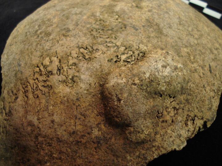
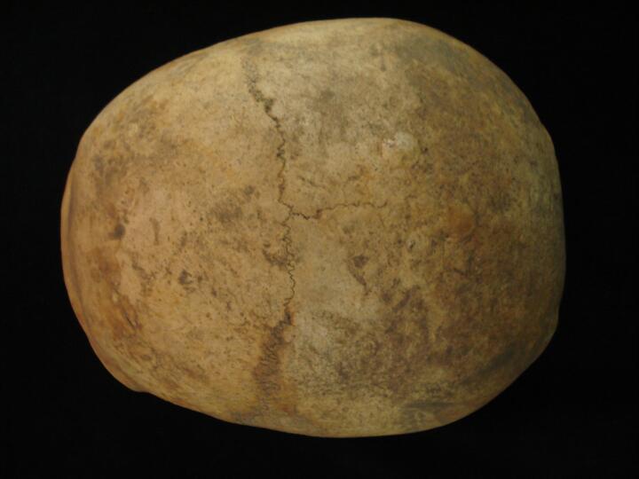
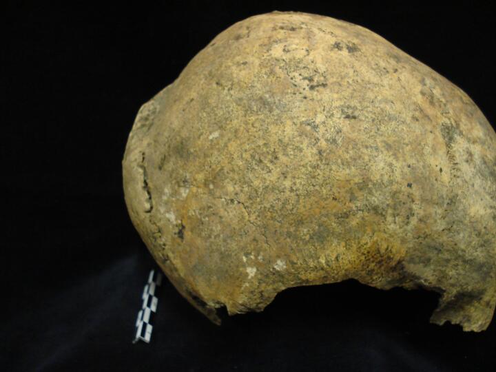
BA84_2640_1.jpg
|
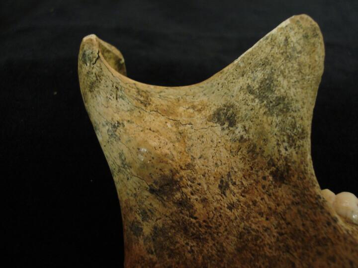
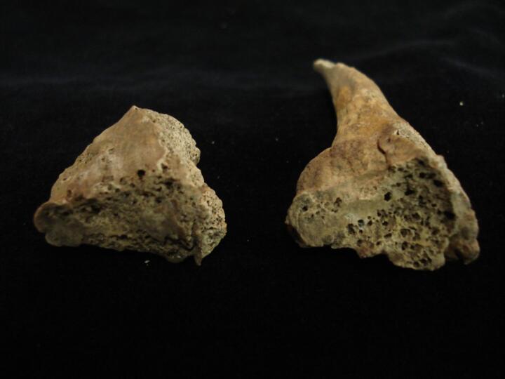
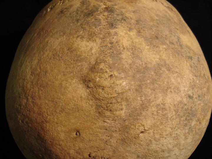
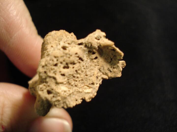
Skull, right parietal button osteoma
|
| BA84
|
2640
|
2
|
BA84_2640_2.jpg
|
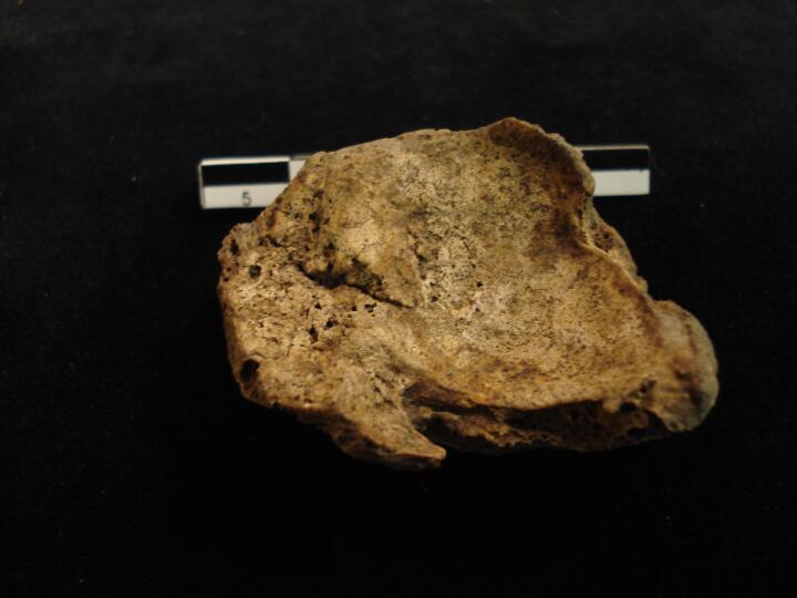
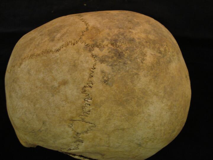
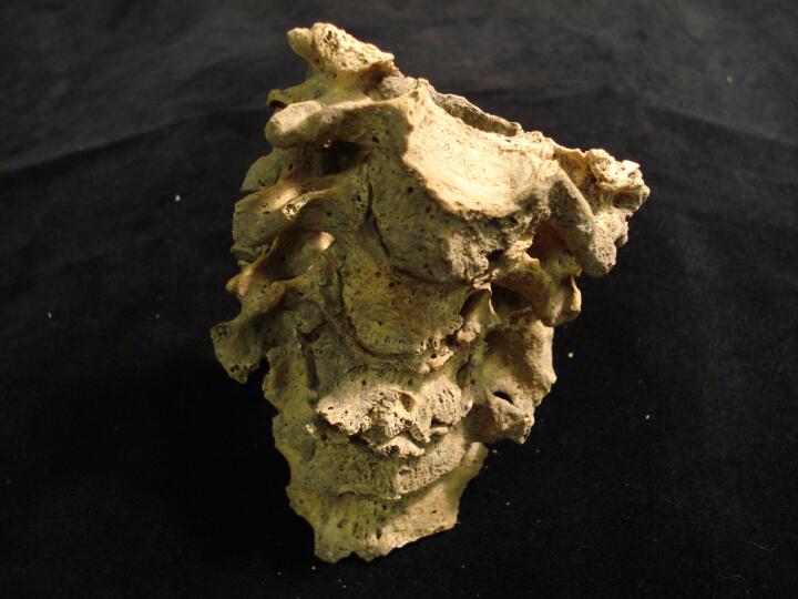
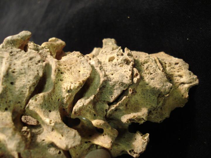
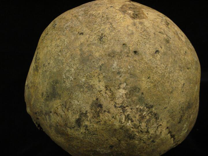
Skull occipital (posterior view) Lambdoid bone & Lambdoid wormians
|
| BA84
|
2640
|
3
|
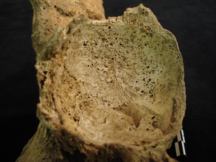
BA84_2640_3.jpg
|
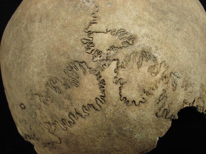
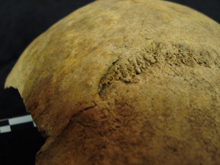
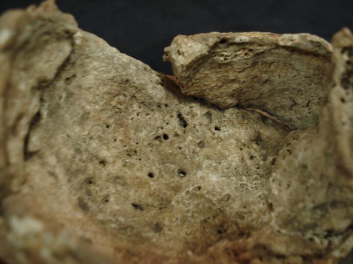
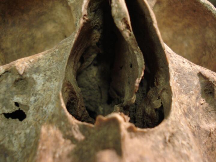
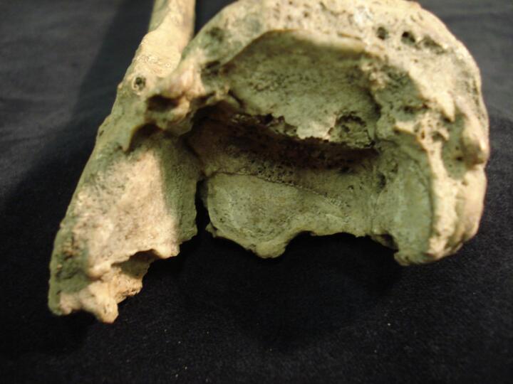
Skull endocranial surface, deep meningeal vessel impressions
|
| BA84
|
2640
|
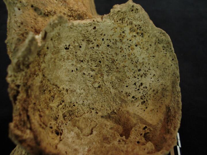
4
|
BA84_2640_4.jpg
|
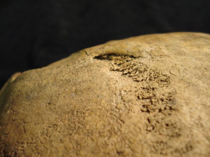
Skull endocranial surface, deep meningeal vessel impressions (left side)
|
| BA84
|
2640
|
5
|
BA84_2640_5.jpg
|
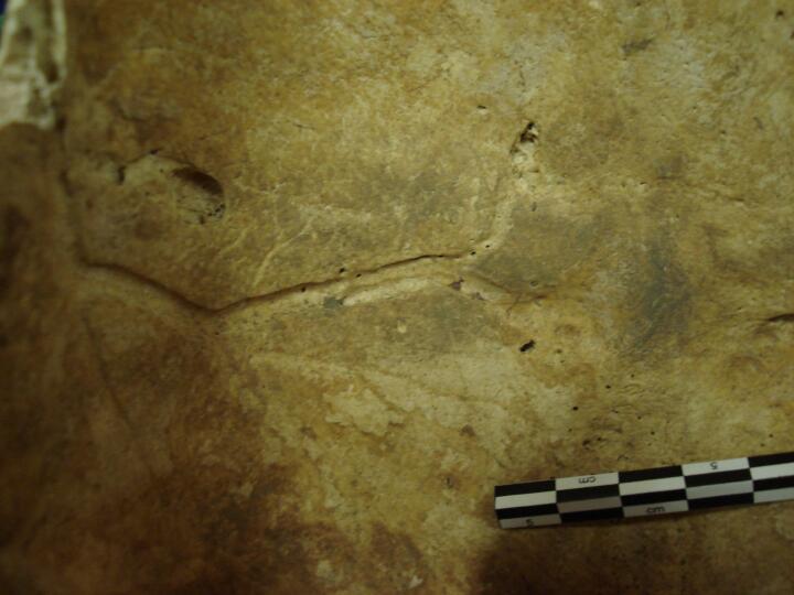
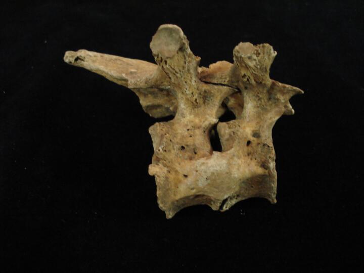
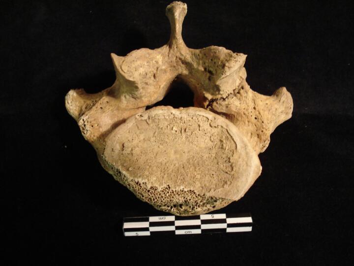
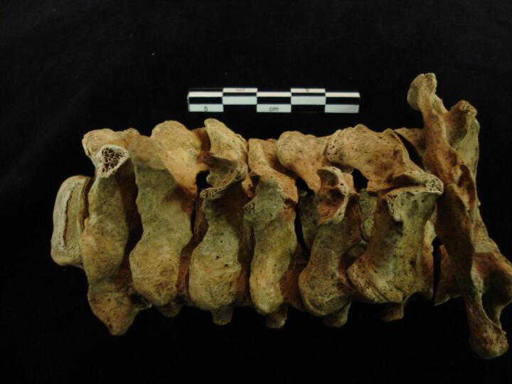
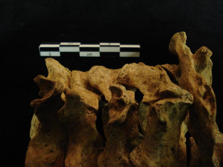
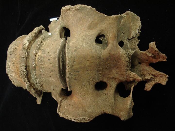
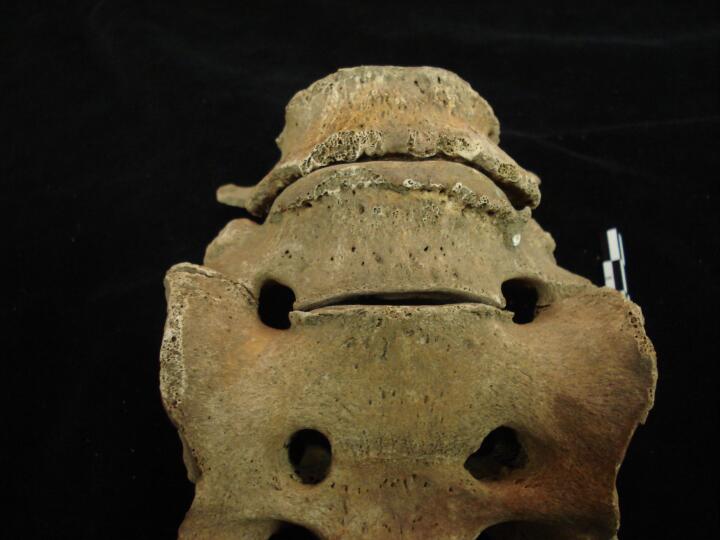
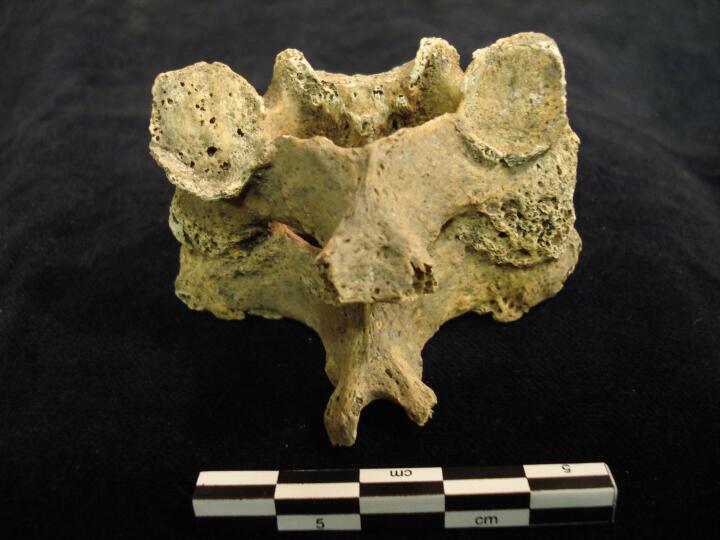
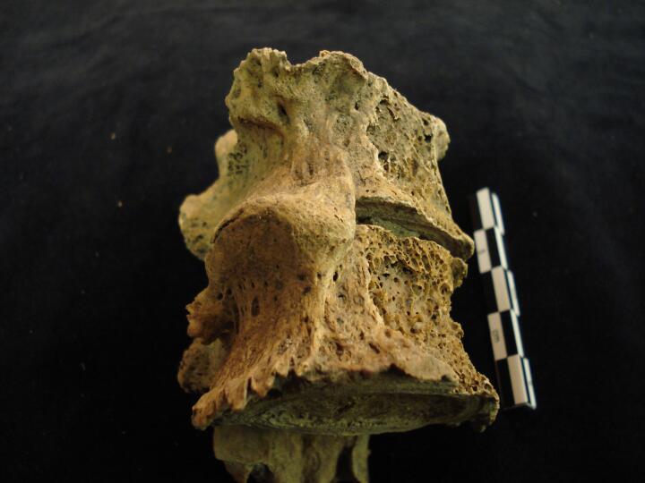
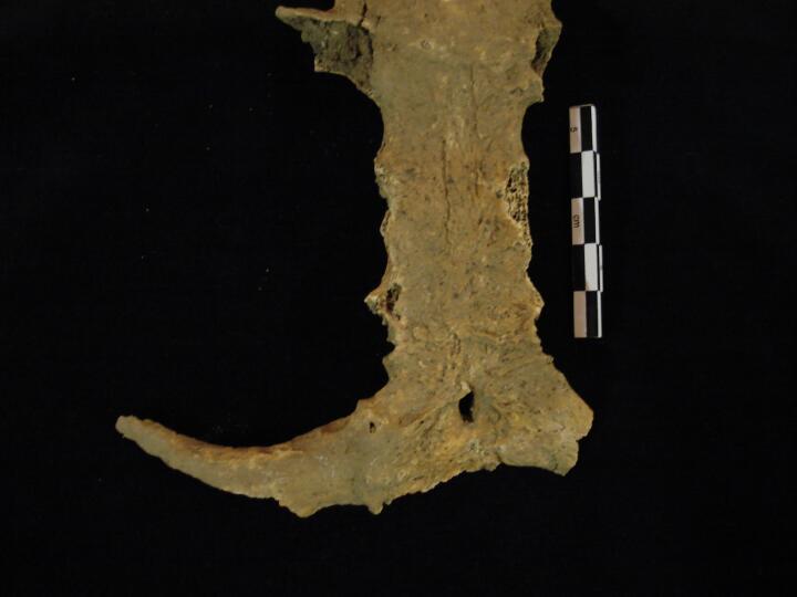
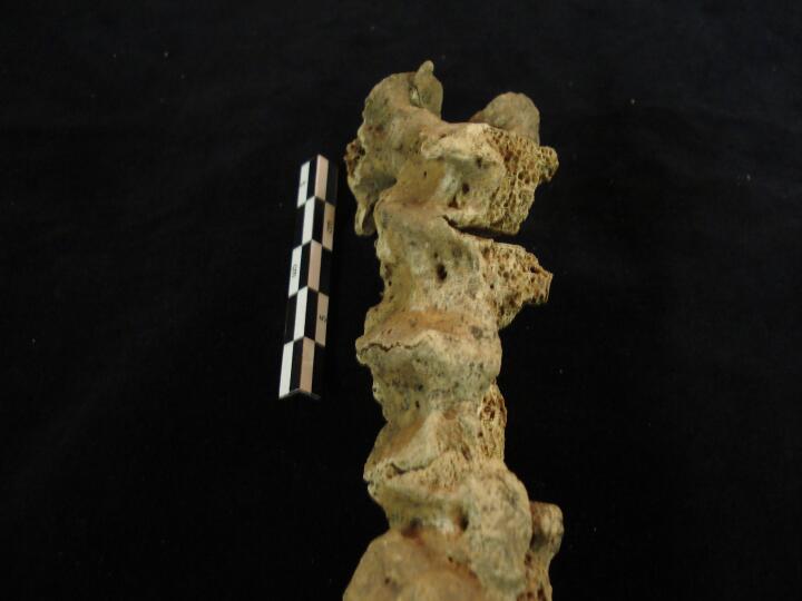
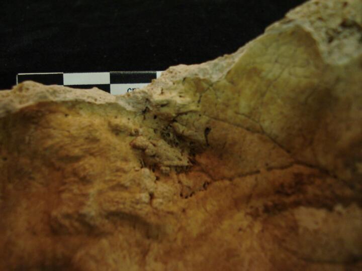
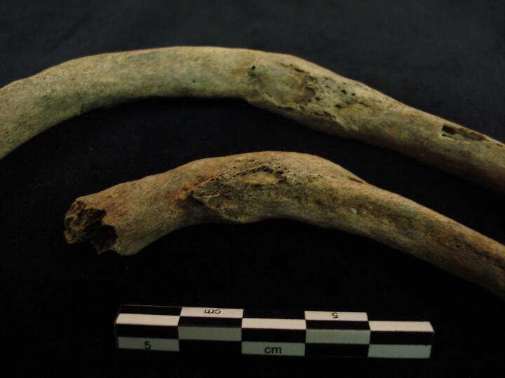
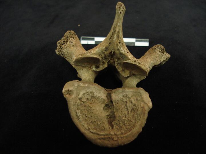
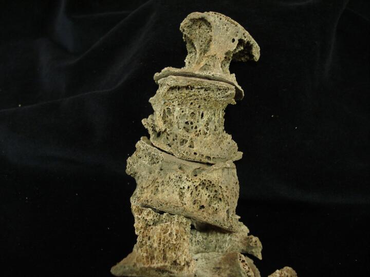
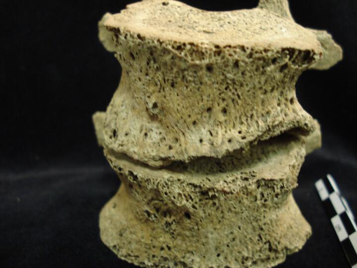
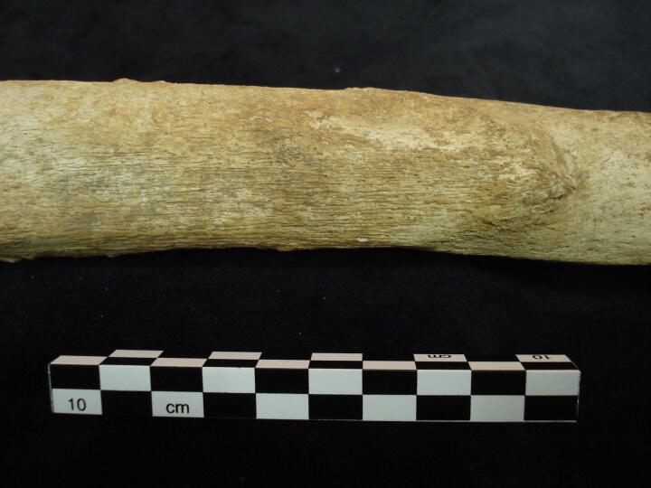
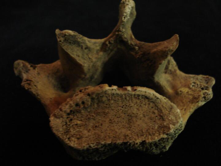
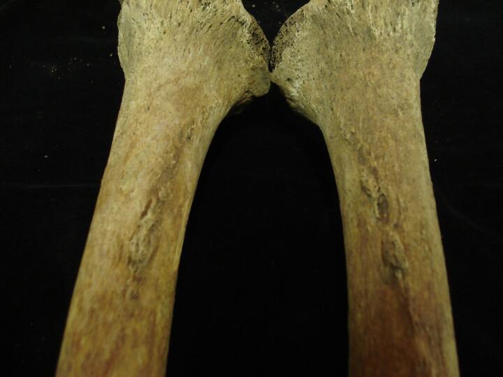
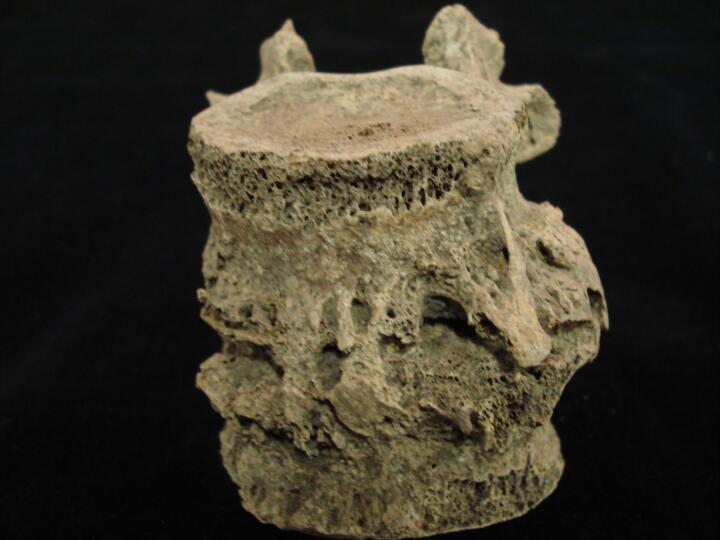
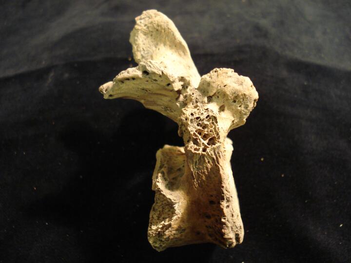
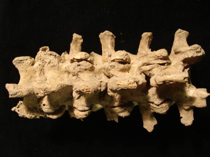
Thoracic vertebrae (right side), some PM damage, 'candlewax' fusion, DISH
|
| BA84
|
2640
|
6
|
BA84_2640_6.jpg
|
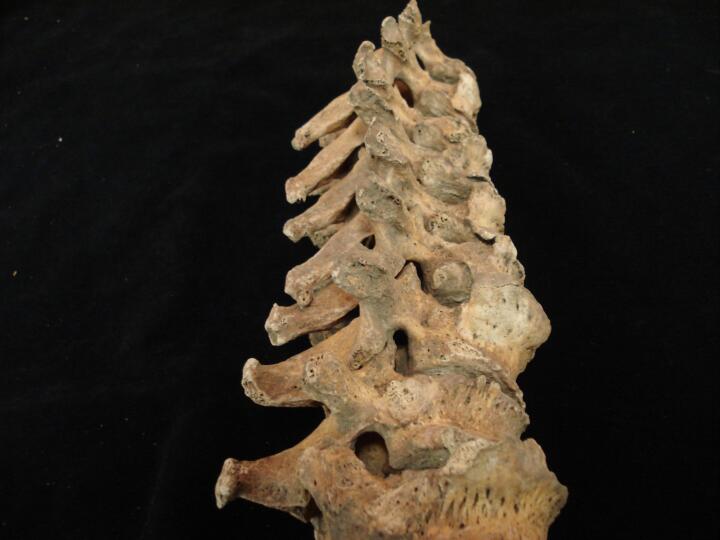
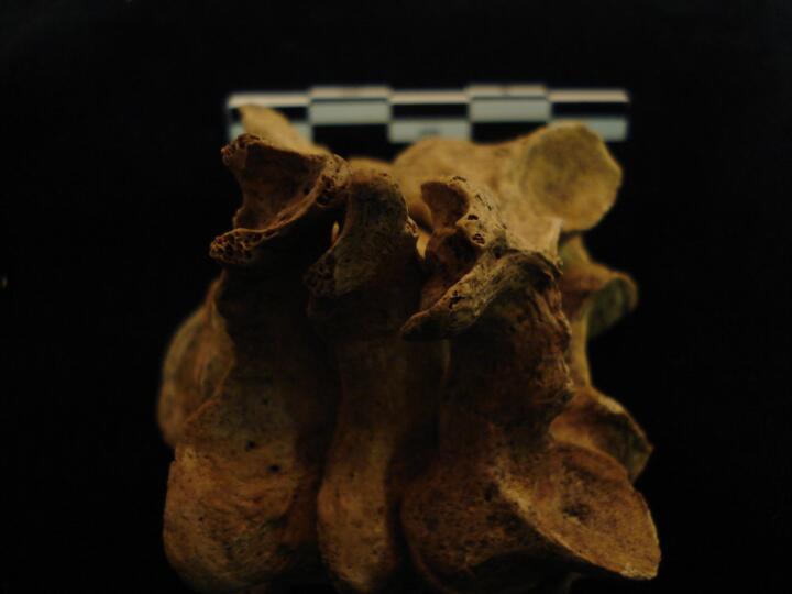
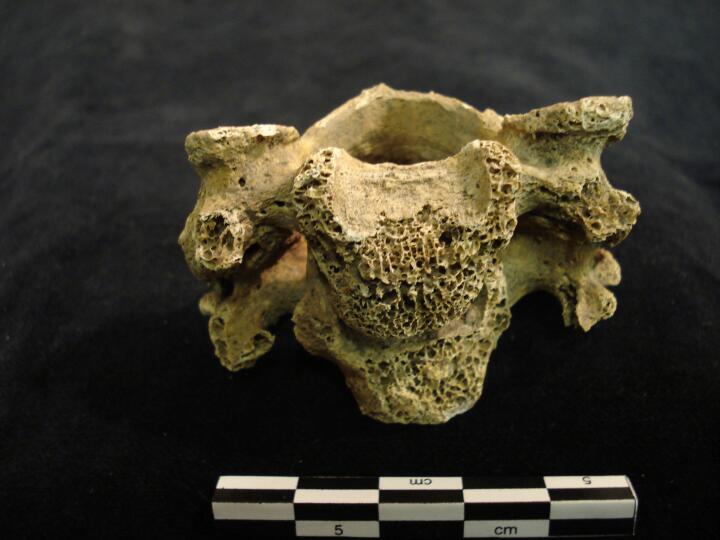
Thoracic vertebrae (right side/posterior view), some PM damage, 'candlewax' fusion, DISH
|
| BA84
|
2640
|
7
|
BA84_2640_7.jpg
|
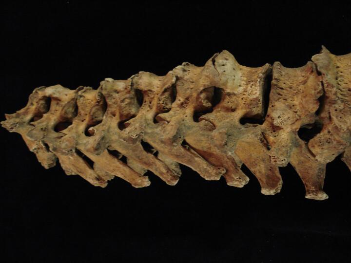
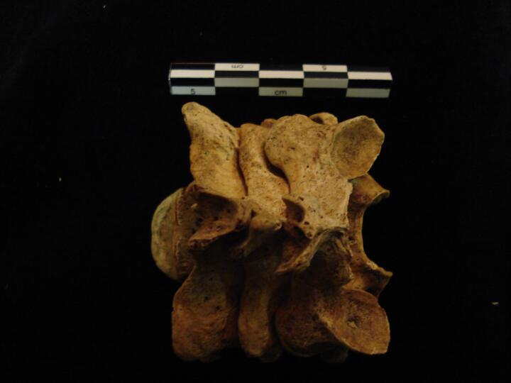
Thoracic vertebrae, Th9 to Th12 (right side/close up), some PM damage, 'candlewax' fusion, DISH
|
| BA84
|
2640
|
8
|
BA84_2640_8.jpg
|
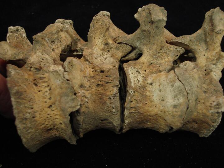
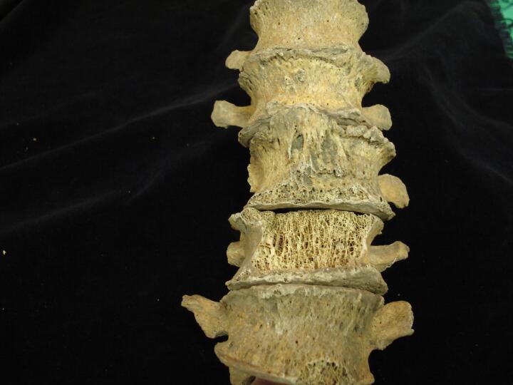
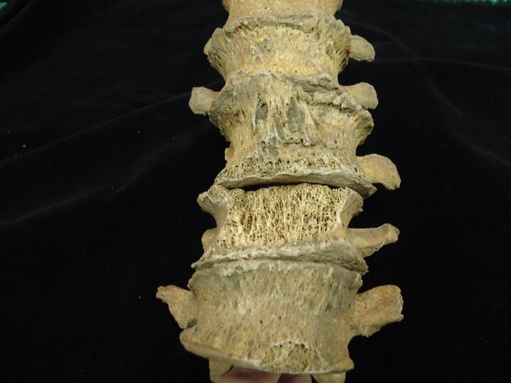
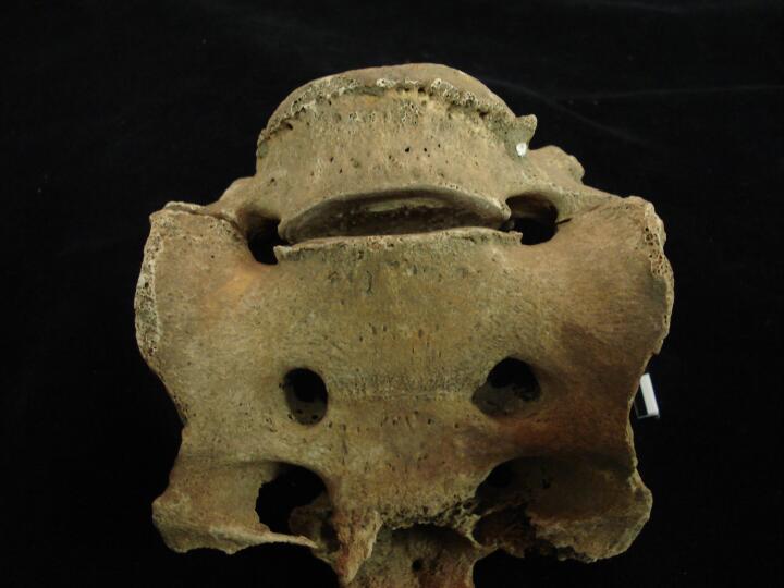
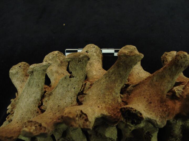
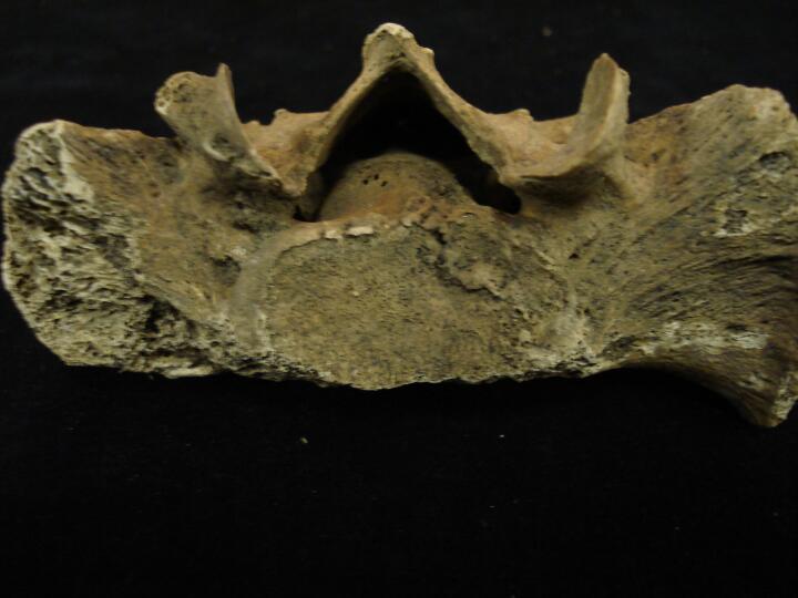
Thoracic vertebrae Th4 & Th5, fused right side, 'candlewax', DISH
|
| BA84
|
2640
|
9
|
BA84_2640_9.jpg
|
Thoracic vertebrae Th4 & Th5, fused right side, 'candlewax', DISH
|
| BA84
|
2640
|
10
|
BA84_2640_10.jpg
|
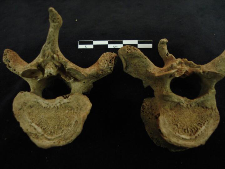
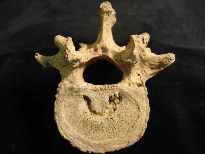
Lumbar vertebrae, articulated (anterior view/close up) severe marginal osteophytic lipping & lateral wedging of vertebral bodies
|
| BA84
|
2640
|
11
|
BA84_2640_11.jpg
|
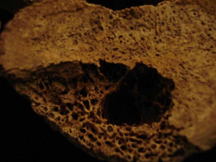
Lumbar vertebra L1 centrum (inferior view) schmorl's node & marginal osteophytic lipping
|
| BA84
|
2640
|
12
|
BA84_2640_12.jpg
|
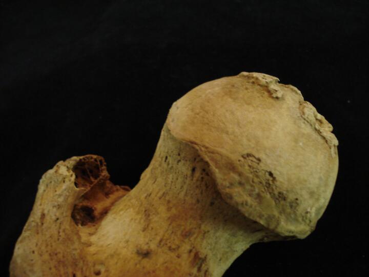
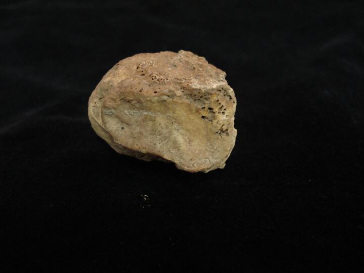
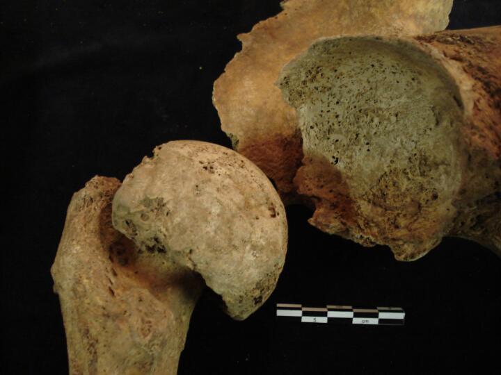
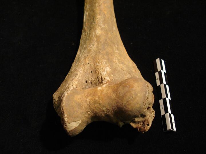
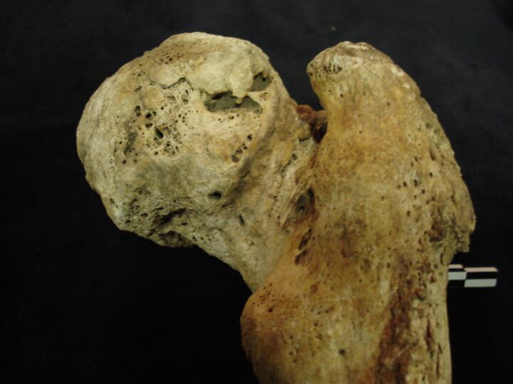
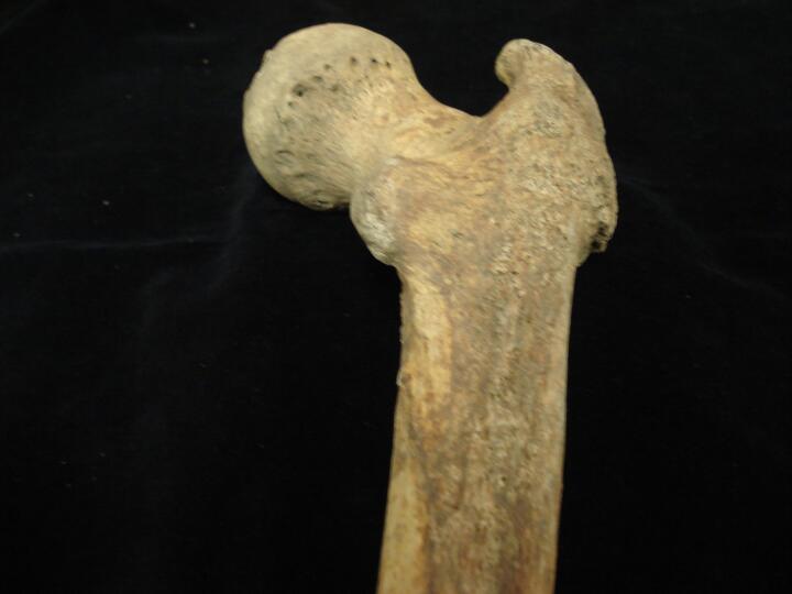
Left femoral head joint surface osteophytes
|
| BA84
|
2640
|
13
|
BA84_2640_13.jpg
|
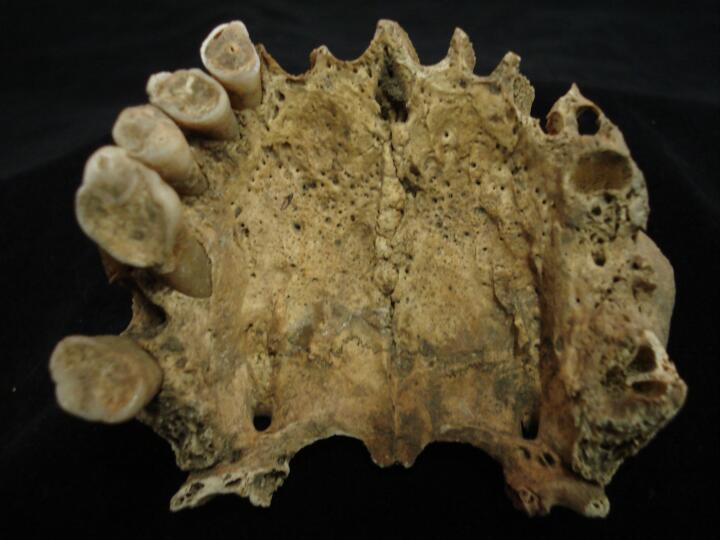
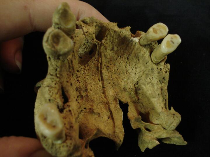
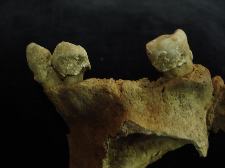
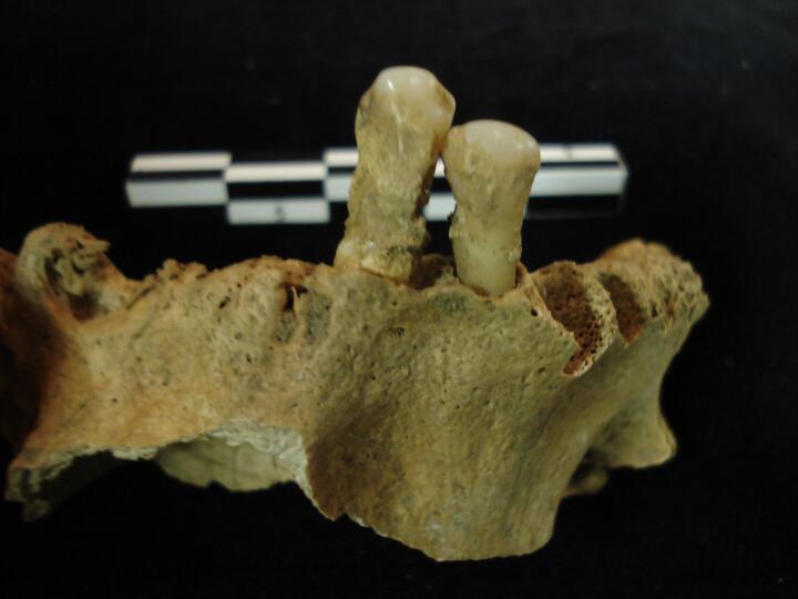
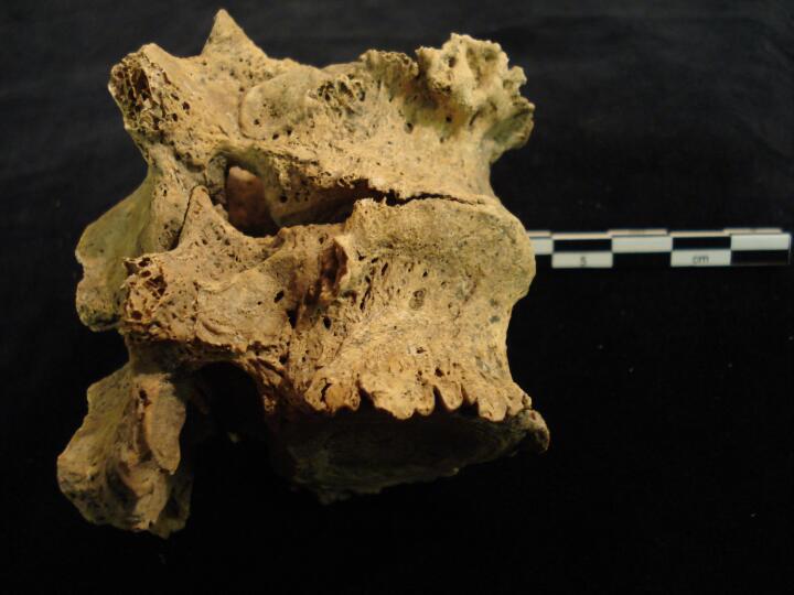
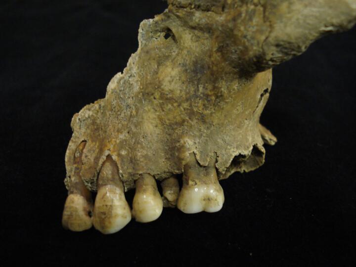
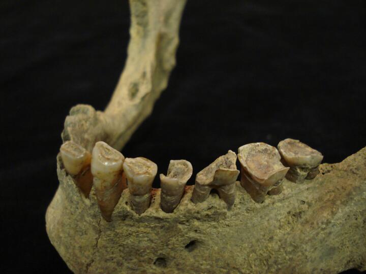
Maxilla (palatal view) heavy wear of dentition & palatine torus
|
| BA84
|
2640
|
14
|
BA84_2640_14.jpg
|
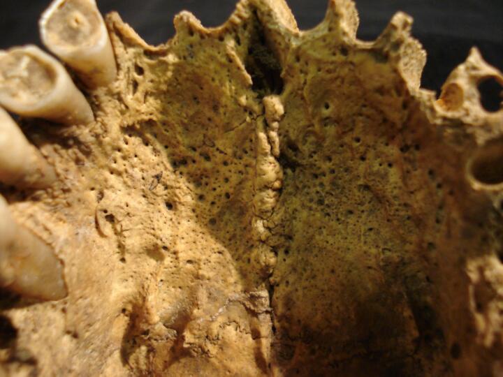
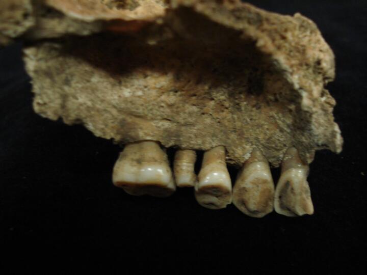
Maxilla (palatal view/close up) palatine torus
|
| BA84
|
2643
|
1
|
BA84_2643_1.jpg
|
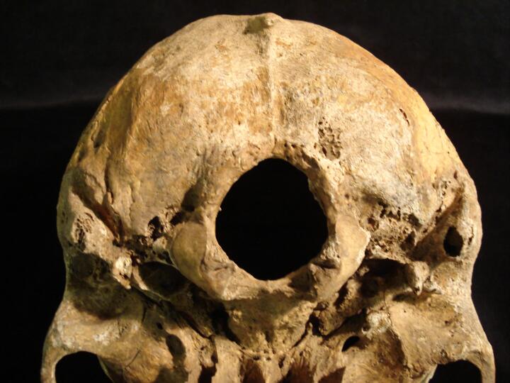
Sternum (anterior view) sternal foramen
|
| BA84
|
2669
|
1
|
BA84_2669_1.jpg
|
Lumbar vertebra L5 (superior view) cleft on the right side, unilateral spondylolysis
|
| BA84
|
2669
|
2
|
BA84_2669_2.jpg
|
Lumbar vertebra L5 (posterior view) cleft on the right side unilateral spondylolysis
|
| BA84
|
2722
|
1
|
BA84_2722_1.jpg
|
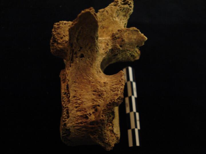
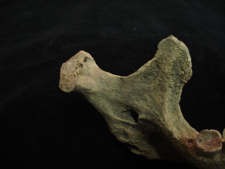
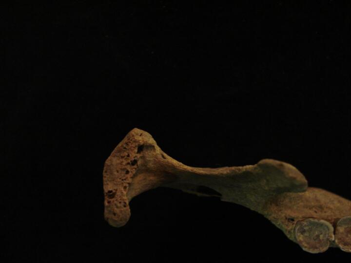
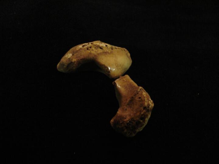
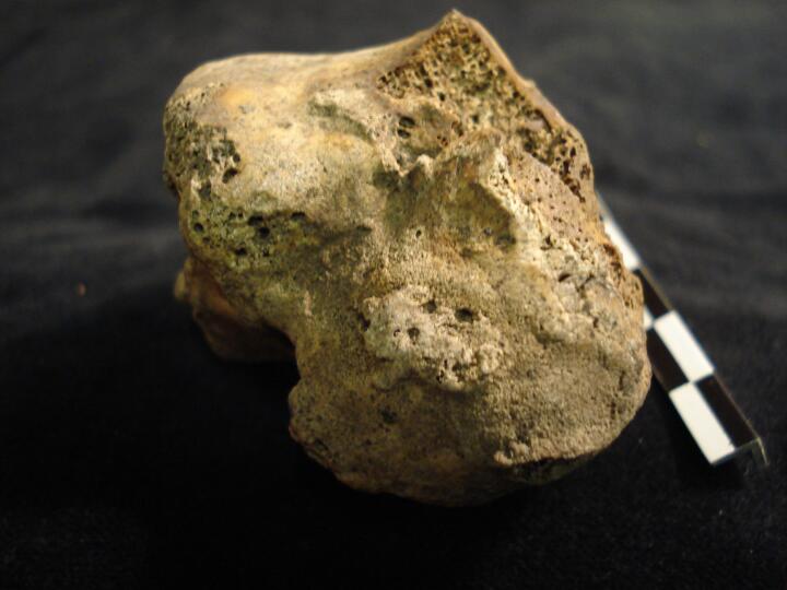
Skull occipital bone (side view) very prominent inion protuberance
|
| BA84
|
2722
|
2
|
BA84_2722_2.jpg
|
Skull occipital bone (posterior view) very prominent inion protuberance
|
| BA84
|
2722
|
3
|
BA84_2722_3.jpg
|
Skull occipital bone ( left side view) very prominent inion protuberance
|
| BA84
|
2758
|
1
|
BA84_2758_1.jpg
|
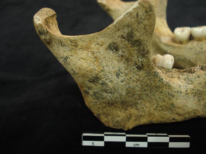
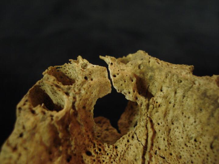
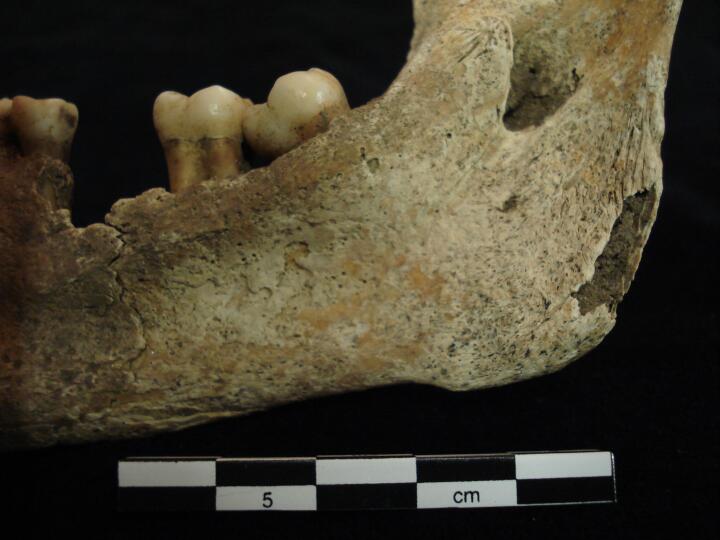
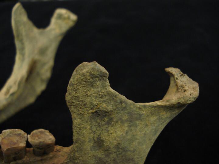
Mandible (right side) ?bone cyst
|
| BA84
|
2758
|
2
|
BA84_2758_2.jpg
|
Mandible (right side/close up) ?bone cyst
|
| BA84
|
2758
|
3
|
BA84_2758_3.jpg
|
Mandible (right side/close up) ?bone cyst
|
| BA84
|
2758
|
4
|
BA84_2758_4.jpg
|
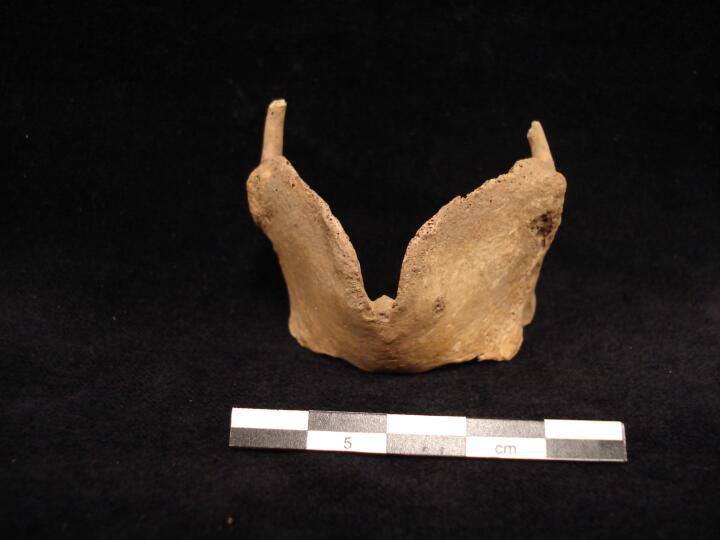
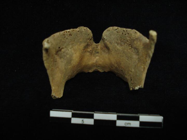
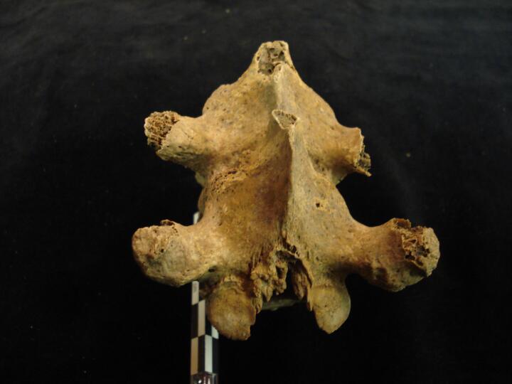
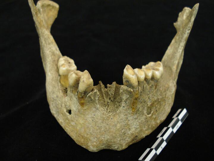
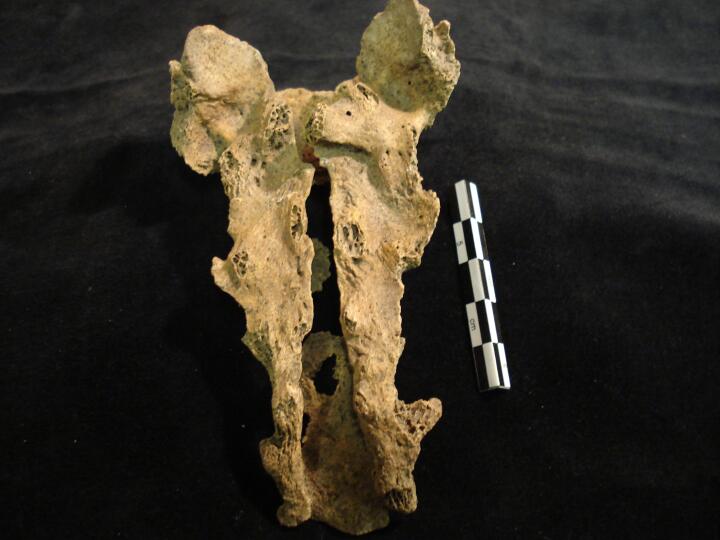
Cervical vertebrae C1 to C7 articulated (posterior view) showing spinous processes, ?congenital abnormality
|
| BA84
|
2758
|
5
|
BA84_2758_5.jpg
|
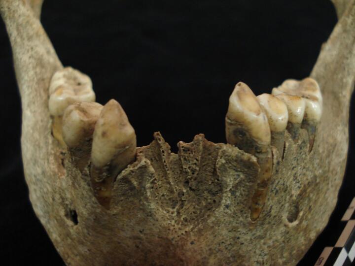
Cervical vertebrae C1 to C7 articulated (posterior view) close up showing spinous processes, ?congenital abnormality
|
| BA84
|
2758
|
6
|
BA84_2758_6.jpg
|
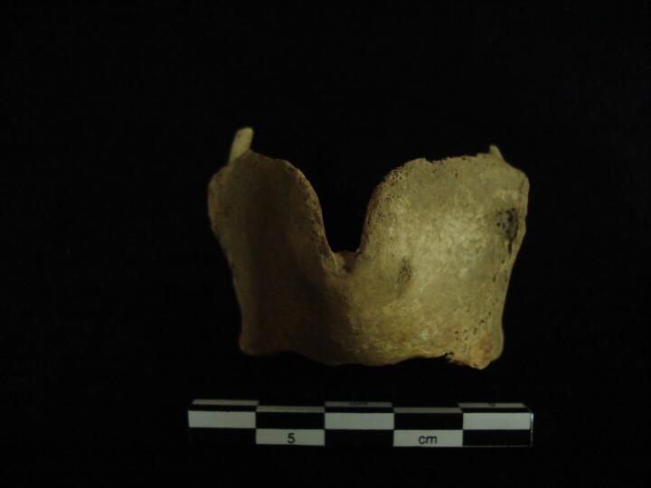
Cervical vertebrae C3 to C5 (superior view) showing spinous processes, ?congenital abnormality
|
| BA84
|
2758
|
7
|
BA84_2758_7.jpg
|
Cervical vertebrae C3 to C5 (superior view) close up showing spinous processes, ?congenital abnormality
|
| BA84
|
2762
|
1
|
BA84_2762_1.jpg
|
Right cuboid degenerative joint changes & eburnation, osteoarthritis
|
| BA84
|
2762
|
2
|
BA84_2762_2.jpg
|
2nd cuneiform & 2nd metatarsal joint destruction & eburnation, osteoarthritis
|
| BA84
|
2762
|
3
|
BA84_2762_3.jpg
|
2nd cuneiform joint destruction & eburnation, osteoarthritis
|
| BA84
|
2762
|
4
|
BA84_2762_4.jpg
|
2nd metatarsal joint destruction & eburnation, osteoarthritis
|
| BA84
|
2762
|
5
|
BA84_2762_5.jpg
|
Right 1st metacarpal & trapezium, eburnation, osteoarthritis
|
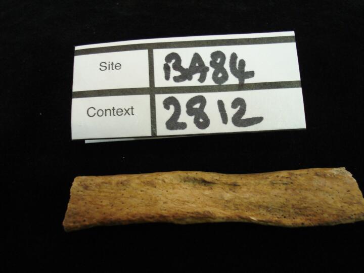
| BA84
|
2812
|
1
|
BA84_2812_1.jpg
|
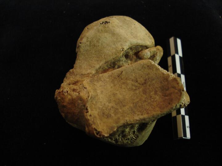
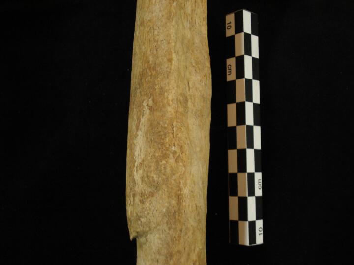
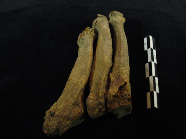
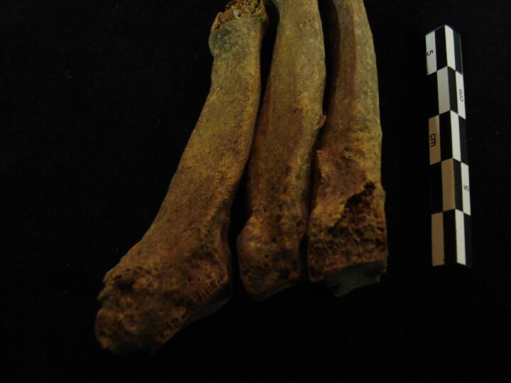
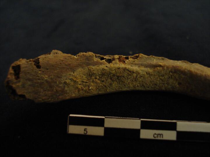
Right rib shaft fragment with healed blunt force trauma (pleural surface)
|
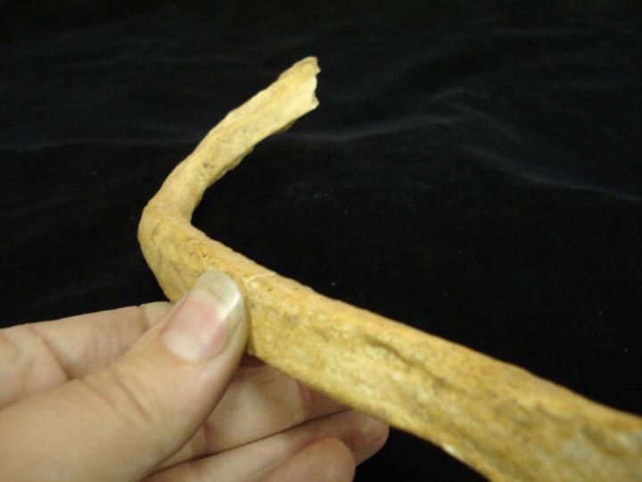
| BA84
|
2812
|
2
|
BA84_2812_2.jpg
|
Right rib shaft fragment with healed blunt force trauma (pleural surface/close up)
|
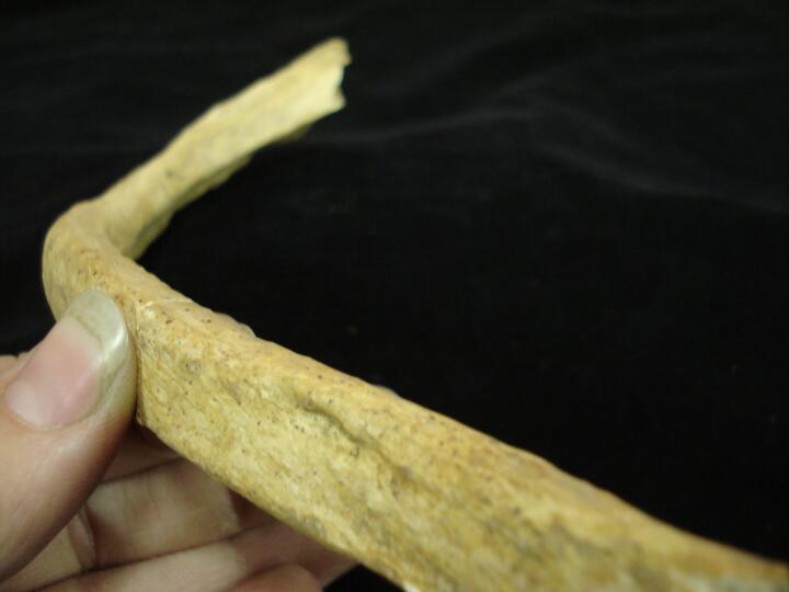
| BA84
|
2812
|
3
|
BA84_2812_3.jpg
|
Complete right rib showing healed blunt force trauma (pleural surface)
|
| BA84
|
2812
|
4
|
BA84_2812_4.jpg
|
Complete right rib showing healed blunt force trauma (pleural surface/close up)
|
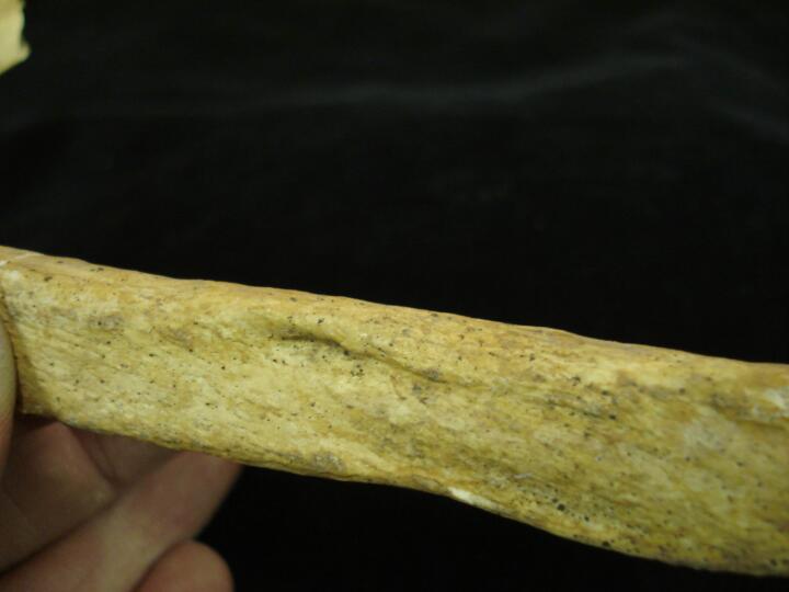
| BA84
|
2812
|
5
|
BA84_2812_5.jpg
|
Right rib mid shaft (close up) of healed blunt force trauma at an angle
|
| BA84
|
2861
|
1
|
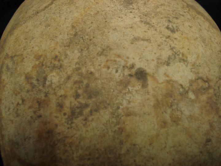
BA84_2861_1.jpg
|
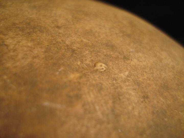
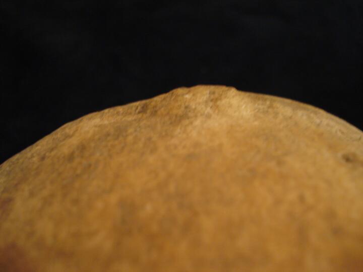
Skull frontal bone (front view) button osteoma
|
| BA84
|
2861
|
2
|
BA84_2861_2.jpg
|
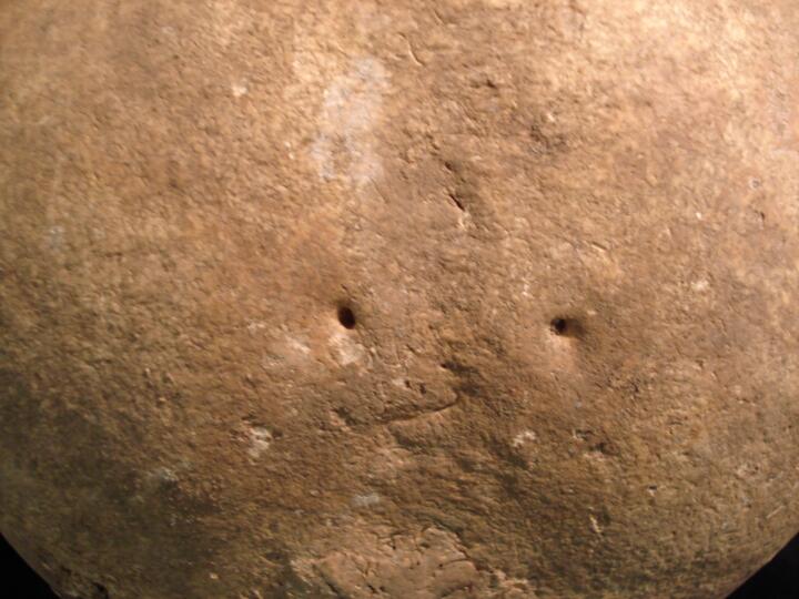
Skull parietals, parietal foramen
|
| BA84
|
2861
|
3
|
BA84_2861_3.jpg
|
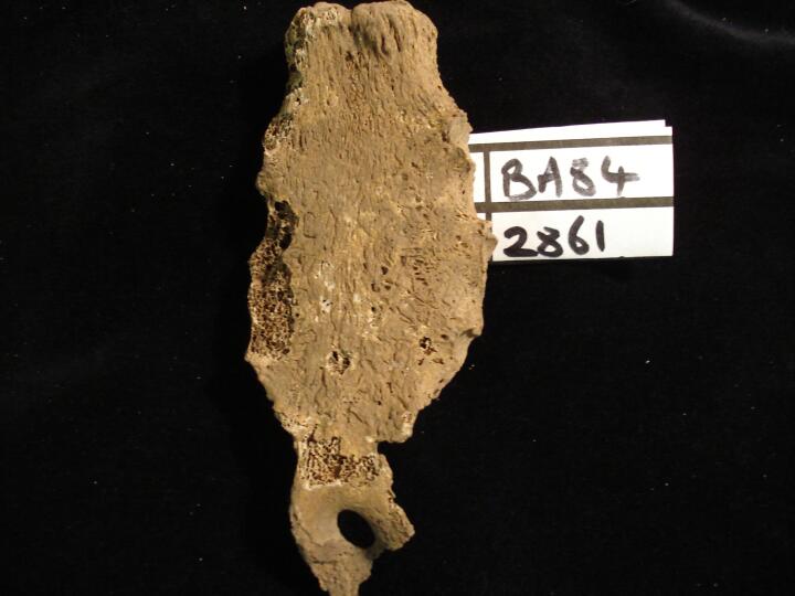
Sternum & xyphoid with aperture (anterior view)
|
| BA84
|
2865
|
1
|
BA84_2865_1.jpg
|
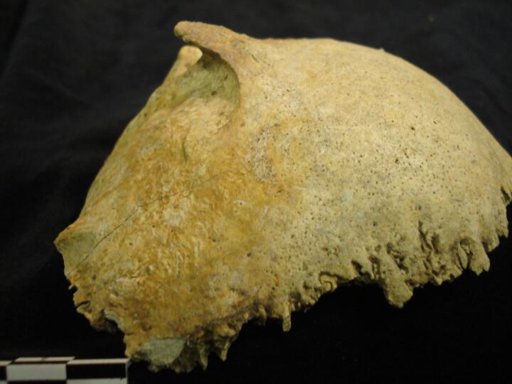
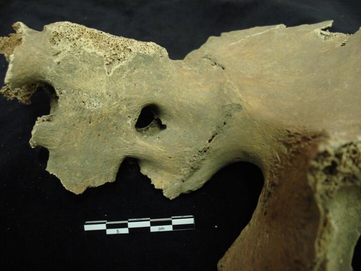
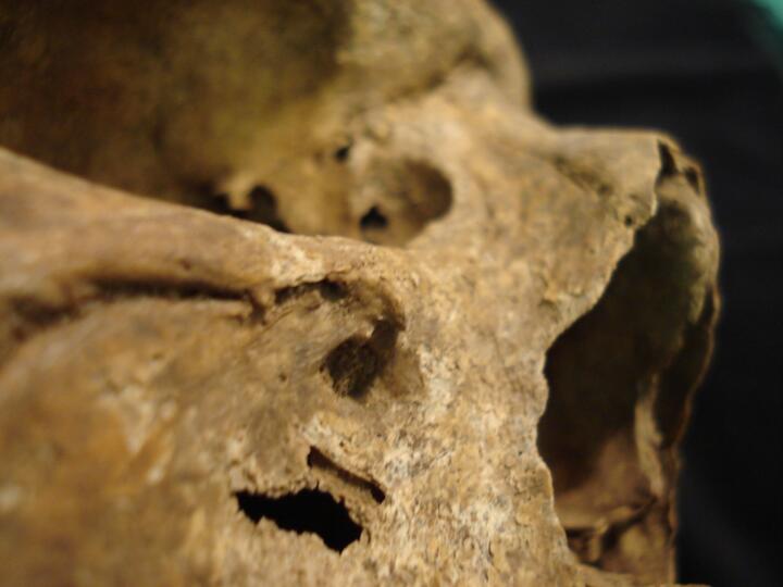
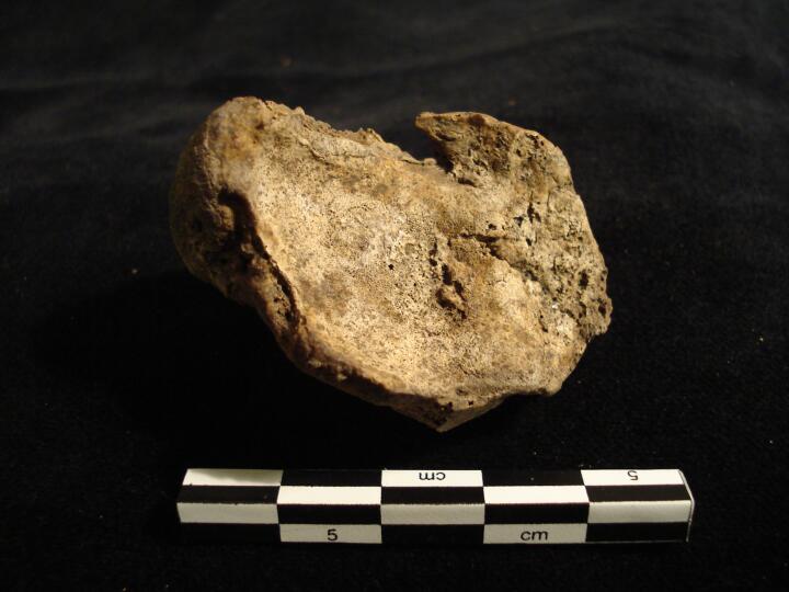
Skull (posterior view/right side) large smooth raised area of bone ?button osteoma, across Lambdoid suture
|
| BA84
|
2865
|
2
|
BA84_2865_2.jpg
|
Skull (posterior view/right side) close up of large smooth raised area of bone ?button osteoma, across Lambdoid suture
|
| BA84
|
2865
|
3
|
BA84_2865_3.jpg
|
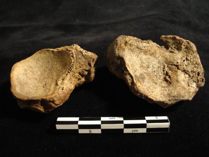
Skull endocranial surface corresponding to the area of raised bone on the ectocranial surface, no apparent pathological change
|
| BA84
|
2865
|
4
|
BA84_2865_4.jpg
|
Skull fragment (side view) close up of raised bone
|
| BA84
|
2865
|
5
|
BA84_2865_5.jpg
|
Skull fragment (side view) close up of raised bone
|
| BA84
|
2865
|
6
|
BA84_2865_6.jpg
|
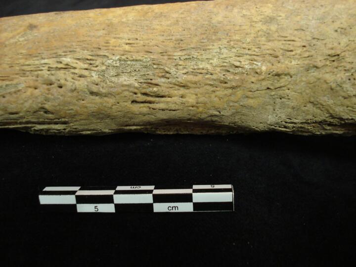
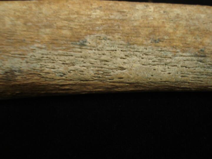
Right fibula (medial view) healed non-specific periosteal infection of shaft
|
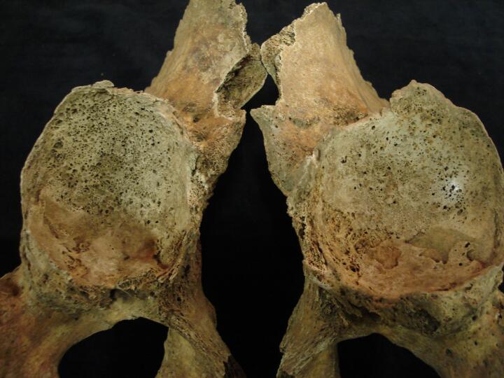
| BA84
|
2876
|
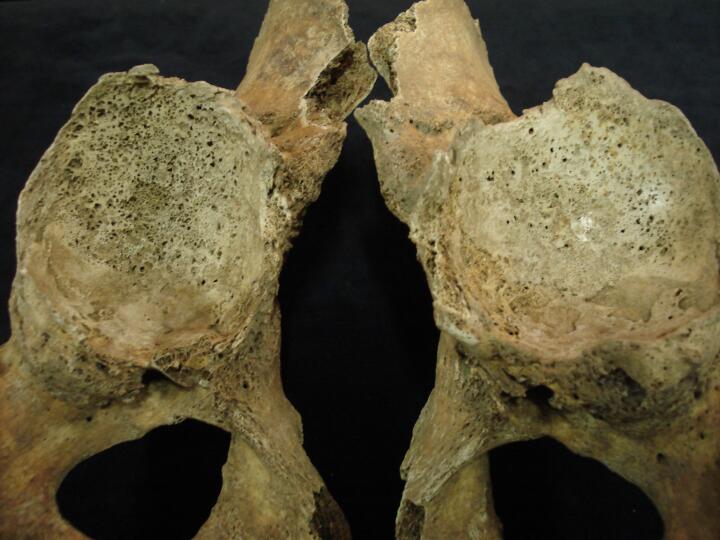
1
|
BA84_2876_1.jpg
|
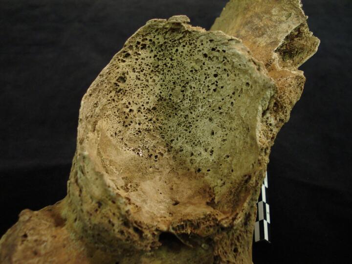
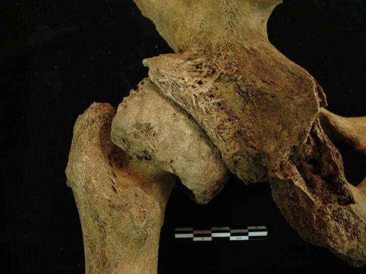
Right pelvis, acetabulum enlarged & gross destruction of joint surface with eburnation, osteoarthritis
|
| BA84
|
2876
|
2
|
BA84_2876_2.jpg
|
Right pelvis, acetabulum (close up) enlarged & gross destruction of joint surface with eburnation, osteoarthritis
|
| BA84
|
2876
|
3
|
BA84_2876_3.jpg
|
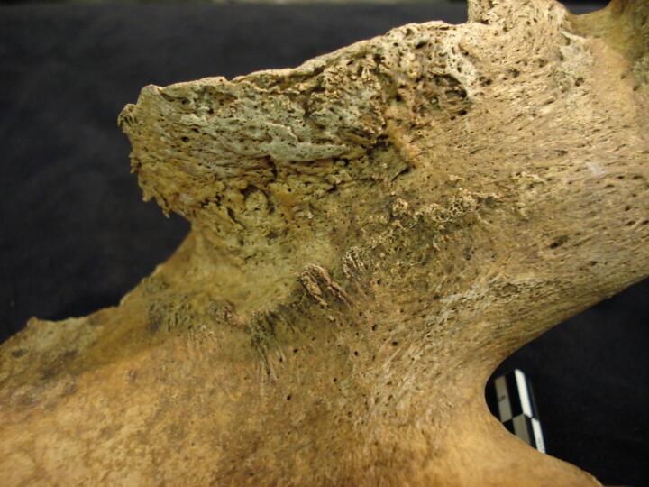
Right pelvis (posterior view) enlargement & osteophytic lipping of the acetabulum
|
| BA84
|
2876
|
4
|
BA84_2876_4.jpg
|
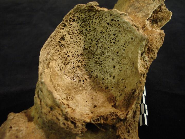
Left pelvis, acetabulum enlarged & gross destruction of joint surface with eburnation, osteoarthritis
|
| BA84
|
2876
|
5
|
BA84_2876_5.jpg
|
Left pelvis, acetabulum (close up) enlarged & gross destruction of joint surface with eburnation, osteoarthritis
|
| BA84
|
2876
|
6
|
BA84_2876_6.jpg
|
Left pelvis (posterior view) enlargement & osteophytic lipping of the acetabulum
|
| BA84
|
2876
|
7
|
BA84_2876_7.jpg
|
Right & left acetabulum joint surface destruction & eburnation, osteoarthritis
|
| BA84
|
2876
|
8
|
BA84_2876_8.jpg
|
Right & left acetabulum gross joint surface destruction & eburnation, osteoarthritis
|
| BA84
|
2876
|
9
|
BA84_2876_9.jpg
|
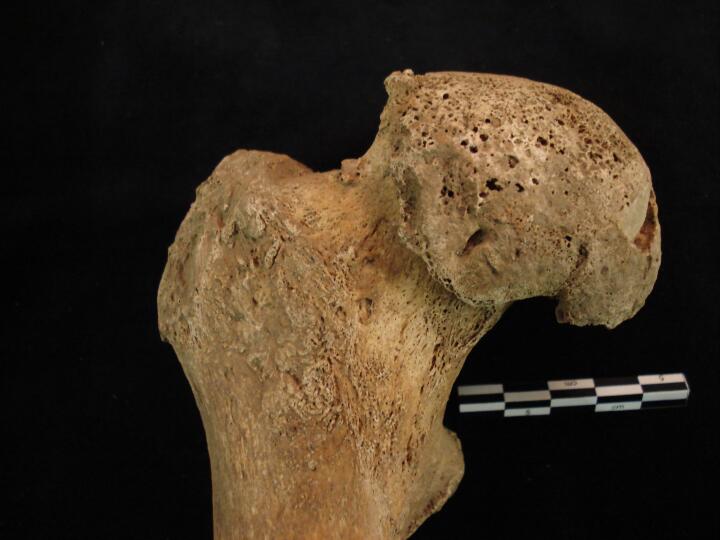
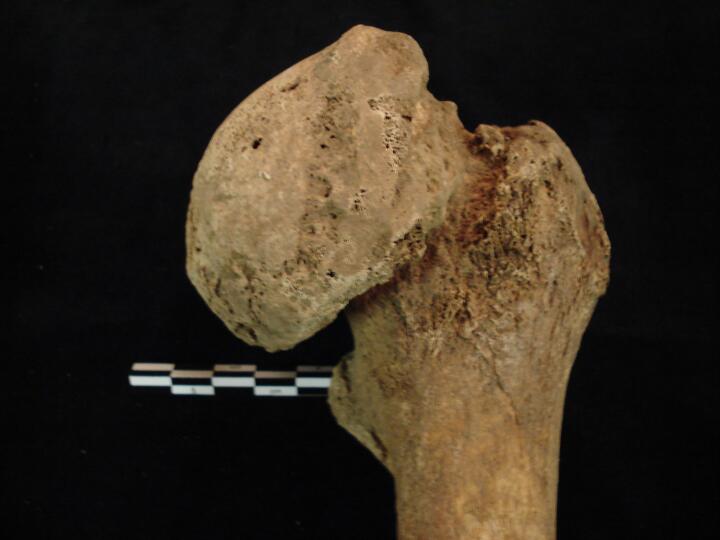
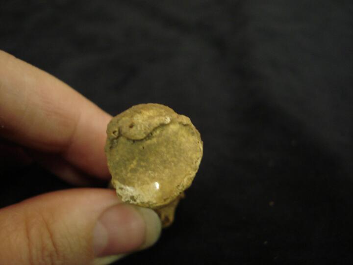
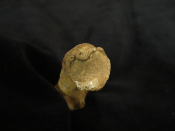
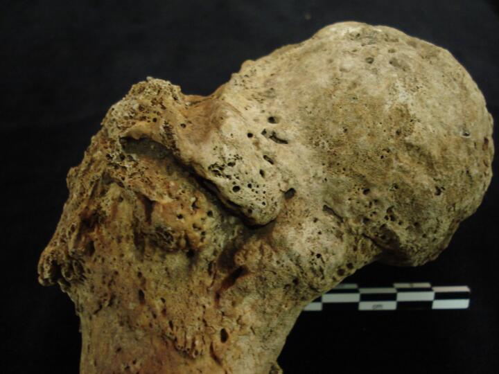
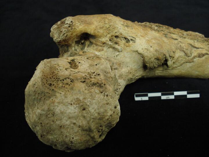
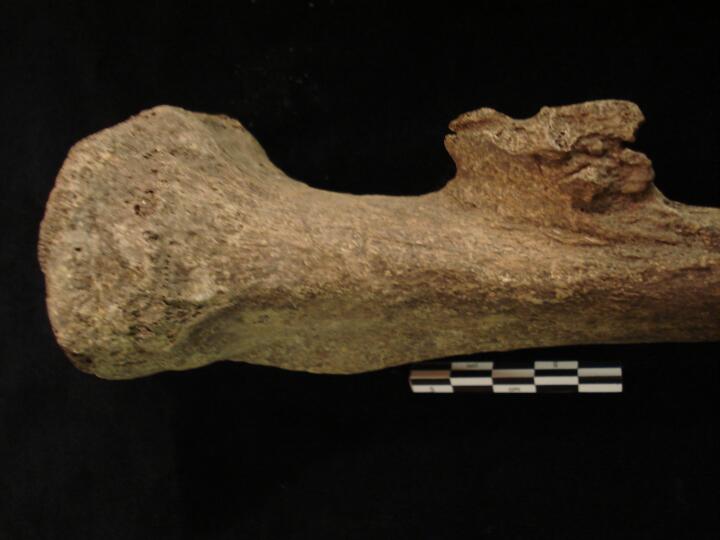
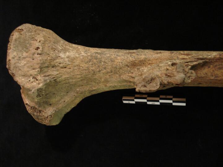
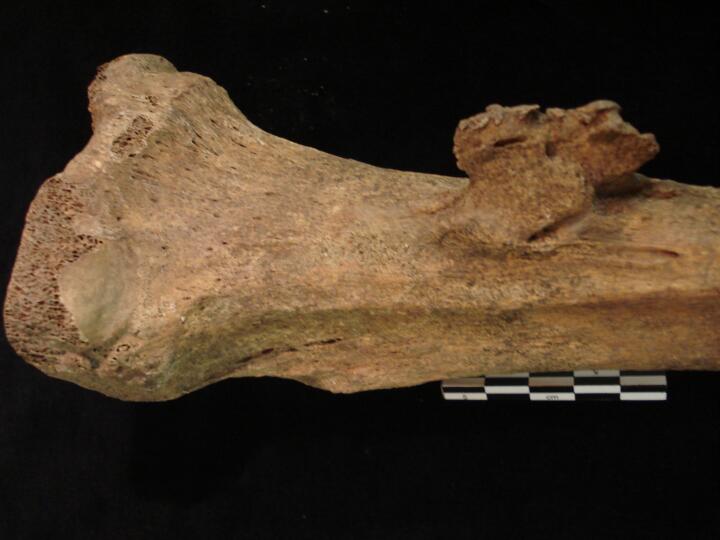
Right femoral head (anterior view) joint changes
|
| BA84
|
2876
|
10
|
BA84_2876_10.jpg
|
Right femoral head (posterior view) joint changes
|
| BA84
|
2876
|
11
|
BA84_2876_11.jpg
|
Left femoral head (anterior view) joint changes
|
| BA84
|
2876
|
12
|
BA84_2876_12.jpg
|
Left femoral head (posterior view) joint changes
|
| BA84
|
2876
|
13
|
BA84_2876_13.jpg
|
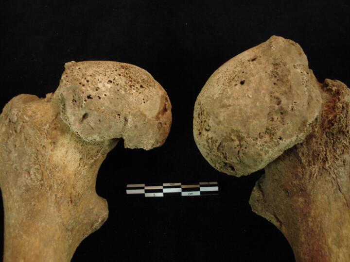
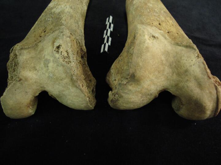
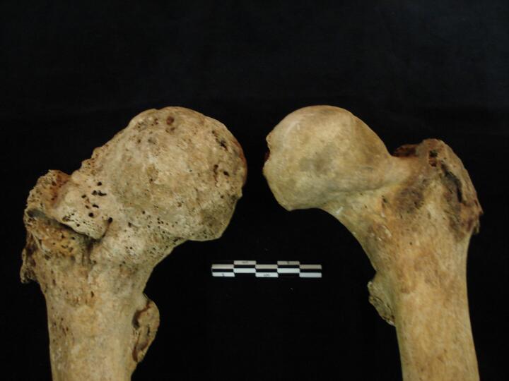
Right & left femoral heads showing changes to the femoral head shape & joint changes, eburnation, osteoarthritis
|
| BA84
|
2876
|
14
|
BA84_2876_14.jpg
|
Right femoral head & right pelvis showing joint surface destruction & eburnation (anterior view)
|
| BA84
|
2876
|
15
|
BA84_2876_15.jpg
|
Right femoral head & right pelvis articulated (posterior view)
|
| BA84
|
2876
|
16
|
BA84_2876_16.jpg
|
Left femoral head & right pelvis showing gross joint surface destruction & eburnation (anterior view)
|
| BA84
|
2876
|
17
|
BA84_2876_17.jpg
|
Left femoral head & right pelvis articulated (posterior view)
|
| BA84
|
2876
|
18
|
BA84_2876_18.jpg
|
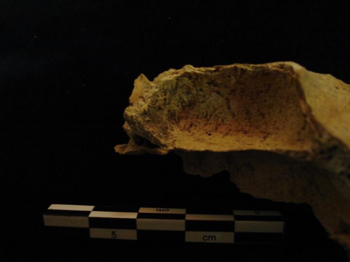
Left maxillary sinus (superior view) remodelled bone indicating healed infection, sinusitis
|
| BA84
|
2876
|
19
|
BA84_2876_19.jpg
|
Left maxillary sinus (superior view/close up) remodelled bone indicating healed infection, sinusitis
|
| BA84
|
2876
|
20
|
BA84_2876_20.jpg
|
Maxilla (right side) palatal view of external draining periapical lesion (abscess) teeth present with calculus
|
| BA84
|
2876
|
21
|
BA84_2876_21.jpg
|
Maxilla (right side) buccal surface heavy calculus & AM tooth loss
|
| BA84
|
2876
|
22
|
BA84_2876_22.jpg
|
Maxilla (left side) buccal surface heavy calculus & AM tooth loss
|
| BA84
|
2876
|
23
|
BA84_2876_23.jpg
|
Maxilla buccal surface (anterior view) large external draining periapical lesion (abscess)
|
| BA84
|
2876
|
24
|
BA84_2876_24.jpg
|
Maxilla lingual surface (palatal view) large external draining periapical lesion (abscess)
|
| BA84
|
2876
|
25
|
BA84_2876_25.jpg
|
Complete thyroid bone (anterior view)
|
| BA84
|
2876
|
26
|
BA84_2876_26.jpg
|
Complete thyroid bone (anterior view)
|
| BA84
|
2876
|
27
|
BA84_2876_27.jpg
|
Complete thyroid bone (posterior view)
|
| BA84
|
2898
|
1
|
BA84_2898_1.jpg
|
Right ulna (full length medial view) healed fracture
|
| BA84
|
2898
|
2
|
BA84_2898_2.jpg
|
Right ulna (medial view) close up of callus of healed fracture
|
| BA84
|
2898
|
3
|
BA84_2898_3.jpg
|
Right ulna (lateral view) close up of callus of healed fracture
|
| BA84
|
2898
|
4
|
BA84_2898_4.jpg
|
Left humerus distal end (posterior view) soft tissue trauma
|
| BA84
|
2898
|
5
|
BA84_2898_5.jpg
|
Left humerus distal end (anterior view) soft tissue trauma
|
| BA84
|
2898
|
6
|
BA84_2898_6.jpg
|
Left humerus distal end (lateral view) soft tissue trauma
|
| BA84
|
2898
|
7
|
BA84_2898_7.jpg
|
Lumbar vertebra L4, bilateral spondylolysis (superior view) with pars interarticularis
|
| BA84
|
2898
|
8
|
BA84_2898_8.jpg
|
Lumbar vertebra L4, bilateral spondylolysis (inferior view) with pars interarticularis
|
| BA84
|
2898
|
9
|
BA84_2898_9.jpg
|
Lumbar vertebra L4, bilateral spondylolysis (inferior view) with pars interarticularis showing eburnation, osteoarthritis
|
| BA84
|
2898
|
10
|
BA84_2898_10.jpg
|
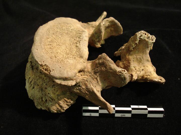
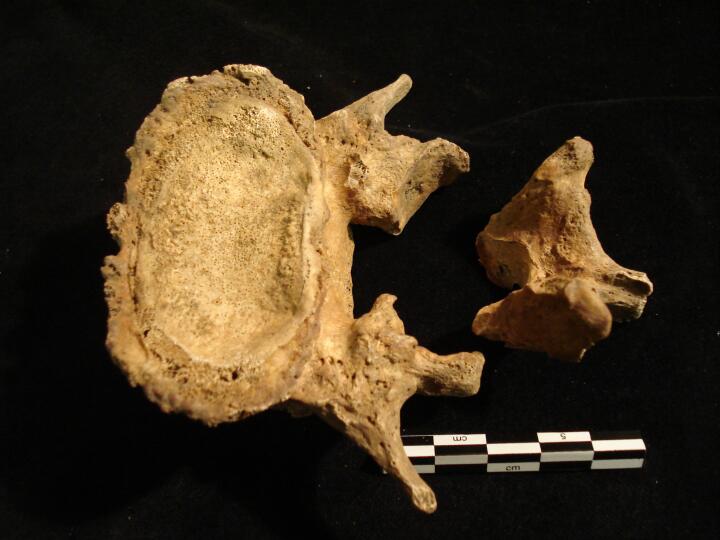
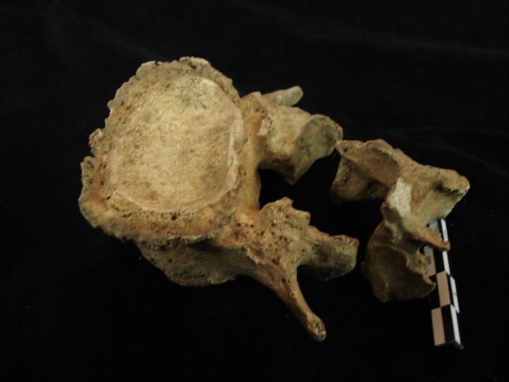
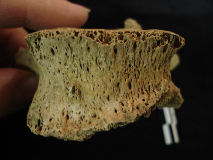
Sacrum (anterior view) with lumber vertebrae L4 & L5 in articulation
|
| BA84
|
2898
|
11
|
BA84_2898_11.jpg
|
Sacrum & Lumbar vertebra L5 (anterior view)
|
| BA84
|
2898
|
12
|
BA84_2898_12.jpg
|
Sacrum & Lumbar vertebra L5 (superior view) showing facets for sacralisation
|
| BA84
|
2898
|
13
|
BA84_2898_13.jpg
|
Sacrum (anterior view) with lumber vertebrae L4 & L5 in articulation
|
| BA84
|
2973
|
1
|
BA84_2973_1.jpg
|
Left & right femora patellar surface (anterior view) jont change & eburnation, osteoarthritis
|
| BA84
|
2973
|
2
|
BA84_2973_2.jpg
|
Left & right femora patellar surface (anterior view) jont change & eburnation, osteoarthritis
|
| BA84
|
2973
|
3
|
BA84_2973_3.jpg
|
Right tibia proximal end (medial shaft surface) healed non-specific periosteal infection
|
| BA84
|
3002
|
1
|
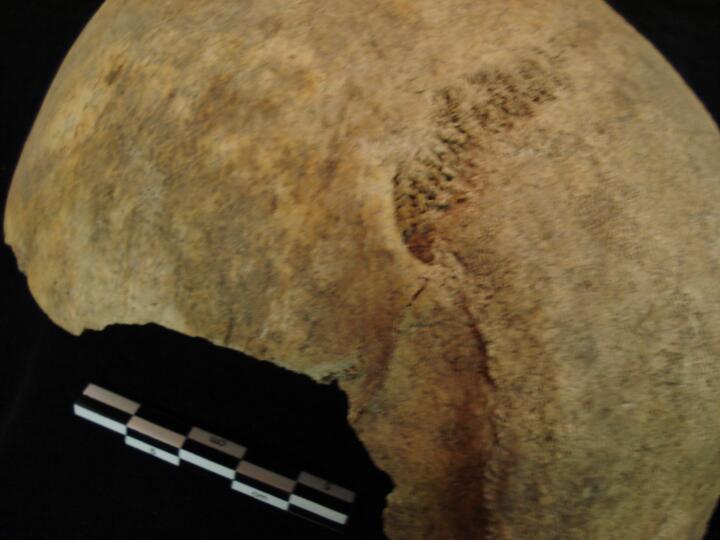
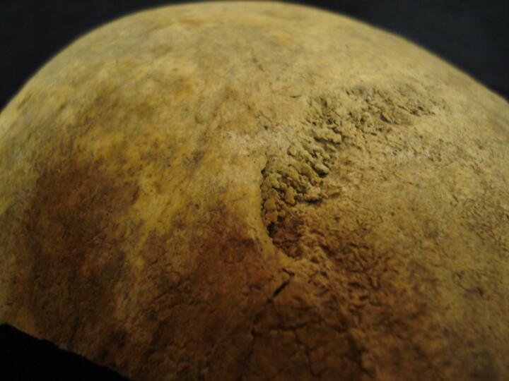
BA84_3002_1.jpg
|
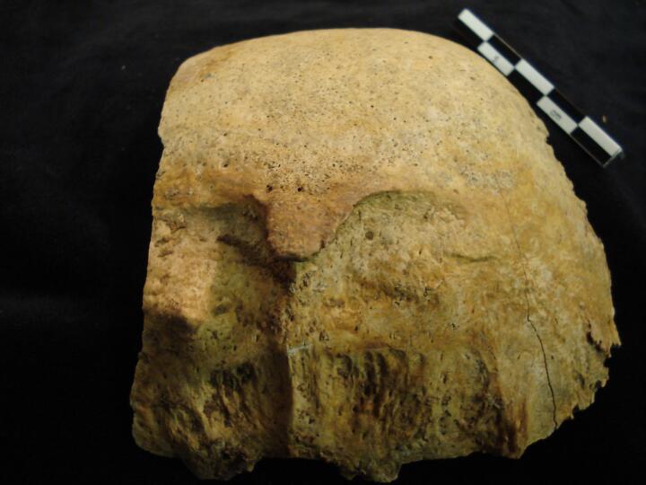
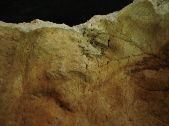
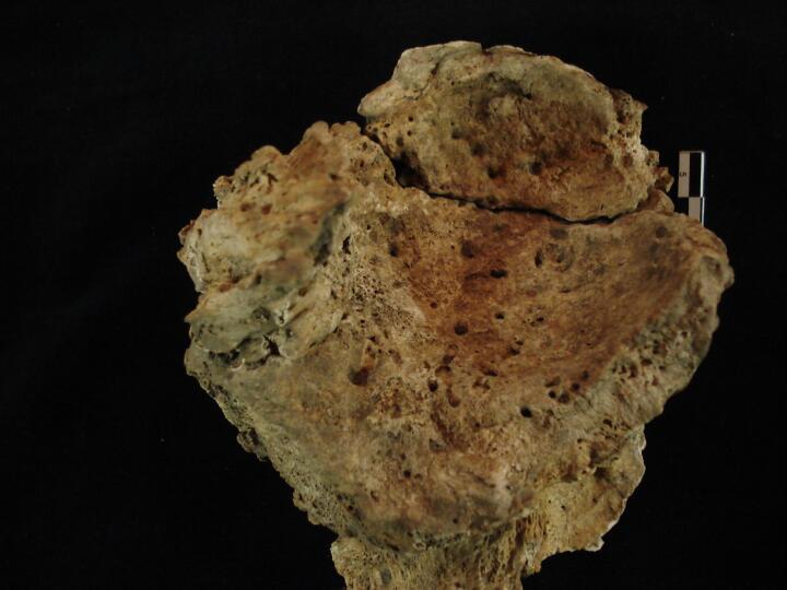
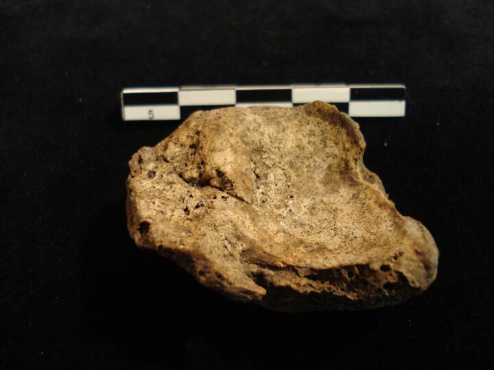
Skull, parietal fragments in cross section, thickened appearance ?Paget's Disease
|
| BA84
|
3002
|
2
|
BA84_3002_2.jpg
|
Skull, parietal fragment in cross section, thickened appearance ?Paget's Disease
|
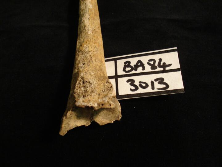
| BA84
|
3013
|
1
|
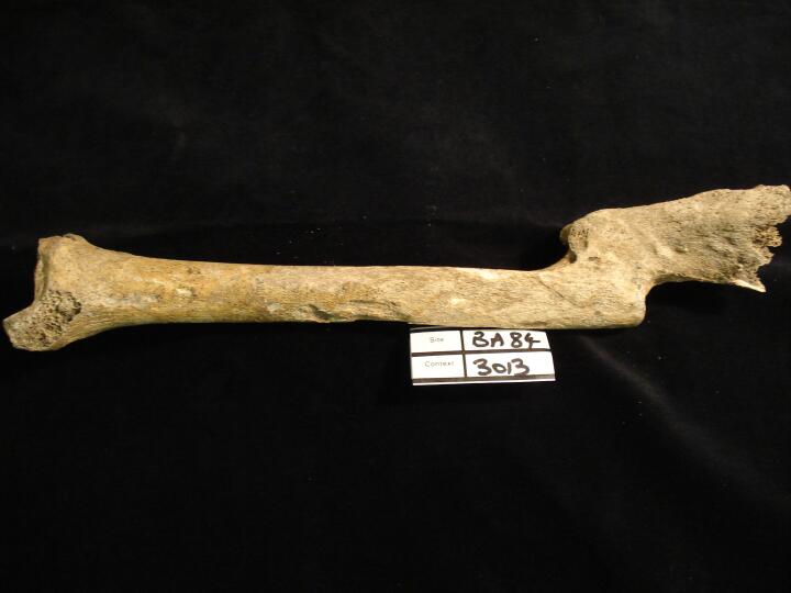
BA84_3013_1.jpg
|
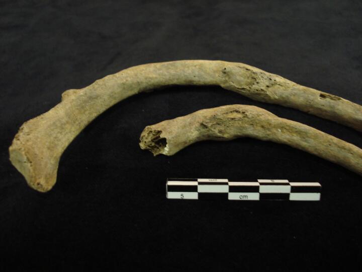
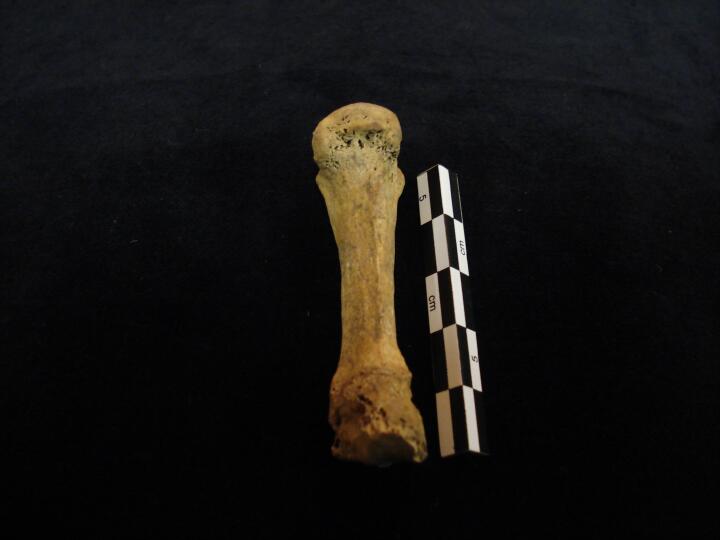
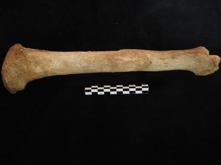
Left radius healed fracture (posterior view)
|
| BA84
|
3013
|
2
|
BA84_3013_2.jpg
|
Left radius healed fracture (anterior view)
|
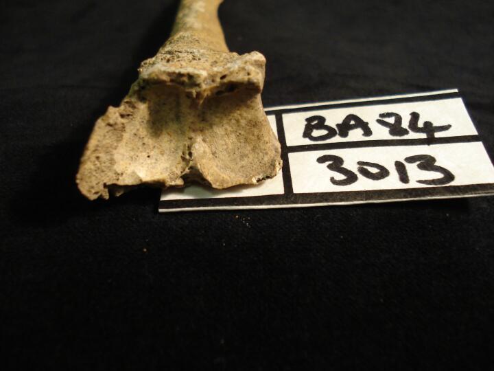
| BA84
|
3013
|
3
|
BA84_3013_3.jpg
|
Left radius view of distal articular surface with eburnation, osteoarthritis (OA)
|
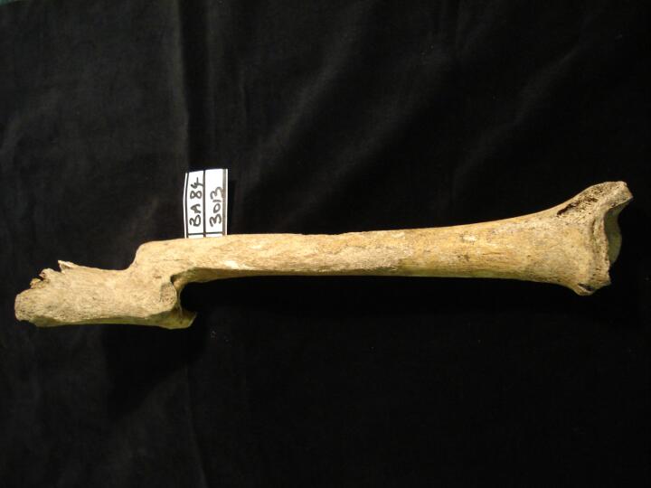
| BA84
|
3013
|
4
|
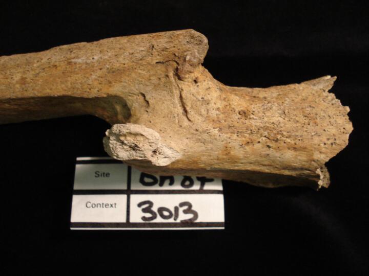
BA84_3013_4.jpg
|
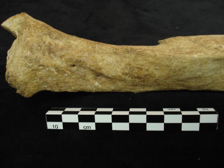
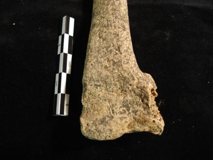
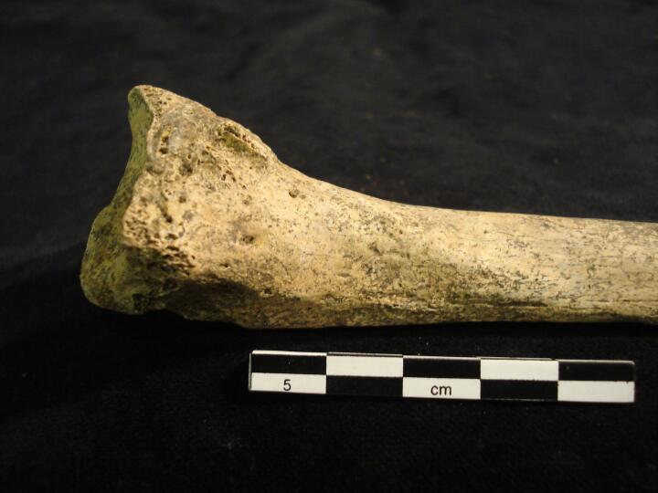
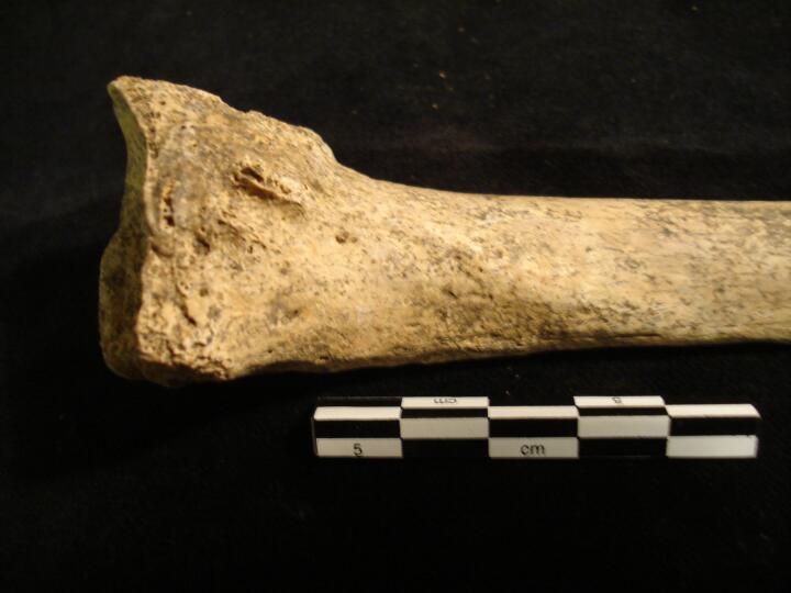
Right tibia healed malaigned fracture (full length anterior view)
|
| BA84
|
3013
|
5
|
BA84_3013_5.jpg
|
Right tibia healed malaligned fracture (close up anterior view)
|
| BA84
|
3013
|
6
|
BA84_3013_6.jpg
|
Right tibia healed malaligned fracture (close up posterior view)
|
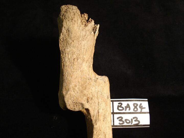
| BA84
|
3013
|
7
|
BA84_3013_7.jpg
|
Right tibia helaed fracture (posterior view/medial)
|
| BA84
|
3013
|
8
|
BA84_3013_8.jpg
|
Right tibia healed malaligned fracture (full length medial view)
|
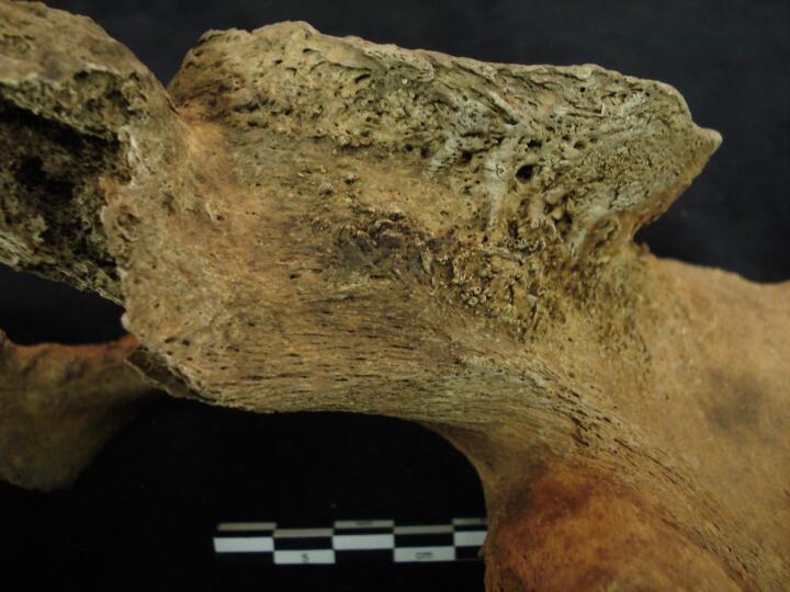
| BA84
|
3031
|
1
|
BA84_3031_1.jpg
|
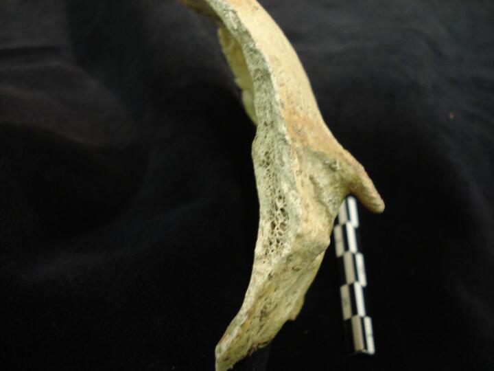
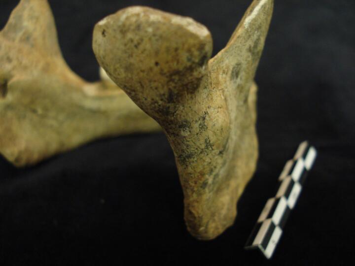
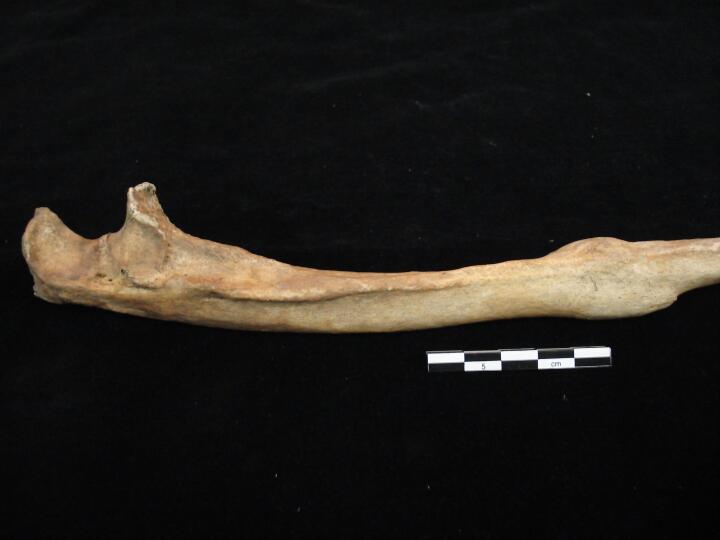
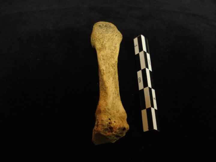
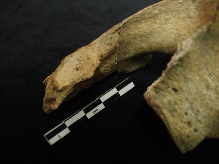
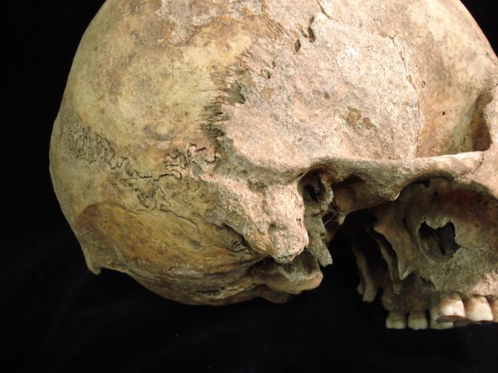
Skull, frontal bone (right side view) junction of coronal suture & temporal bone, healed blunt force trauma
|
| BA84
|
3031
|
2
|
BA84_3031_2.jpg
|
Skull, frontal bone (right side view/close up) junction of coronal suture & temporal bone, healed blunt force trauma
|
| BA84
|
3031
|
3
|
BA84_3031_3.jpg
|
Skull, frontal bone (front view/close up) junction of coronal suture & temporal bone, healed blunt force trauma
|
| BA84
|
3031
|
4
|
BA84_3031_4.jpg
|
Skull, frontal bone (looking across suture/close up) directly towards healed blunt force trauma
|
| BA84
|
3031
|
5
|
BA84_3031_5.jpg
|
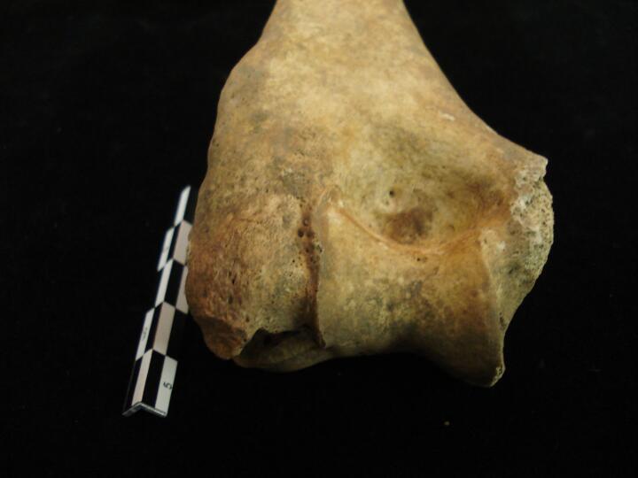
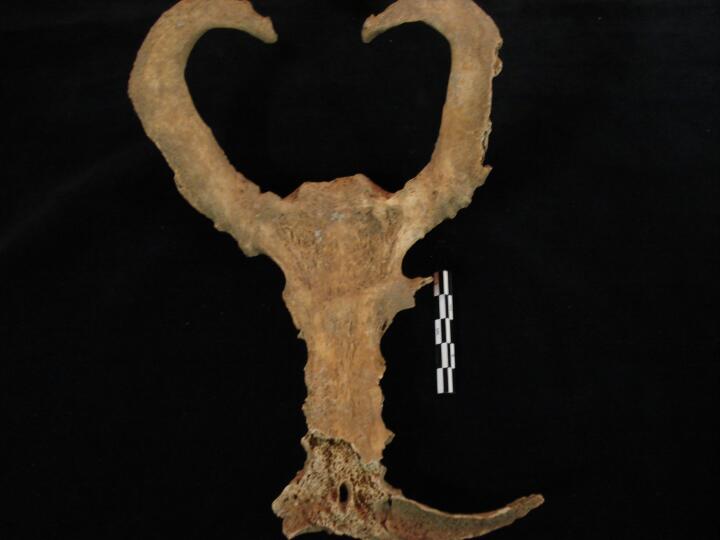
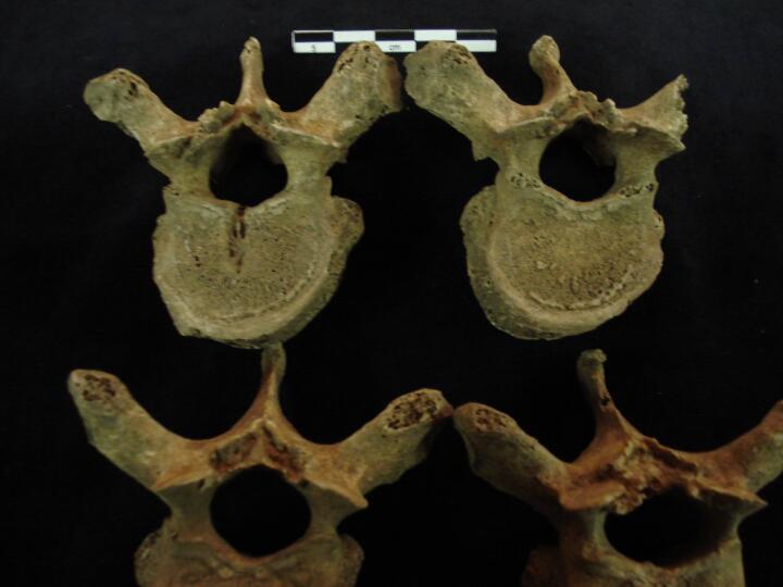
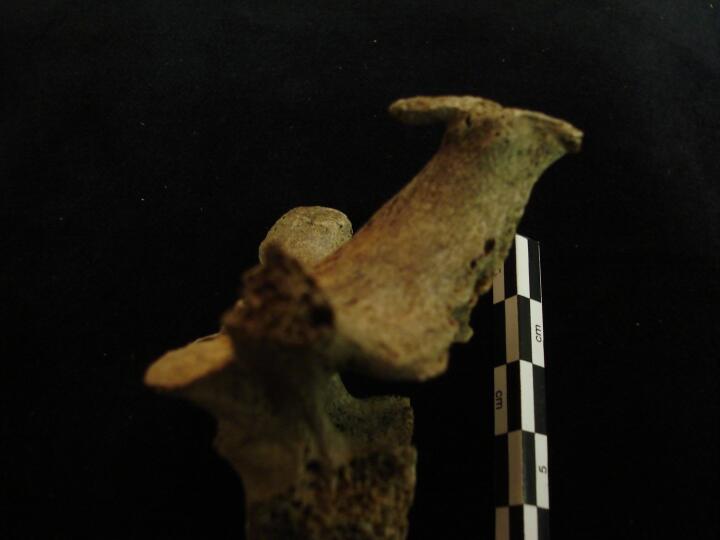
Cervical vertebrae C3 & C4 (posterior view) fusion of apophyseal joints & on C3 superior facets destruction of joint surface & eburnation
|
| BA84
|
3031
|
6
|
BA84_3031_6.jpg
|
Cervical vertebrae C3 & C4 (anterior view) fusion of apophyseal joints
|
| BA84
|
3031
|
7
|
BA84_3031_7.jpg
|
Cervical vertebrae C3 & C4 (rights side view) fusion of apophyseal joints
|
| BA84
|
3031
|
8
|
BA84_3031_8.jpg
|
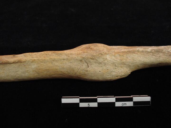
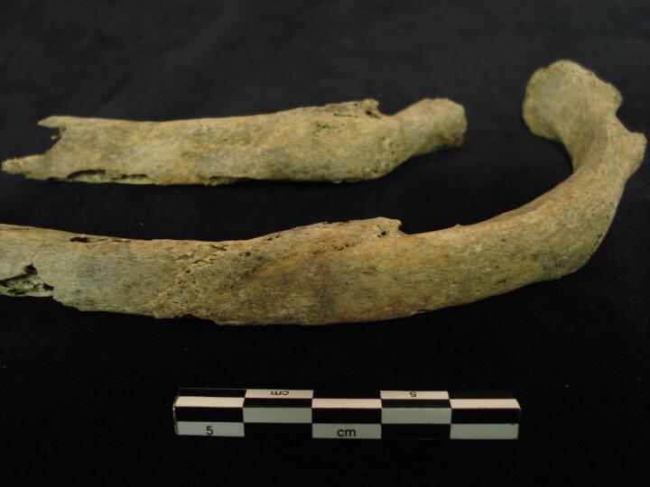
Thoracic vertebrae (right side view) fusion of anterior vertebral bodies
|
| BA84
|
3031
|
9
|
BA84_3031_9.jpg
|
Thoracic vertebrae (anterior view) fusion 'candlewax' of anterior vertebral bodies on right side
|
| BA84
|
3031
|
10
|
BA84_3031_10.jpg
|
Thoracic vertebrae, upper (posterior view) fusion of the apophyseal joint
|
| BA84
|
3031
|
11
|
BA84_3031_11.jpg
|
Thoracic vertebrae, upper (anterior view) fusion across the whole centrum
|
| BA84
|
3048
|
1
|
BA84_3048_1.jpg
|
Sacrolilac fusion (ankylosis) of the left side (anterior view)
|
| BA84
|
3048
|
2
|
BA84_3048_2.jpg
|
Sacrolilac fusion (ankylosis) of the left side (anterior view/close up)
|
| BA84
|
3048
|
3
|
BA84_3048_3.jpg
|
Sacrolilac fusion (ankylosis) of the left side (superior view/close up)
|
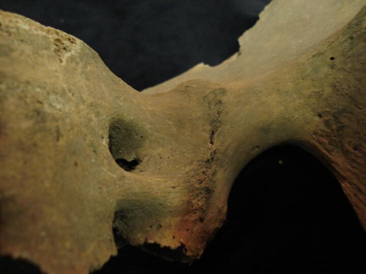
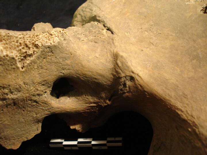
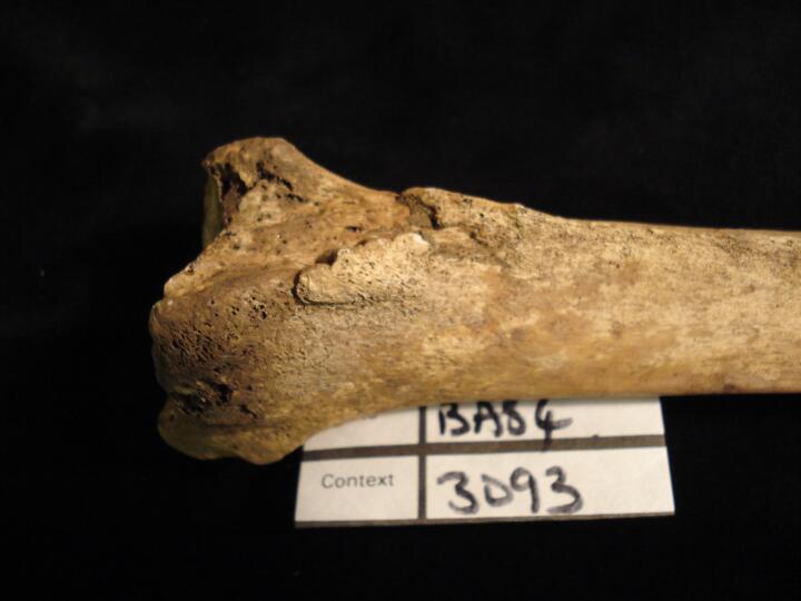
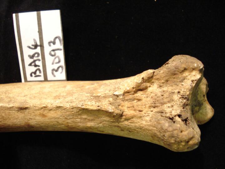
| BA84
|
3093
|
1
|
BA84_3093_1.jpg
|
Left tibia soft tissue trauma (distal end lateral aspect)
|
| BA84
|
3093
|
2
|
BA84_3093_2.jpg
|
Left tibia soft tissue trauma (distal end/lateral aspect towards interosseous border)
|
| BA84
|
3119
|
1
|
BA84_3119_1.jpg
|
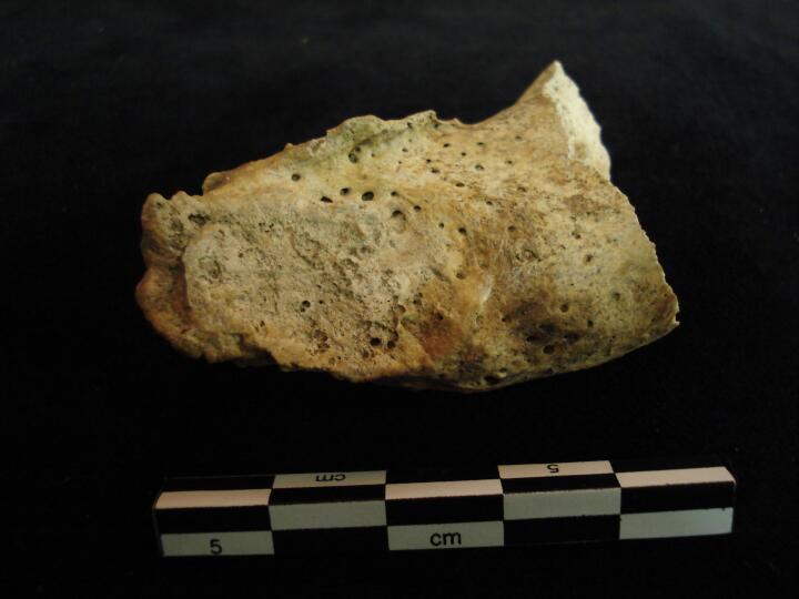
Right calcaneus, talus & navicular (superior view) destructive changes, ? Septic arthropathy
|
| BA84
|
3119
|
2
|
BA84_3119_2.jpg
|
Right talus head & navicular destructive changes, ? Septic arthropathy
|
| BA84
|
3119
|
3
|
BA84_3119_3.jpg
|
Right talus & navicular (posterior view) destructive changes, ? Septic arthropathy
|
| BA84
|
3119
|
4
|
BA84_3119_4.jpg
|
Right talus & navicular (anterior view) destructive changes, ? Septic arthropathy
|
| BA84
|
3127
|
1
|
BA84_3127_1.jpg
|
Manubrium, sternum & 1st ribs all fused (anterior view)
|
| BA84
|
3127
|
2
|
BA84_3127_2.jpg
|
Manubrium & 1st ribs fused (anterior view)
|
| BA84
|
3127
|
3
|
BA84_3127_3.jpg
|
Sternum & lower rib fused (anterior view) & sternal foramen
|
| BA84
|
3127
|
4
|
BA84_3127_4.jpg
|
Manubrium, sternum & 1st ribs all fused (posterior view)
|
| BA84
|
3175
|
1
|
BA84_3175_1.jpg
|
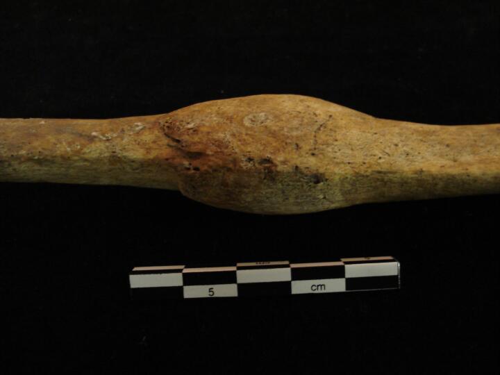
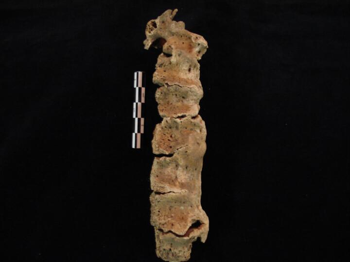
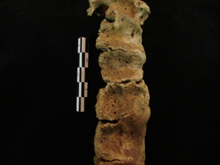
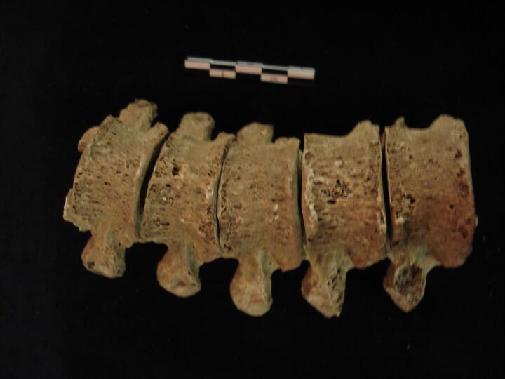
Thoracic vertebrae (right side view) showing 'candewax' fusion, Diffuse Idiopathic Skeletal Hyperostosis (DISH)
|
| BA84
|
3175
|
2
|
BA84_3175_2.jpg
|
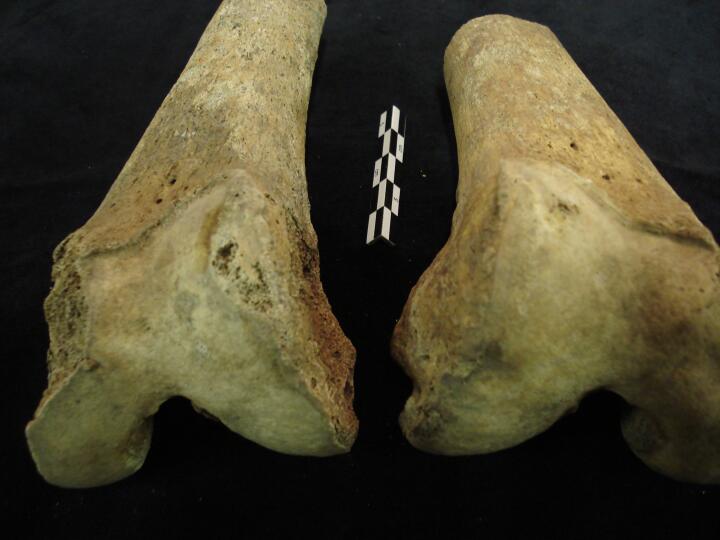
Thoracic vertebrae (right side view/close up) showing 'candlewax' fusion, Diffuse Idiopathic Skeletal Hyperostosis (DISH)
|
| BA84
|
3175
|
3
|
BA84_3175_3.jpg
|
Thoracic vertebrae (anterior view) showing 'candlewax' fusion, Diffuse Idiopathic Skeletal Hyperostosis (DISH)
|
| BA84
|
3181
|
1
|
BA84_3181_1.jpg
|
Left 1st metacarpal (proximal end) joint changes & eburnation, osteoarthritis
|
| BA84
|
3181
|
2
|
BA84_3181_2.jpg
|
Left 1st metacarpal (proximal end) joint changes & eburnation, osteoarthritis
|
| BA84
|
3190
|
1
|
BA84_3190_1.jpg
|
Skull, left frontal fragment (endocranial surface) raised nodules of bone, Hperostosis Frontalis Interna (Stage 3)
|
| BA84
|
3190
|
2
|
BA84_3190_2.jpg
|
Skull, left frontal fragment (endocranial surface/close up) raised nodules of bone, Hperostosis Frontalis Interna (Stage 3)
|
| BA84
|
3200
|
1
|
BA84_3200_1.jpg
|
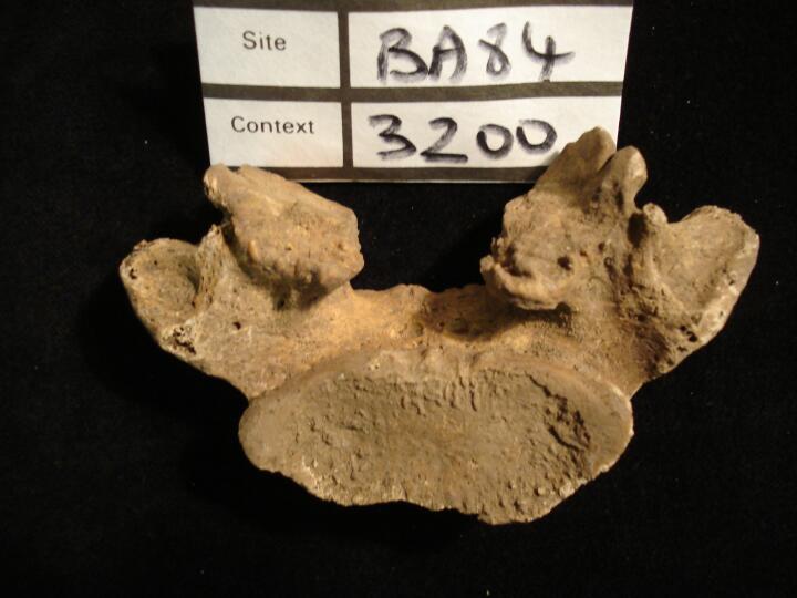
Lumbar L5 bilateral spondylosisis (inferior view) (pars interarticularis not present)
|
| BA84
|
3200
|
2
|
BA84_3200_2.jpg
|
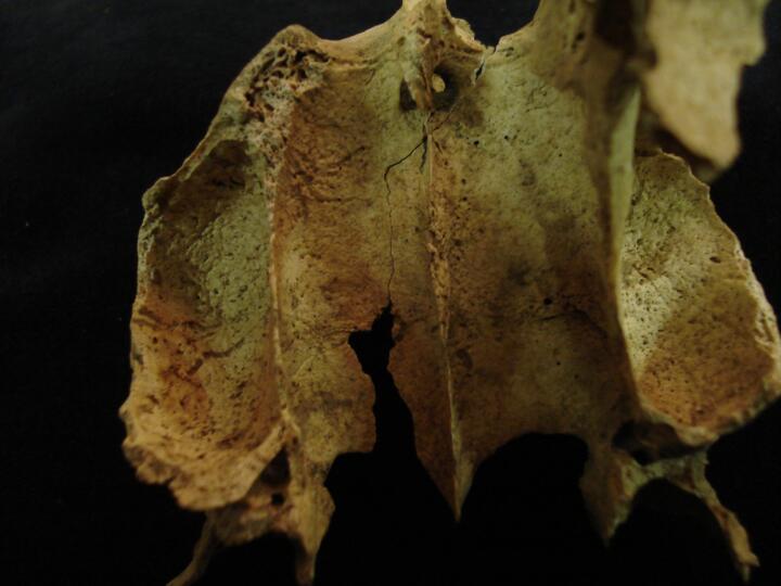
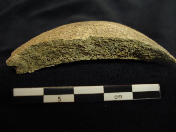
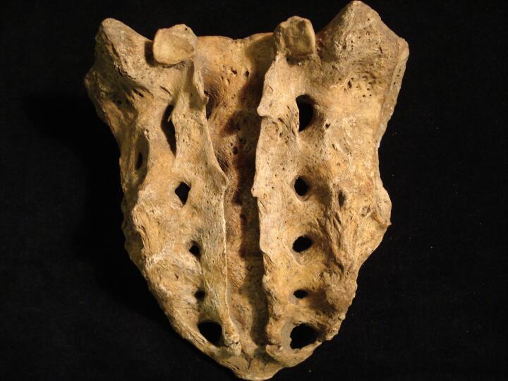
Sacrum (posterior view) spina bifida occulta (cleft from S1 to S5) & sacralisation of 1st coccygeal vertebra
|
| BA84
|
3203
|
1
|
BA84_3203_1.jpg
|
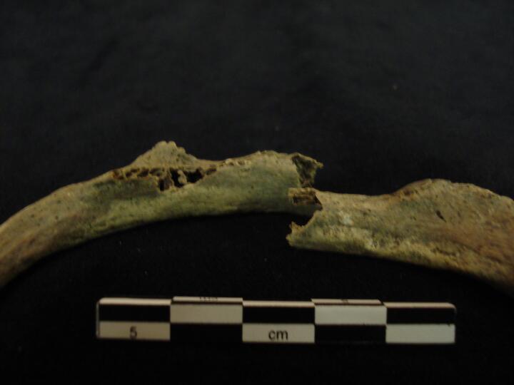
Two left ribs (visceral surface) healed fracture
|
| BA84
|
3203
|
2
|
BA84_3203_2.jpg
|
Two left ribs (visceral surface/close up) healed fracture
|
| BA84
|
3203
|
3
|
BA84_3203_3.jpg
|
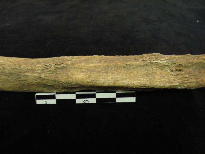
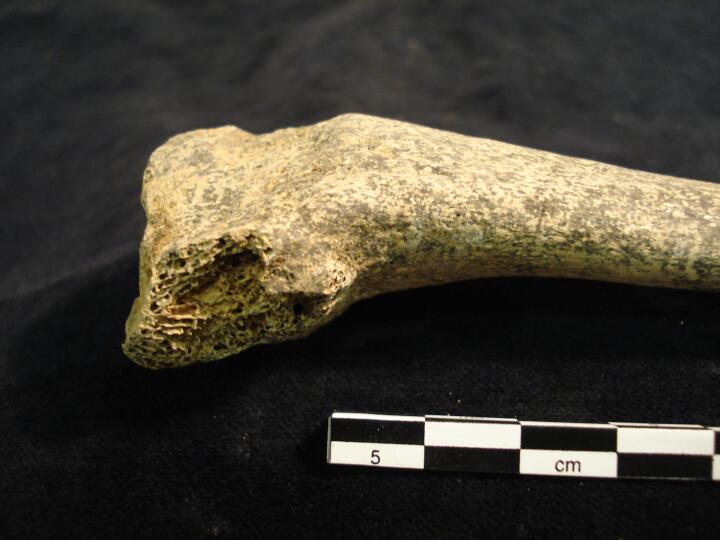
Two left ribs (pleural surface) healed fracture
|
| BA84
|
3203
|
4
|
BA84_3203_4.jpg
|
Thoracic vertebra Th5 to TH9 (anterior view) lateral wedging of vertebral bodies, scoliosis
|
| BA84
|
3203
|
5
|
BA84_3203_5.jpg
|
Thoracic vertebrae, various (superior view) marked 'V' shaped impression on the supeior aspect of the spinous processes
|
| BA84
|
3203
|
6
|
BA84_3203_6.jpg
|
Thoracic vertebra Th9 (superior view) centrum with Schmorl's node & marked 'V' shape impression on spinous process
|
| BA84
|
3203
|
7
|
BA84_3203_7.jpg
|
Thoracic vertebra Th9 (inferior view) centrum with Schmorl's node
|
| BA84
|
3203
|
8
|
BA84_3203_8.jpg
|
Thoracic vertebrae, articulated (posterior view) bony extensions eminating from end of spinous process
|
| BA84
|
3203
|
9
|
BA84_3203_9.jpg
|
Thoracic vertebrae Th5 & Th6 showing the corresponding marked 'V' shaped impressions on the spinous process
|
| BA84
|
3203
|
10
|
BA84_3203_10.jpg
|
Thoracic vertebra Th3 (side view) 'hook' bony extension from end of spinous process
|
| BA84
|
3220
|
1
|
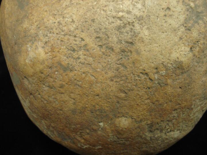
BA84_3220_1.jpg
|
Skull (superior view) multiple button osteoma's
|
| BA84
|
3220
|
2
|
BA84_3220_2.jpg
|
Skull, frontal bone close up of three button osteoma's
|
| BA84
|
3220
|
3
|
BA84_3220_3.jpg
|
Skull, parietal bones close up of two button osteoma's
|
| BA84
|
3220
|
4
|
BA84_3220_4.jpg
|
Skull, occipital bone close up of two button osteoma's
|
| BA84
|
3220
|
5
|
BA84_3220_5.jpg
|
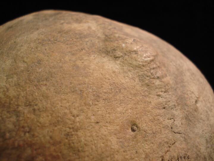
Skull, left parietal close up of one button osteoma
|
| BA84
|
3220
|
6
|
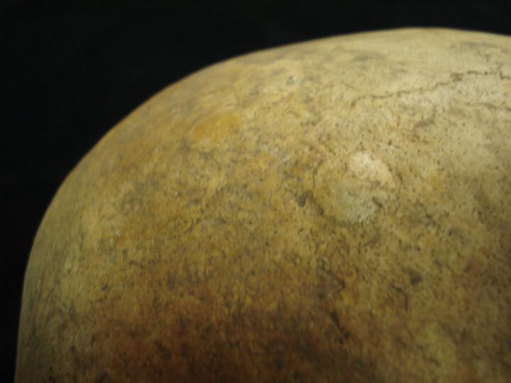
BA84_3220_6.jpg
|
Skull, side view of button osteoma
|
| BA84
|
3231
|
1
|
BA84_3231_1.jpg
|
Left maxilla (palatal/lingual view) 'peg' 2nd premolar
|
| BA84
|
3231
|
2
|
BA84_3231_2.jpg
|
Left maxilla (buccal view) 'peg' 2nd premolar
|
| BA84
|
3231
|
3
|
BA84_3231_3.jpg
|
Left maxilla (lingual view) 'peg' 2nd premolar
|
| BA84
|
3231
|
4
|
BA84_3231_4.jpg
|
Mandible (buccal view) external draining periapical lesion (abscess) anterior teeth
|
| BA84
|
3231
|
5
|
BA84_3231_5.jpg
|
Mandible (buccal view/close up) external draining periapical lesion (abscess) anterior teeth
|
| BA84
|
3231
|
6
|
BA84_3231_6.jpg
|
Right side of mandible (lingual view) new bone growth associated with infection
|
| BA84
|
3231
|
7
|
BA84_3231_7.jpg
|
Right side of mandible (lingual view/close up) new bone growth associated with infection
|
| BA84
|
3231
|
8
|
BA84_3231_8.jpg
|
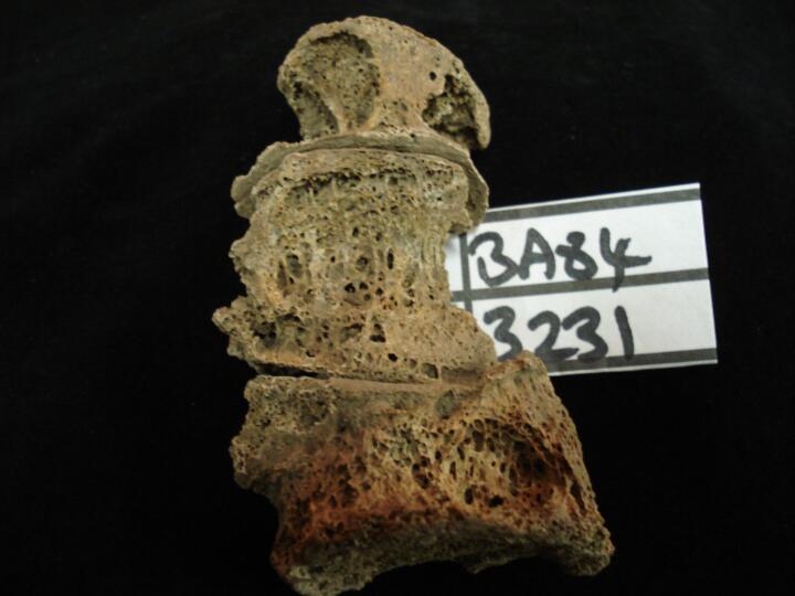
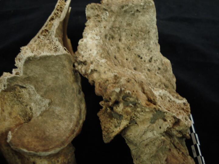
Lumbar vertebrae L3, l4 & L5 articulating (anterior view) gross destruction of anterior bodies from specific infection, Tuberculosis
|
| BA84
|
3231
|
9
|
BA84_3231_9.jpg
|
Lumbar vertebrae L3, l4 & L5 articulating (anterior view) destruction of anteriro bodies from specific infection, Tuberculosis
|
| BA84
|
3231
|
10
|
BA84_3231_10.jpg
|
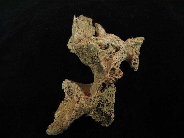
Lumbar vertebra ?L5 & L4 (side view) showing fusion of spinous process & apophyseal joint, tuberculosis
|
| BA84
|
3231
|
11
|
BA84_3231_11.jpg
|
Lumbar vertebra ?L5 & L4 (side view/close up) showing fusion of spinous process & apophyseal joint, tuberculosis
|
| BA84
|
3231
|
12
|
BA84_3231_12.jpg
|
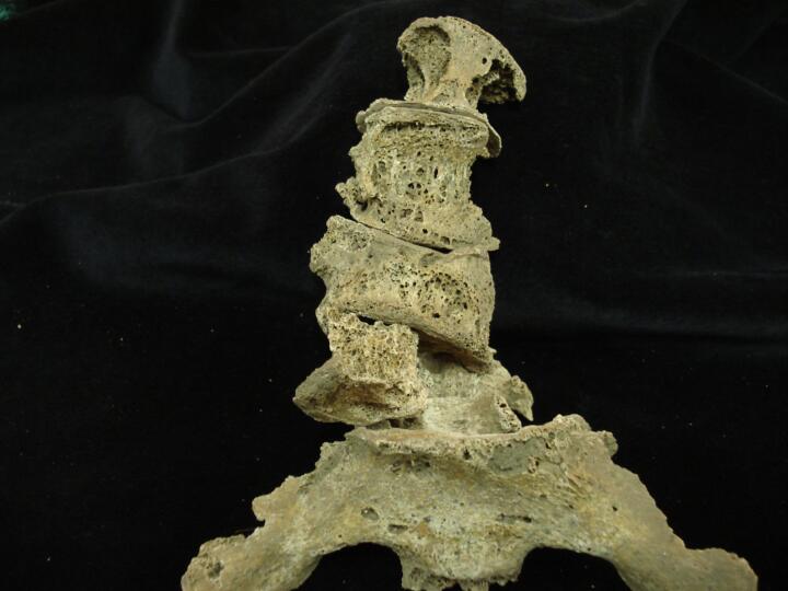
Articulation of lumbar vertebrae & sacrum (some PM damage) showing gross destruction to the vertebral bodies from tuberculosis & collapse of vertebrae (anterior view)
|
| BA84
|
3231
|
13
|
BA84_3231_13.jpg
|
Articulation of lumbar vertebrae L2 to L5 (some PM damage) showing gross destruction to the vertebral bodies from tuberculosis & collapse of vertebrae (anterior view)
|
| BA84
|
3259
|
1
|
BA84_3259_1.jpg
|
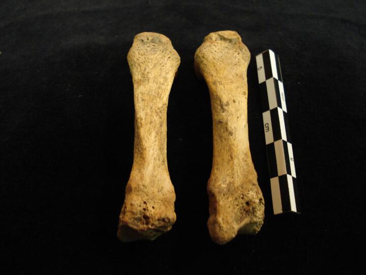
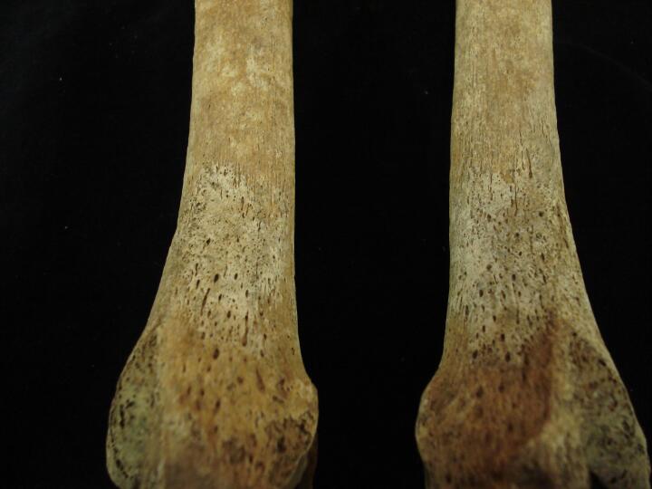
Left 3rd metacarpal (dorsal view) absent styloid process
|
| BA84
|
3259
|
2
|
BA84_3259_2.jpg
|
Left 3rd metacarpal (palmar view) absent styloid process
|
| BA84
|
3259
|
3
|
BA84_3259_3.jpg
|
Left & Right 3rd metacarpal to show comparison of styloid process
|
| BA84
|
3259
|
4
|
BA84_3259_4.jpg
|
Left 3rd metacarpal, distal articular end, & absent styloid process
|
| BA84
|
3259
|
5
|
BA84_3259_5.jpg
|
Lumbar vertebrae, L3 & L4 (anterior view) probable indication with destruction of anterior vertebral bodies of specific infection, Tuberculosis
|
| BA84
|
3259
|
6
|
BA84_3259_6.jpg
|
Lumbar vertebrae, L3 & L4 (anterior view/close up) probable indication with destruction of anterior vertebral bodies of specific infection, ?Tuberculosis
|
| BA84
|
3259
|
7
|
BA84_3259_7.jpg
|
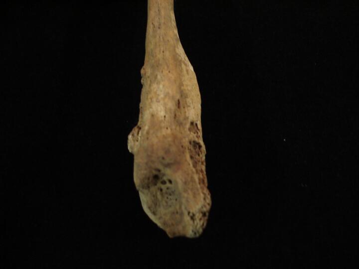
Lumbar vertebra L3 (inferior view) destruction of centrum from infection, ?Tuberculosis
|
| BA84
|
3259
|
8
|
BA84_3259_8.jpg
|
Lumbar vertebra L3 (inferior view/close up) destruction of centrum from infection, ?Tuberculosis (some PM damage)
|
| BA84
|
3259
|
9
|
BA84_3259_9.jpg
|
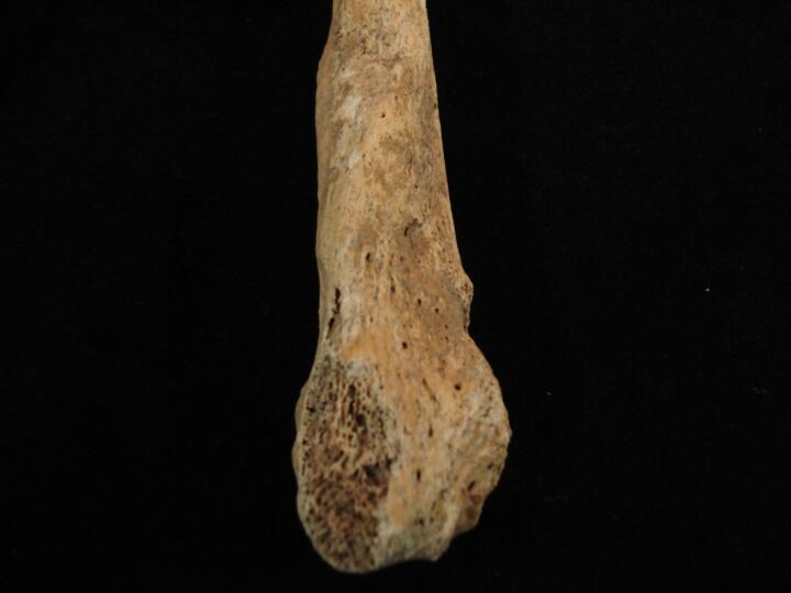
Lumber vertebra L3 (left side view) destruction of vertebral body from infection, ?Tuberculosis
|
| BA84
|
3259
|
10
|
BA84_3259_10.jpg
|
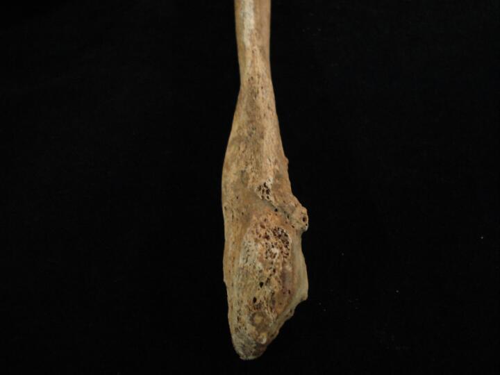
Lumber vertebra L3 (anterior view/close) destruction of vertebral body from infection, ?Tuberculosis
|
| BA84
|
3274
|
1
|
BA84_3274_1.jpg
|
Right scapula acromial end (posterior view) degenerative joint changes
|
| BA84
|
3274
|
2
|
BA84_3274_2.jpg
|
Right acromial joint (broken PM) degenerative joint changes & eburnation, osteoarthrits (inferior view)
|
| BA84
|
3274
|
3
|
BA84_3274_3.jpg
|
Right clavicle, acromial end degenerative joint changes
|
| BA84
|
3274
|
4
|
BA84_3274_4.jpg
|
Maxilla,left (buccal view) exteranl draining periapical lesion (abscess)
|
| BA84
|
3274
|
5
|
BA84_3274_5.jpg
|
Left & right calcanea Os calcis, non-metric trait calcaneal notch
|
| BA84
|
3274
|
6
|
BA84_3274_6.jpg
|
Right calcaneus, os calcis, non-metric trait calcaneal notch (close up)
|
| BA84
|
3274
|
7
|
BA84_3274_7.jpg
|
Right talus (plantar view) bony nodules in the sulcus
|
| BA84
|
3274
|
8
|
BA84_3274_8.jpg
|
Left tibia healed fracture, full length (anterior view)
|
| BA84
|
3274
|
9
|
BA84_3274_9.jpg
|
Left tibia healed fracture, full length (anterior view/close up)
|
| BA84
|
3274
|
10
|
BA84_3274_10.jpg
|
Left tibia healed fracture (medial view) close up of fracture site
|
| BA84
|
3274
|
11
|
BA84_3274_11.jpg
|
Left tibia healed fracture, mid shaft (anterior view/close up)
|
| BA84
|
3274
|
12
|
BA84_3274_12.jpg
|
Left tibia healed fracture, mid shaft (anterior view/close up)
|
| BA84
|
3274
|
13
|
BA84_3274_13.jpg
|
Left tibia distal end (lateral view) showing healed fracture
|
| BA84
|
3274
|
14
|
BA84_3274_14.jpg
|
Right femoral head (anterior view) joint changes
|
| BA84
|
3274
|
15
|
BA84_3274_15.jpg
|
Right femoral head (posterior view) joint changes
|
| BA84
|
3274
|
16
|
BA84_3274_16.jpg
|
Right femoral head (medial view) joint changes
|
| BA84
|
3274
|
17
|
BA84_3274_17.jpg
|
Right femoral head (medial view) joint changes
|
| BA84
|
3274
|
18
|
BA84_3274_18.jpg
|
Left & right femoral heads for comparison (anterior view)
|
| BA84
|
3274
|
19
|
BA84_3274_19.jpg
|
Right pelvis, acetabulum two fragments separate, healed but non united fracture
|
| BA84
|
3274
|
20
|
BA84_3274_20.jpg
|
Right pelvis, acetabulum two fragments (placed anatomically together), healed but non united fracture
|
| BA84
|
3274
|
21
|
BA84_3274_21.jpg
|
Right pelvis, acetabulum three fragments (placed anatomically together), healed but non united fracture
|
| BA84
|
3274
|
22
|
BA84_3274_22.jpg
|
Right pelvis medial view of internal surface of acetabulum
|
| BA84
|
3274
|
23
|
BA84_3274_23.jpg
|
Right pelvis, acetabulum five fragments separate, healed but non united fracture
|
| BA84
|
3274
|
24
|
BA84_3274_24.jpg
|
Five fragments of right acetabulum from fracture, healed but non united
|
| BA84
|
3274
|
25
|
BA84_3274_25.jpg
|
Right pelvis (posterior view)
|
| BA84
|
3274
|
26
|
BA84_3274_26.jpg
|
Right pelvis (medial/posterior view) of acetabulum with one fragment from non united fracture
|
| BA84
|
3274
|
27
|
BA84_3274_27.jpg
|
Right pelvis with one fragment & left pelvis for comparison
|
| BA84
|
3274
|
28
|
BA84_3274_28.jpg
|
Right pelvis (posterior view of non united fracture of the acetabulum
|
| BA84
|
3274
|
29
|
BA84_3274_29.jpg
|
Left & right pelvis comparing the acetabulae (posterior view)
|
| BA84
|
3274
|
30
|
BA84_3274_30.jpg
|
Right pelvis & one fragment from non united fracture (anterior view)
|
| BA84
|
3385
|
1
|
BA84_3385_1.jpg
|
Right fibula distal end (posterior/lateral view) healed fracture
|
| BA84
|
3385
|
2
|
BA84_3385_2.jpg
|
Right fibula distal end (posterior view) healed fracture
|
| BA84
|
3385
|
3
|
BA84_3385_3.jpg
|
Right fibula distal end (posterior/anterior view) healed fracture
|
| BA84
|
3385
|
4
|
BA84_3385_4.jpg
|
Lumbar vertebra L5 centrum (inferior view) ?infection
|
| BA84
|
3385
|
5
|
BA84_3385_5.jpg
|
Sacrum S1 body (superior view) ?infection
|
| BA84
|
3385
|
6
|
BA84_3385_6.jpg
|
Sacrum S1 body (superior view/close up) ?infection
|
| BA84
|
3385
|
7
|
BA84_3385_7.jpg
|
Sacrum & lumbar L5 showing corresponding area of ?infection
|
| BA84
|
3400
|
1
|
BA84_3400_1.jpg
|
Skull (base view) torticolis
|
| BA84
|
3400
|
2
|
BA84_3400_2.jpg
|
Skull (base view/close up) torticolis
|
| BA84
|
3400
|
3
|
BA84_3400_3.jpg
|
Right orbit, inferior margin bony 'hook' like projection (side view)
|
| BA84
|
3400
|
4
|
BA84_3400_4.jpg
|
Right orbit, inferior margin bony 'hook' like projection (front view)
|
| BA84
|
3400
|
5
|
BA84_3400_5.jpg
|
Skull (front view) uneven nasal aperture
|
| BA84
|
3400
|
6
|
BA84_3400_6.jpg
|
Skull (side view) occipital bone prominent inion protuberance
|
| BA84
|
3458
|
1
|
BA84_3458_1.jpg
|
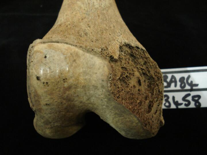
Right femur distal end (anterior view) joint change & eburnation, osteoarthritis of patellar surface
|
| BA84
|
3458
|
2
|
BA84_3458_2.jpg
|
Right femur proximal end (posterior view) marked muscle attachments
|
| BA84
|
3458
|
3
|
BA84_3458_3.jpg
|
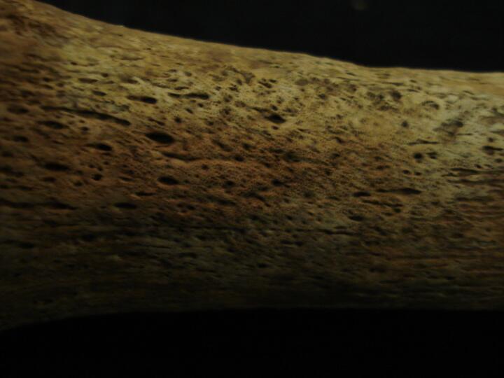
Right tibia distal end (lateral view) healed non-specific periosteal infection
|
| BA84
|
3458
|
4
|
BA84_3458_4.jpg
|
Right tibia distal end (medial view) healed non-specific periosteal infection
|
| BA84
|
3458
|
5
|
BA84_3458_5.jpg
|
Right tibia distal end (medial view/close up) healed non-specific periosteal infection
|
| BA84
|
3458
|
6
|
BA84_3458_6.jpg
|
Right & left tibia distal end (medial view) active & healed non-specific periosteal infection
|
| BA84
|
3458
|
7
|
BA84_3458_7.jpg
|
Right & left tibia proximal end (posterior view) marked muscle attachment for Soleus
|
| BA84
|
3515
|
1
|
BA84_3515_1.jpg
|
Left metatarsals MT 3, 4 & 5 (dorsal view) ridge on dorsal surface of the bones, ?trauma
|
| BA84
|
3515
|
2
|
BA84_3515_2.jpg
|
Left metatarsals MT 3, 4 & 5 (dorsal view/close up) ridge on dorsal surface of the bones, ?trauma
|
| BA84
|
3515
|
3
|
BA84_3515_3.jpg
|
Metatarsal MT4, (medial view) MT3 (lateral view) enlarged areas for articualtion asociated with ?trauma
|
| BA84
|
3515
|
4
|
BA84_3515_4.jpg
|
Two right ribs (visceral surface) area of healed non-specific infection
|
| BA84
|
3515
|
5
|
BA84_3515_5.jpg
|
One right rib (visceral surface/close up) area of healed non-specific infection
|
| BA84
|
3515
|
6
|
BA84_3515_6.jpg
|
One left rib (visceral surface/close up) area of healed non-specific infection
|
| BA84
|
3541
|
1
|
BA84_3541_1.jpg
|
Right tibia (posterior view) soft tissue trauma proximal end, myostisis ossificans traumatica
|
| BA84
|
3541
|
2
|
BA84_3541_2.jpg
|
Right tibia (medial view) soft tissue trauma proximal end, myostisis ossificans traumatica
|
| BA84
|
3541
|
3
|
BA84_3541_3.jpg
|
Left tibia (posterior view) soft tissue trauma proximal end, myostisis ossificans traumatica
|
| BA84
|
3541
|
4
|
BA84_3541_4.jpg
|
Left tibia (medial view) soft tissue trauma proximal end, myostisis ossificans traumatica
|
| BA84
|
3541
|
5
|
BA84_3541_5.jpg
|
Left & right tibia (posterior view) soft tissue trauma proximal end, myostisis ossificans traumatica
|
| BA84
|
3541
|
6
|
BA84_3541_6.jpg
|
Left & right tibia (side view) soft tissue trauma proximal end, myostisis ossificans traumatica
|
| BA84
|
3541
|
7
|
BA84_3541_7.jpg
|
Right tibia (medial view/close up) soft tissue trauma proximal end, myostisis ossificans traumatica
|
| BA84
|
3541
|
8
|
BA84_3541_8.jpg
|
Left tibia (medial view/close up) soft tissue trauma proximal end, myostisis ossificans traumatica
|
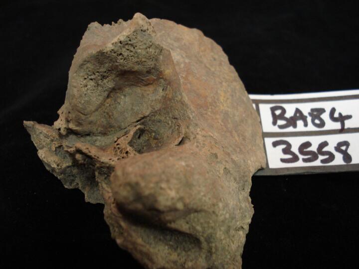
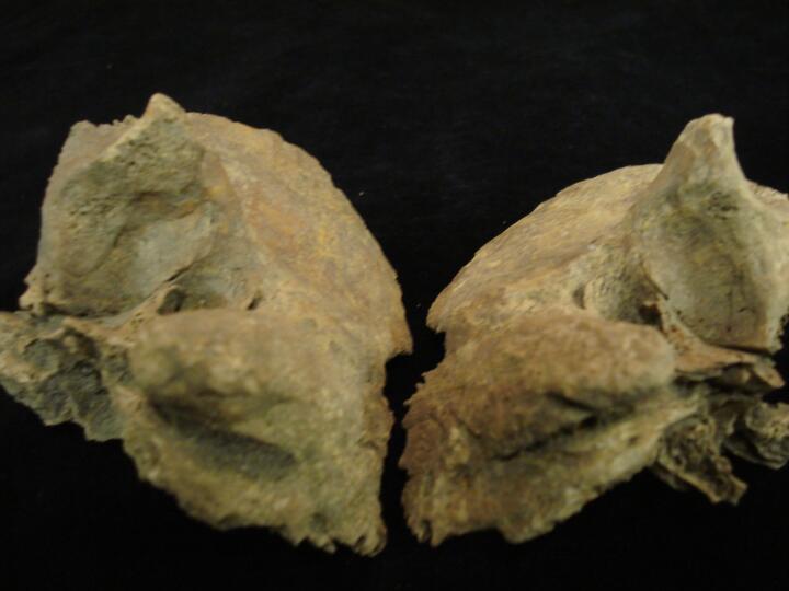
| BA84
|
3558
|
1
|
BA84_3558_1.jpg
|
Mandible (buccal view) very heavy wear of dentition & destruction of left tempro mandibular joint
|
| BA84
|
3558
|
2
|
BA84_3558_2.jpg
|
Mandible (buccal view) close up of very heavy wear of dentition (left side)
|
| BA84
|
3558
|
3
|
BA84_3558_3.jpg
|
Mandible (buccal view) close up of destruction of left tempro mandibular joint
|
| BA84
|
3558
|
4
|
BA84_3558_4.jpg
|
Mandible (lingual view) close up of destruction of left tempro mandibular joint
|
| BA84
|
3558
|
5
|
BA84_3558_5.jpg
|
Mandible (superior view) close up of destruction of left tempro mandibular joint
|
| BA84
|
3558
|
6
|
BA84_3558_6.jpg
|
Skull left tempro mandibular joint surface (posterior view)
|
| BA84
|
3558
|
7
|
BA84_3558_7.jpg
|
Skull left & right tempro mandibular joint surfaces (posterior view)
|
| BA84
|
3558
|
8
|
BA84_3558_8.jpg
|
Lumbar vertebrae L2 & L3 (anterior view) ? Vertebral compression fracture
|
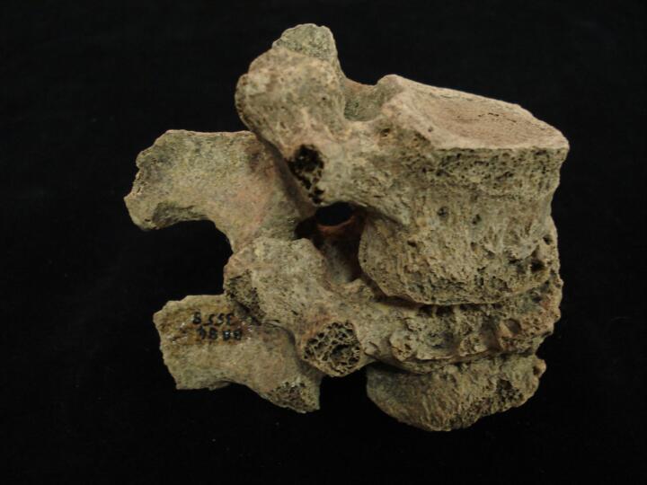
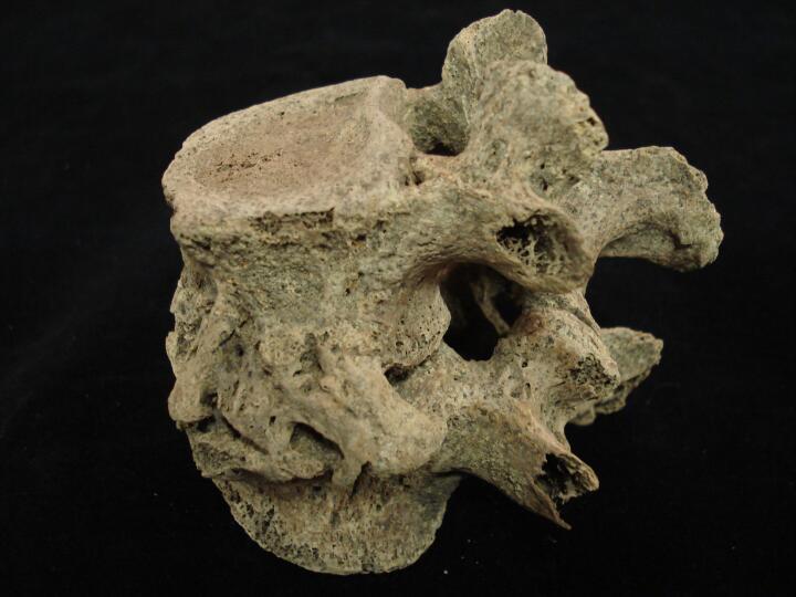
| BA84
|
3558
|
9
|
BA84_3558_9.jpg
|
Lumbar vertebrae L2 & L3 (side/posterior view) ? Vertebral compression fracture
|
| BA84
|
3558
|
10
|
BA84_3558_10.jpg
|
Lumbar vertebrae L2 & L3 (left side view) ? Vertebral compression fracture
|
| BA84
|
3558
|
11
|
BA84_3558_11.jpg
|
Lumbar vertebrae L2 & L3 (anterior view/close up) ? Vertebral compression fracture with fusion across the vertebral bodies
|
| BA84
|
3575
|
1
|
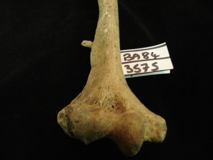
BA84_3575_1.jpg
|
Right humerus distal 1/3 of shaft bony spur/projection at a 90 degree angle (anterior view) soft tissue trauma
|
| BA84
|
3575
|
2
|
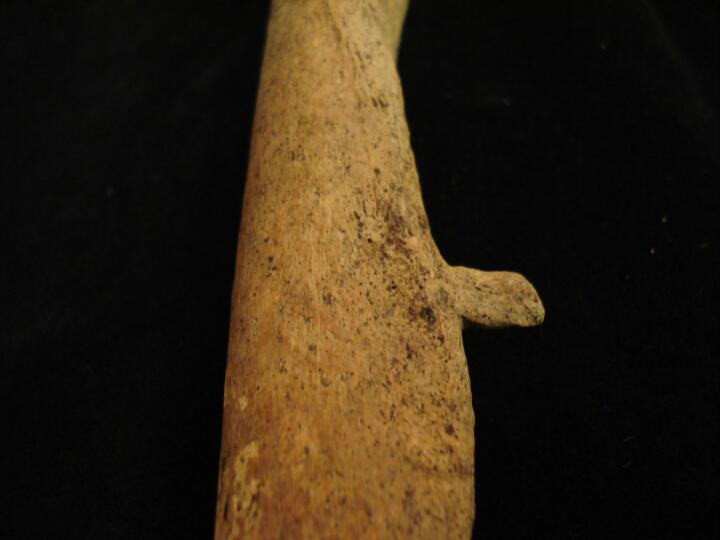
BA84_3575_2.jpg
|
Right humerus distal 1/3 of shaft close up of bony spur/projection at a 90 degree angle (anterior view) soft tissue trauma
|
| BA84
|
3575
|
3
|
BA84_3575_3.jpg
|
Right humerus distal 1/3 of shaft close up of bony spur/projection at a 90 degree angle (posterior view) soft tissue trauma
|
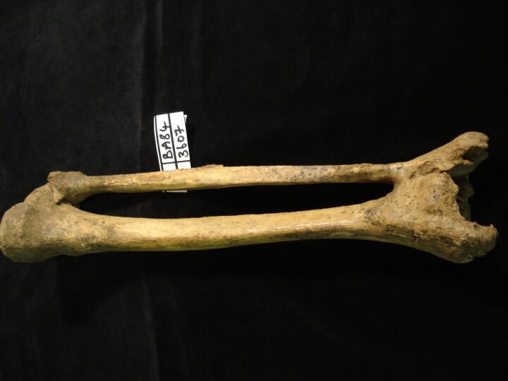
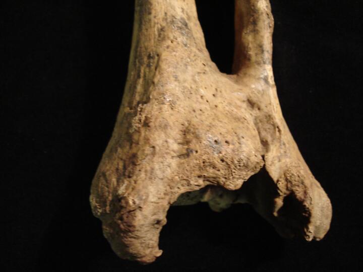
| BA84
|
3607
|
1
|
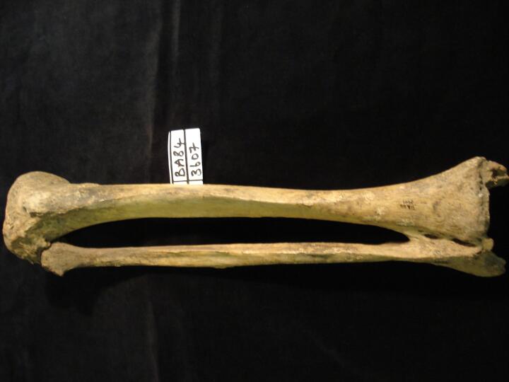
BA84_3607_1.jpg
|
Right tibai & fibula healed fracture with ankylosis between the two bones distal end (anterior view)
|
| BA84
|
3607
|
2
|
BA84_3607_2.jpg
|
Right tibia & fibula healed fracture with ankylosis between the two bones distal end (posterior view)
|
| BA84
|
3607
|
3
|
BA84_3607_3.jpg
|
Right tibia & fibula distal end close up of healed fracture (anterior view)
|
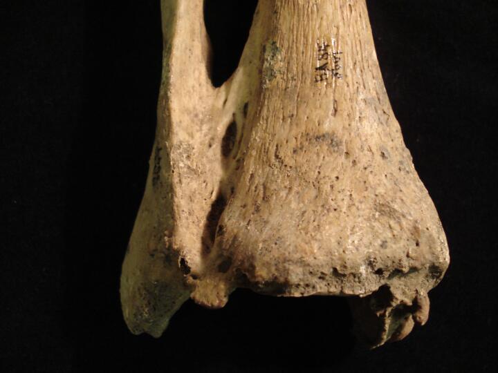
| BA84
|
3607
|
4
|
BA84_3607_4.jpg
|
Right tibia & fibula distal end close up of healed fracture (posterior view)
|
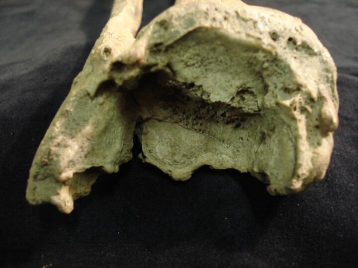
| BA84
|
3607
|
5
|
BA84_3607_5.jpg
|
Right tibia & fibula (superior view) of distal articular surface with intra articular trauma & joint changes
|
| BA84
|
3607
|
6
|
BA84_3607_6.jpg
|
Right tibia & fibula (superior view/close up) of distal articular surface with intra articular trauma & joint changes
|
| BA84
|
3607
|
7
|
BA84_3607_7.jpg
|
Right carpal bones, scaphoid & trapezoid with eburnated surfaces, osteoarthritis
|
| BA84
|
3732
|
1
|
BA84_3732_1.jpg
|
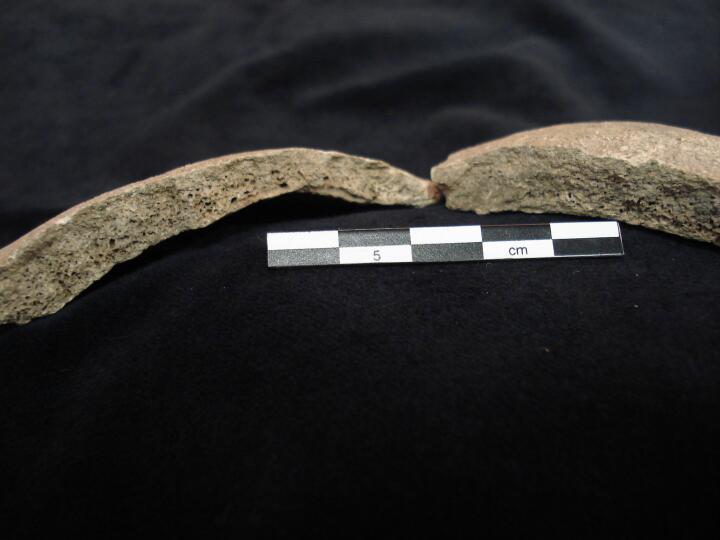
Sacrum (posterior view) spina bifida occulta (cleft from S1 to S5)
|
| BA84
|
3732
|
2
|
BA84_3732_2.jpg
|
Right navicular intra articular fracture & secondary joint changes (posterior view)
|
| BA84
|
3732
|
3
|
BA84_3732_3.jpg
|
Right navicular intra articular fracture & secondary joint changes (posterior view)
|
| BA84
|
3732
|
4
|
BA84_3732_4.jpg
|
Right talus & navicular showing joint changes & intra articular fracture
|
| BA84
|
3732
|
5
|
BA84_3732_5.jpg
|
Right talus (superior view) showing joint changes, asociated with trauma to navicular
|
| BA84
|
3732
|
6
|
BA84_3732_6.jpg
|
Right navicular intra articular fracture & secondary joint changes (posterior view)
|
| BA84
|
3732
|
7
|
BA84_3732_7.jpg
|
Left & right navicular to show comparison (posterior view)
|
| BA84
|
3798
|
1
|
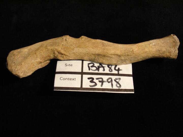
BA84_3798_1.jpg
|
Right clavicle heealed fracture (anterior view)
|
| BA84
|
3798
|
2
|
BA84_3798_2.jpg
|
Right clavicle heealed fracture (posterior view)
|
| BA84
|
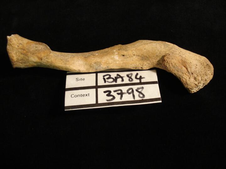
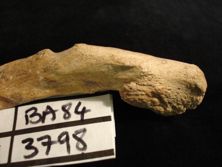
3798
|
3
|
BA84_3798_3.jpg
|
Right clavicle healed fracture (close up acromial end/posterior view)
|
| BA84
|
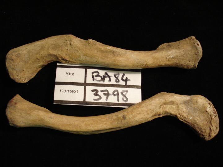
3798
|
4
|
BA84_3798_4.jpg
|
Right & left clavicle to show comparison (anterior view)
|
| BA84
|
3798
|
5
|
BA84_3798_5.jpg
|
Right & left clavicle to show comparison (posterior view)
|
| BA84
|
3798
|
6
|
BA84_3798_6.jpg
|
Skull (front view) frontal bone with metopic suture
|
| BA84
|
3798
|
7
|
BA84_3798_7.jpg
|
Skull (sideview) left coronal suture with coronal wormian bone
|
| BA84
|
3798
|
8
|
BA84_3798_8.jpg
|
Skull (sideview) right coronal suture with coronal wormian bone
|
| BA84
|
3798
|
9
|
BA84_3798_9.jpg
|
Skull showing saggital suture with healed trauma
|
| BA84
|
3798
|
10
|
BA84_3798_10.jpg
|
Skull showing saggital suture with healed trauma (close up)
|
| BA84
|
3798
|
11
|
BA84_3798_11.jpg
|
Skull looking at healed trauma (raised bone) from the frontal bone
|
| BA84
|
3798
|
12
|
BA84_3798_12.jpg
|
Skull (superior view) showing coronal wormian bones & healed trauma on saggital suture
|
| BA84
|
3798
|
13
|
BA84_3798_13.jpg
|
Skull (left side view) healed trauma saggital suture & parietal foramen
|
| BA84
|
3798
|
14
|
BA84_3798_14.jpg
|
Skull (superior/frontal view) showing coronal wormian bones & healed trauma on saggital
|
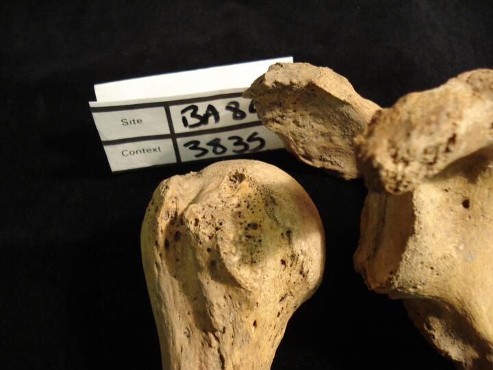
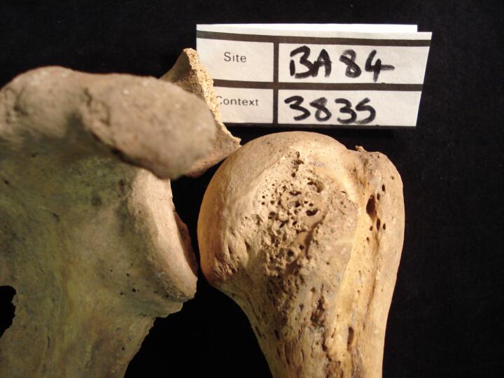
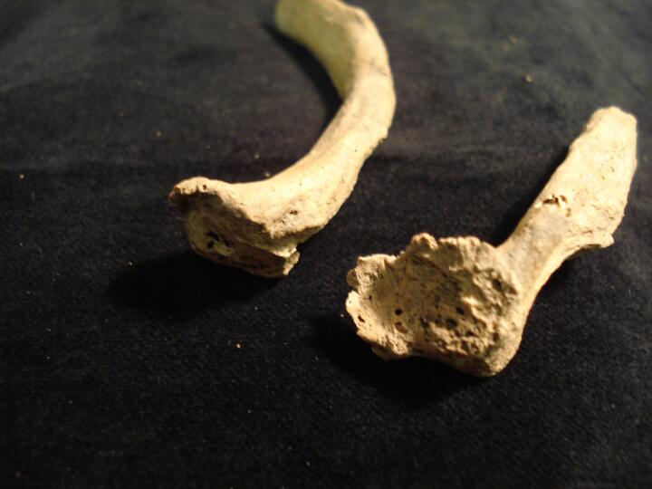
| BA84
|
3835
|
1
|
BA84_3835_1.jpg
|
Right hunmerus & scapula (anterior view) rotator cuff injury
|
| BA84
|
3835
|
2
|
BA84_3835_2.jpg
|
Right scapula (medial view) margianl osteophytic lipping of glenoid cavity
|
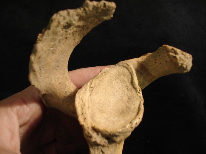
| BA84
|
3835
|
3
|
BA84_3835_3.jpg
|
Left humerus & scapula (anterior view) rotator cuff injury
|
| BA84
|
3835
|
4
|
BA84_3835_4.jpg
|
Unside rib fragments ossified cartilage (visceral view)
|
| BA84
|
3835
|
5
|
BA84_3835_5.jpg
|
Right ribs articular head with joint destruction & osteoarthritis (medial view)
|
| BA84
|
3835
|
6
|
BA84_3835_6.jpg
|
Right rib articular head with joint destruction & osteoarthritis (medial view/close up)
|
| BA84
|
3835
|
7
|
BA84_3835_7.jpg
|
Cervical vertebrae articulated (anterior view) showing degenerative changes
|
| BA84
|
3835
|
8
|
BA84_3835_8.jpg
|
Cervical vertebrae articulated (right side view) showing degenerative changes, osteophytic lipping
|
| BA84
|
3835
|
9
|
BA84_3835_9.jpg
|
Cervical vertebrae (various) inferior & superior view of centrums showing intervertebral disc disease (IVD)
|
| BA84
|
3835
|
10
|
BA84_3835_10.jpg
|
Thoracic vertebrae (various) inferior &superior view of centrums showing intervertebral disc disease (IVD)
|
| BA84
|
3835
|
11
|
BA84_3835_11.jpg
|
Thoracic vertebra (inferior view/close up) of centrum showing intervertebral disc disease (IVD)
|
| BA84
|
3835
|
12
|
BA84_3835_12.jpg
|
Lumber vertebra L5, inferior articular facet enlarged & eburnated (OA) (left side view)
|
| BA84
|
3835
|
13
|
BA84_3835_13.jpg
|
Lumber vertebra L5, inferior articular facet enlarged & eburnated (OA) (left side view/close up)
|
| BA84
|
3835
|
14
|
BA84_3835_14.jpg
|
Lumbar vertebra L1 centrum (superior view) schmorl's node
|
| BA84
|
3835
|
15
|
BA84_3835_15.jpg
|
Lumbar vertebrae, articulated (posteior view) showing facets
|
| BA84
|
3883
|
1
|
BA84_3883_1.jpg
|
Left radius healed fracture, 'Colles', distal end (anterior view)
|
| BA84
|
3883
|
2
|
BA84_3883_2.jpg
|
Left radius healed fracture, 'Colles', distal end (posterior view/close up)
|
| BA84
|
3883
|
3
|
BA84_3883_3.jpg
|
Left radius healed fracture, 'Colles', distal end (anterior view/close up)
|
| BA84
|
3883
|
4
|
BA84_3883_4.jpg
|
Left radius healed fracture, 'Colles', distal end (superior view)
|
| BA84
|
3883
|
5
|
BA84_3883_5.jpg
|
Maxillae, (palatal view) very heavy wear of dentition
|
| BA84
|
3886
|
1
|
BA84_3886_1.jpg
|
Skull, (right side view) possible indication of Bathrocephaly
|
| BA84
|
3886
|
2
|
BA84_3886_2.jpg
|
Skull, (right side view/close up) occipital, possible indication of Bathrocephaly
|
| BA84
|
3886
|
3
|
BA84_3886_3.jpg
|
Skull, (posterior view) possible indication of Bathrocephaly
|


