| Site code
|
Context
|
Frame number
|
Photo
|
Description
|
| MPY86
|
2381
|
2
|
MPY86_2381_2.jpg
|
Osteophytic lipping and eburnation of the right trapezium (view from the scaphoid)
|
| MPY86
|
2381
|
1
|
MPY86_2381_1.jpg
|
Eburnation on the right scaphoid (View from the radius)
|
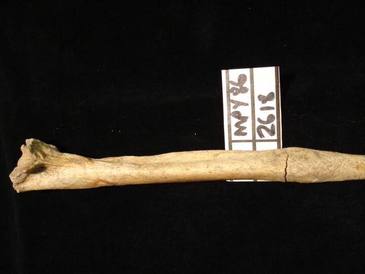
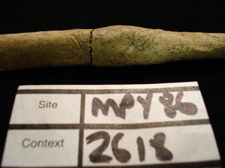
| MPY86
|
2618
|
1
|
MPY86_2618_1.jpg
|
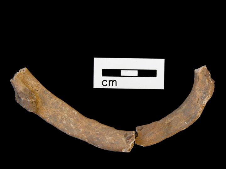
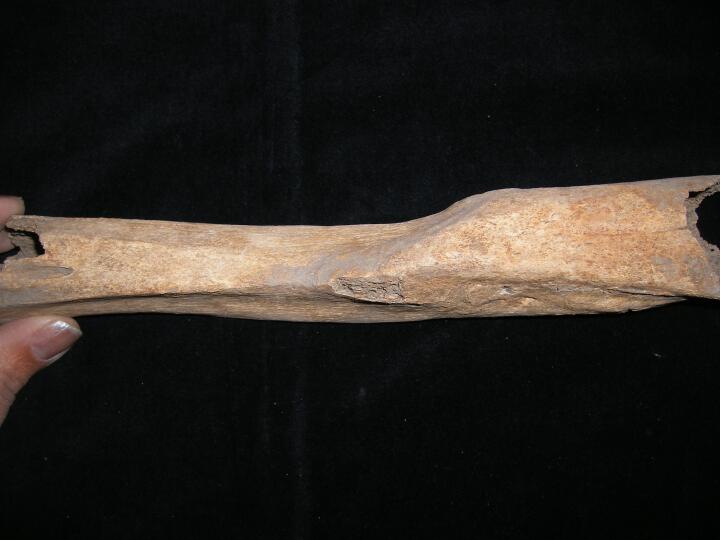
Left ulna 'Parry' fracture (anterior view) very well remodelled
|
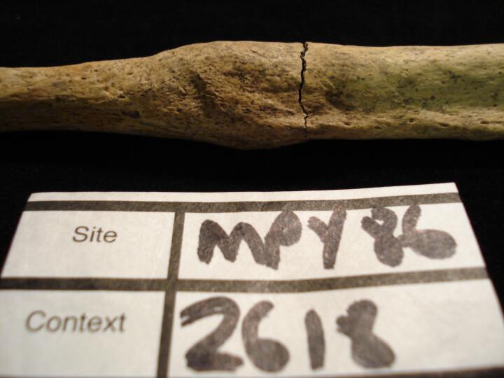
| MPY86
|
2618
|
2
|
MPY86_2618_2.jpg
|
Left ulna 'Parry' fracture (medial view) very well remodelled, close up of callus
|
| MPY86
|
2618
|
3
|
MPY86_2618_3.jpg
|
Left ulna 'Parry' fracture (lateral view) very well remodelled, close up of callus
|
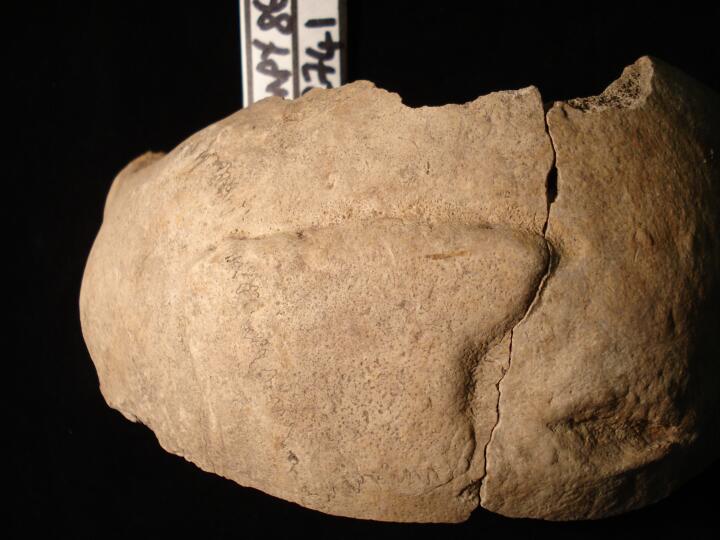
| MPY86
|
2741
|
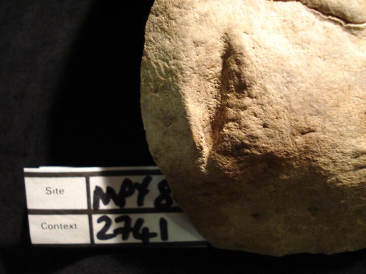
1
|
MPY86_2741_1.jpg
|
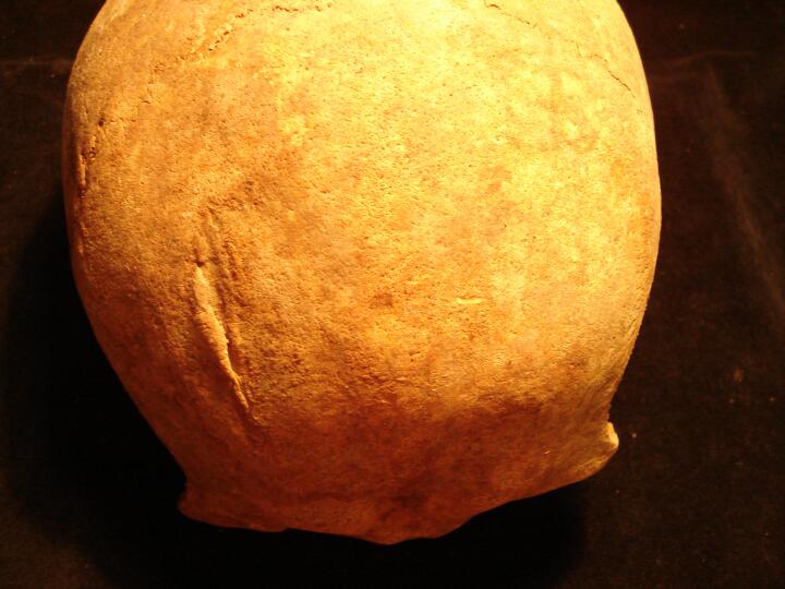
Skull, right frontal & parietal (ectocranial surface) healed sharp force trauma & blunt force trauma (superior view)
|
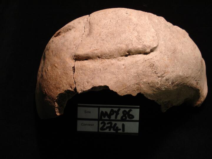
| MPY86
|
2741
|
2
|
MPY86_2741_2.jpg
|
Skull, right frontal & parietal (ectocranial surface) healed sharp force trauma & blunt force trauma (right side view)
|
| MPY86
|
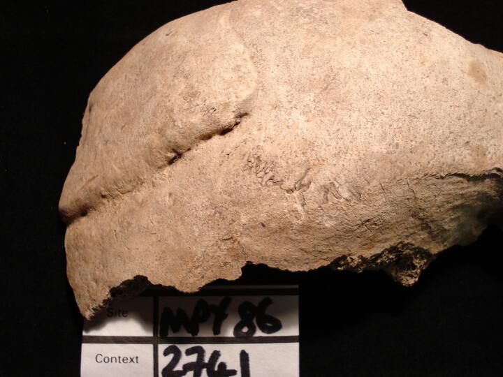
2741
|
3
|
MPY86_2741_3.jpg
|
Skull, right parietal close up of healed blunt force trauma (ectocranial surface)
|
| MPY86
|
2741
|
4
|
MPY86_2741_4.jpg
|
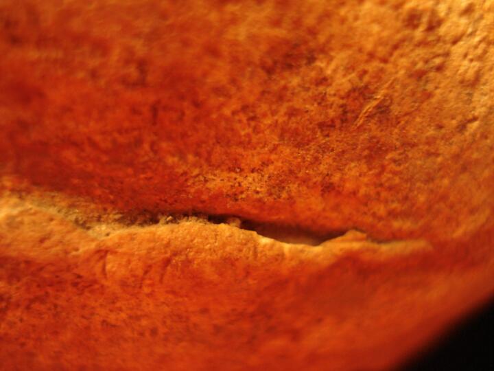
Skull, right frontal bone close up of healed sharp force trauma (right side view)
|
| MPY86
|
2741
|
5
|
MPY86_2741_5.jpg
|
Skull endocranial surface
|
| MPY86
|
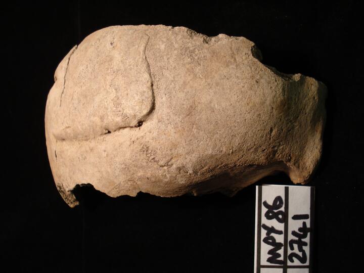
2741
|
6
|
MPY86_2741_6.jpg
|
Skull (anterior view) showing two types of healed trauma blunt & sharp force
|
| MPY86
|
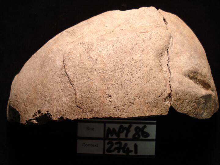
2741
|
7
|
MPY86_2741_7.jpg
|
Skull (left side view) showing two types of healed trauma blunt & sharp force(some post mortem damage)
|
| MPY86
|
2741
|
8
|
MPY86_2741_8.jpg
|
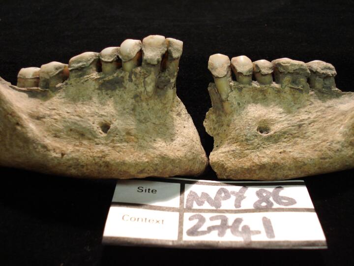
Mandible (buccal view) heavy calculus
|
| MPY86
|
2741
|
9
|
MPY86_2741_9.jpg
|
Mandible (lingual view) heavy calculus
|
| MPY86
|
2741
|
10
|
MPY86_2741_10.jpg
|
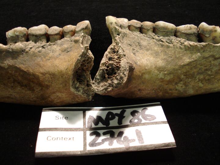
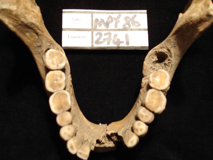
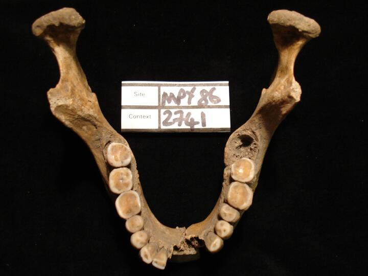
Mandible (occlusal view) heavy wear of teeth
|
| MPY86
|
2741
|
11
|
MPY86_2741_11.jpg
|
Mandible (occlusal view) heavy wear of teeth
|
| MPY86
|
2741
|
12
|
MPY86_2741_12.jpg
|
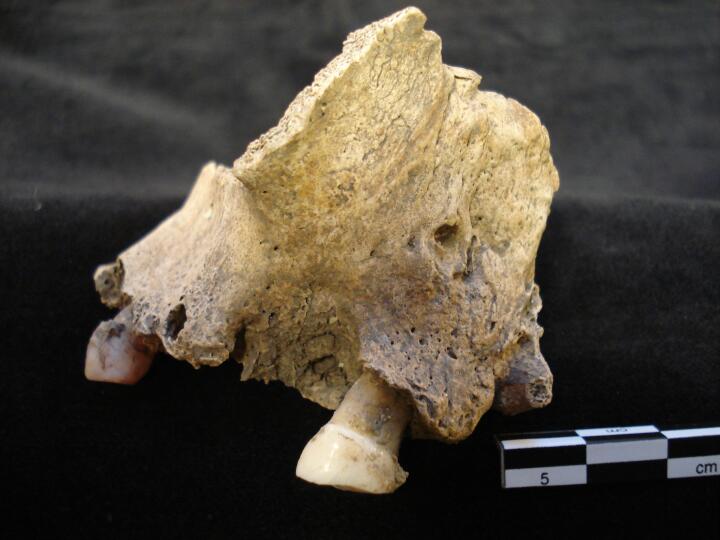
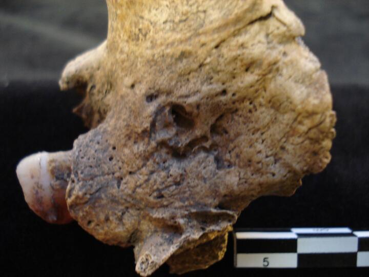
Left maxilla (buccal view) external draining periapical lesion (abcess)and AM & PM loss of teeth
|
| MPY86
|
2741
|
13
|
MPY86_2741_13.jpg
|
Left maxilla (buccal view) severe but healed infection of bone, associated with dentition & periapical lesion (abcess)
|
| MPY86
|
2741
|
14
|
MPY86_2741_14.jpg
|
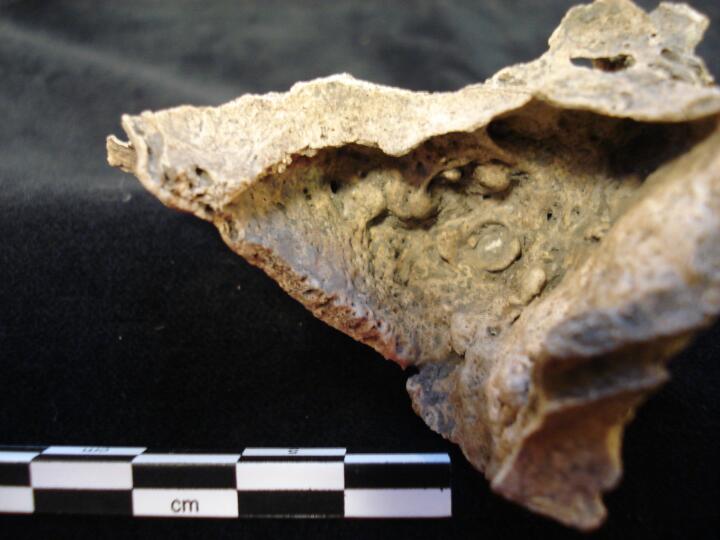
Left maxillary sinus (internal) severe healed non-specific infection of sinus with remodelled nodules of bone
|
| MPY86
|
2741
|
15
|
MPY86_2741_15.jpg
|
Left maxillary sinus (internal) severe healed non-specific infection of sinus with remodelled nodules of bone
|
| MPY86
|
2741
|
16
|
MPY86_2741_16.jpg
|
Left maxillary sinus (internal) severe healed non-specific infection of sinus with remodelled nodules of bone
|
| MPY86
|
2741
|
17
|
MPY86_2741_17.jpg
|
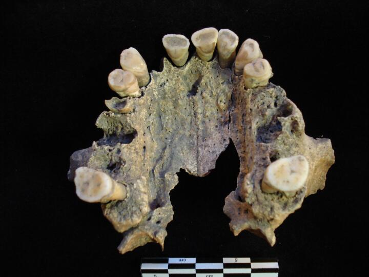
Maxilla (palatal view) showing AM & PM loss of teeth, gross carious lesion of right PM3 & periapical lesion
|
| MPY86
|
2741
|
18
|
MPY86_2741_18.jpg
|
Right maxilla caries related external draining periapical lesion (buccal view)
|
| MPY86
|
2758
|
1
|
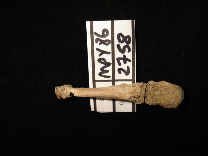
MPY86_2758_1.jpg
|
Right foot ankylosis of lateral cuneiform & 3rd metatarsal (dorsal view)
|
| MPY86
|
2758
|
2
|
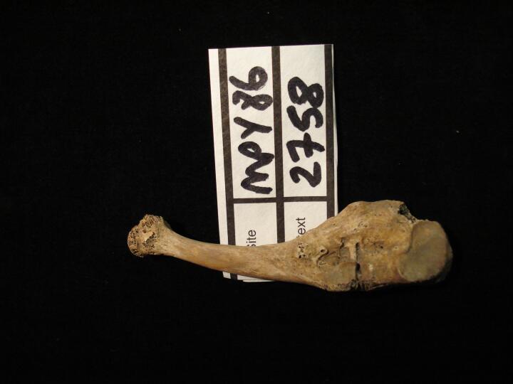
MPY86_2758_2.jpg
|
Right foot ankylosis of lateral cuneiform & 3rd metatarsal (medial view)
|
| MPY86
|
2758
|
3
|
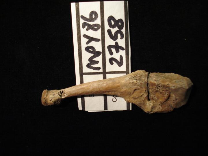
MPY86_2758_3.jpg
|
Right foot ankylosis of lateral cuneiform & 3rd metatarsal (lateral view)
|
| MPY86
|
2858
|
1
|
MPY86_2858_1.jpg
|
DISH. 'Dripping candle wax' like osteophytes causing fusion of T8-12 down the right anterior vertebral bodies. Disc spaces retained.
|
| MPY86
|
2858
|
2
|
MPY86_2858_2.jpg
|
Well healed fracture to the proximal right femur showing an irregular bone surface at the point of fracture.
|
| MPY86
|
2858
|
3
|
MPY86_2858_3.jpg
|
Irregular bone surface and overdevelopment of the linea aspera of the midshaft of the right femur due to a healed fracture.
|
| MPY86
|
2895
|
1
|
MPY86_2895_1.jpg
|
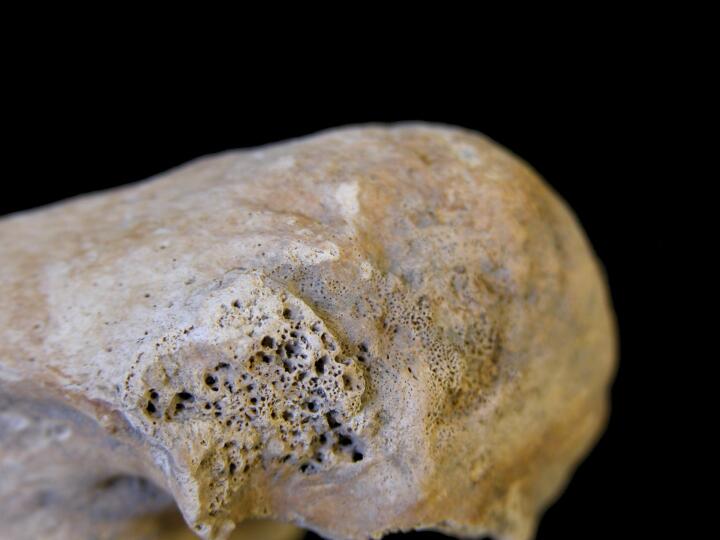
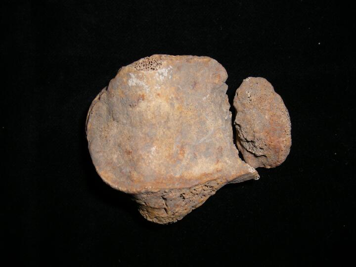
HFI on the inner table of the frontal bone in the region of the frontal crest.
|
| MPY86
|
2995
|
1
|
MPY86_2995_1.jpg
|
Thickening of the anterior neural arch of the atlas (inferior view)
|
| MPY86
|
2995
|
2
|
MPY86_2995_2.jpg
|
Thickening of the anterior neural arch of the atlas (superior view)
|
| MPY86
|
3172
|
1
|
MPY86_3172_1.jpg
|
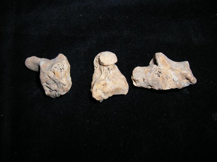
Erosive arthropathy?? Rounded lesions and deformation of carpals
|
| MPY86
|
3548
|
1
|
MPY86_3548_1.jpg
|
Bone cyst in the inferior vertebral body of L3 (inferior view) IVD also visible.
|
| MPY86
|
3548
|
2
|
MPY86_3548_2.jpg
|
Close up of a bone cyst in the inferior vertebral body of L3 (inferior view) IVD also visible.
|
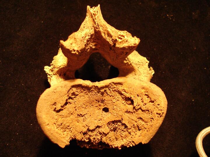
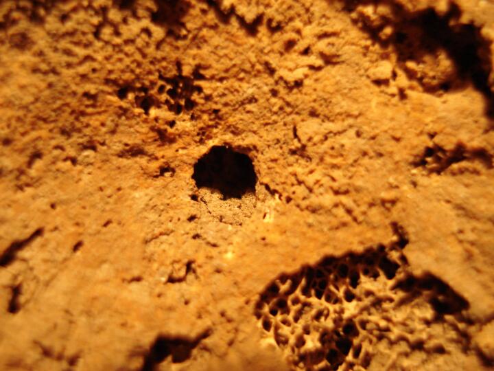
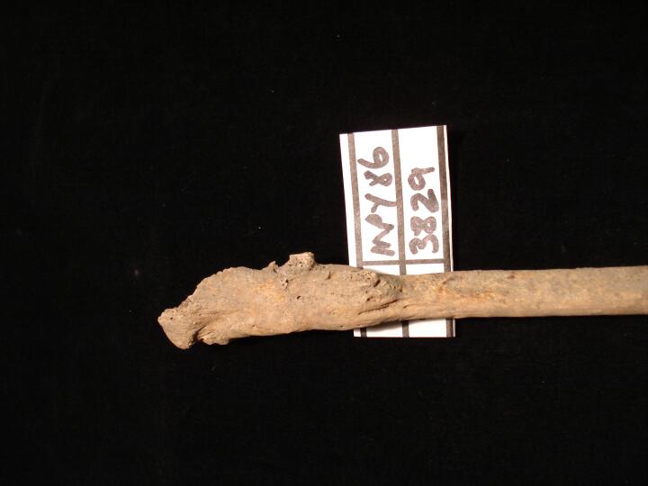
| MPY86
|
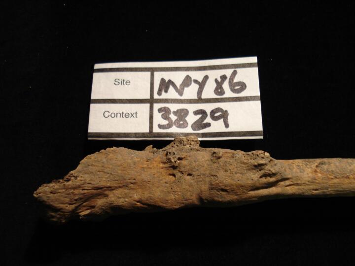
3829
|
1
|
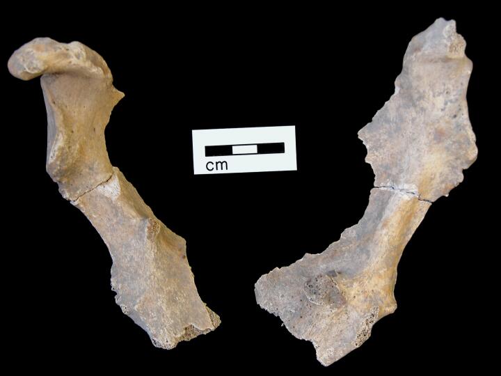
MPY86_3829_1.jpg
|
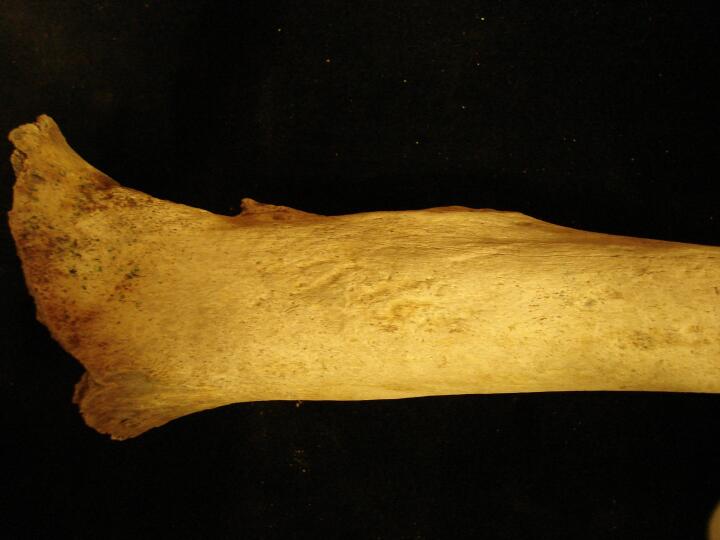
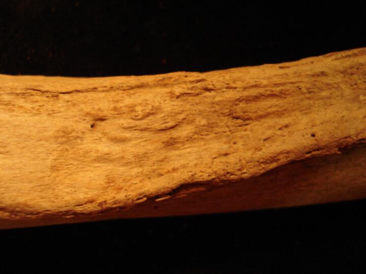
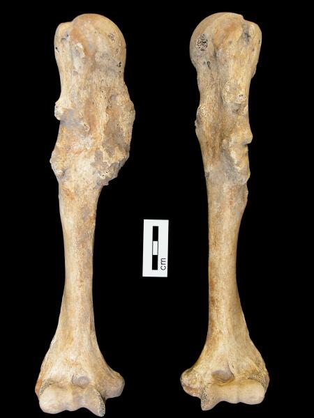
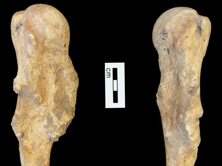
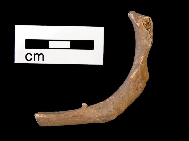
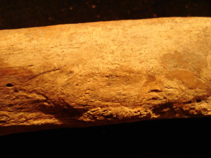
Left fibula healed fracture proximal end (posterior view)
|
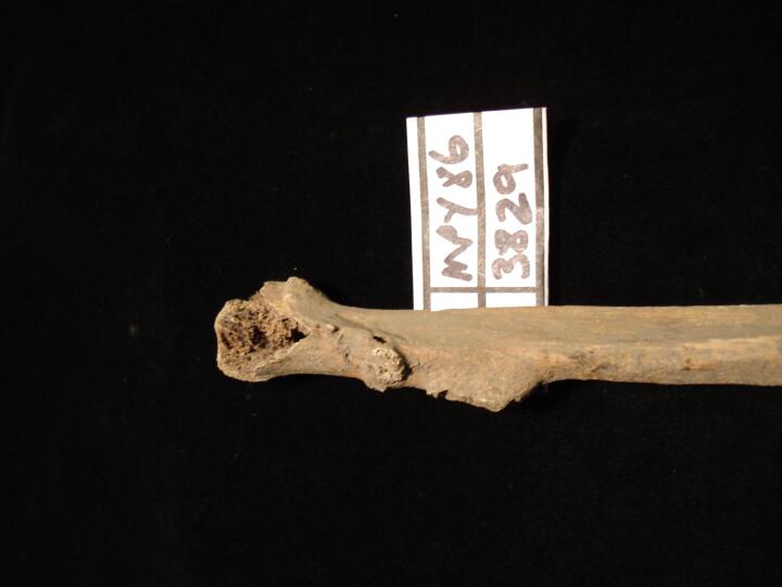
| MPY86
|
3829
|
2
|
MPY86_3829_2.jpg
|
Left fibula healed fracture proximal end (anterior view)
|
| MPY86
|
3829
|
3
|
MPY86_3829_3.jpg
|
Left fibula healed fracture proximal end (medial view)
|
| MPY86
|
3829
|
4
|
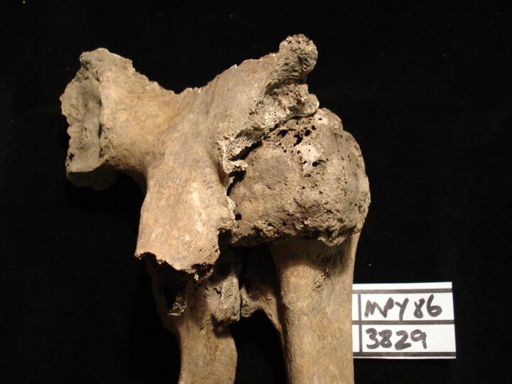
MPY86_3829_4.jpg
|
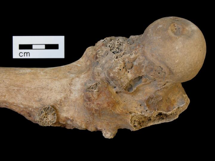
Left acetabulum trauma with secondary osteoarthritis
|
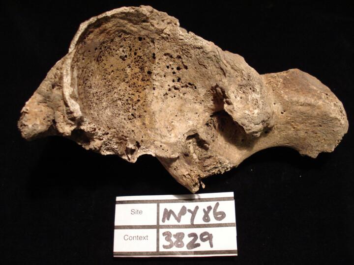
| MPY86
|
3829
|
5
|
MPY86_3829_5.jpg
|
Left acetabulum trauma & bone fragments with secondary osteoarthritis
|
| MPY86
|
3829
|
6
|
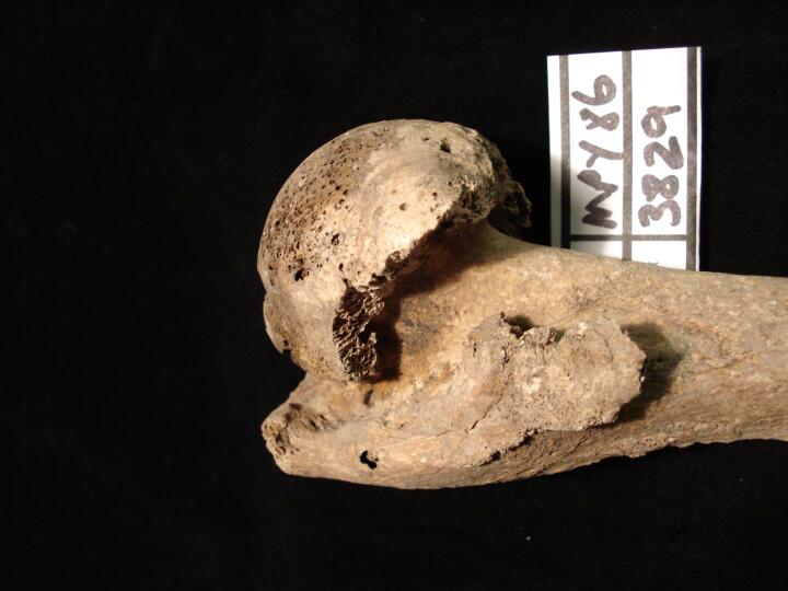
MPY86_3829_6.jpg
|
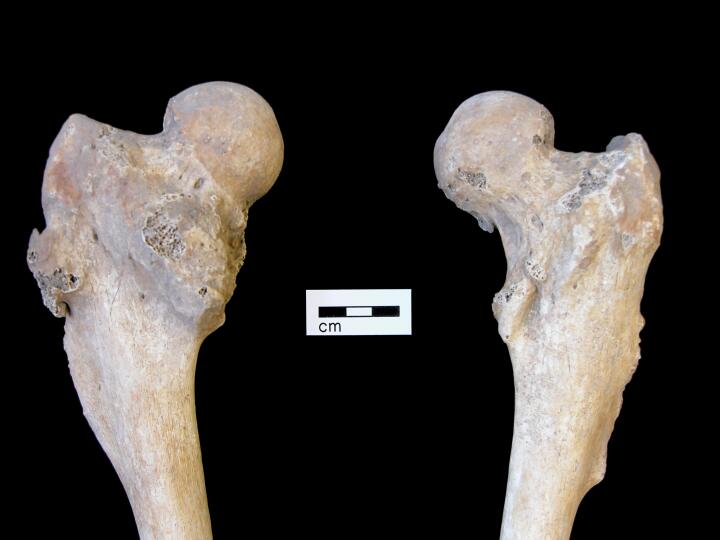
Left femoral head gross secondary osteoarthritic changes from trauma (superior view)
|
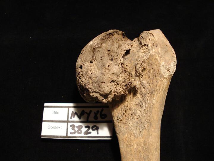
| MPY86
|
3829
|
7
|
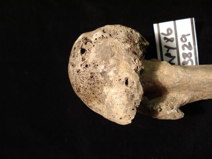
MPY86_3829_7.jpg
|
Left femoral head gross secondary osteoarthritic changes from trauma (anterior view)
|
| MPY86
|
3829
|
8
|
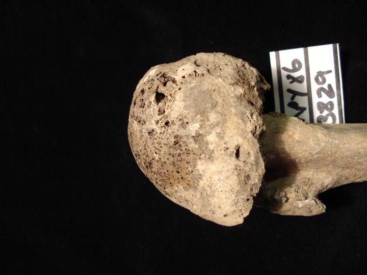
MPY86_3829_8.jpg
|
Left femoral head gross secondary osteoarthritic changes from trauma (posterior view)
|
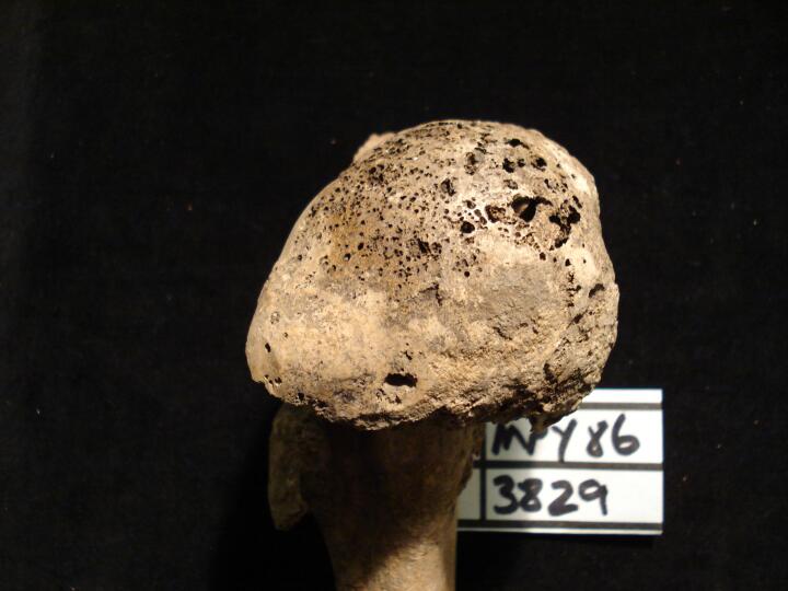
| MPY86
|
3829
|
9
|
MPY86_3829_9.jpg
|
Left femoral head gross secondary osteoarthritic changes from trauma (medial view)
|
| MPY86
|
3829
|
10
|
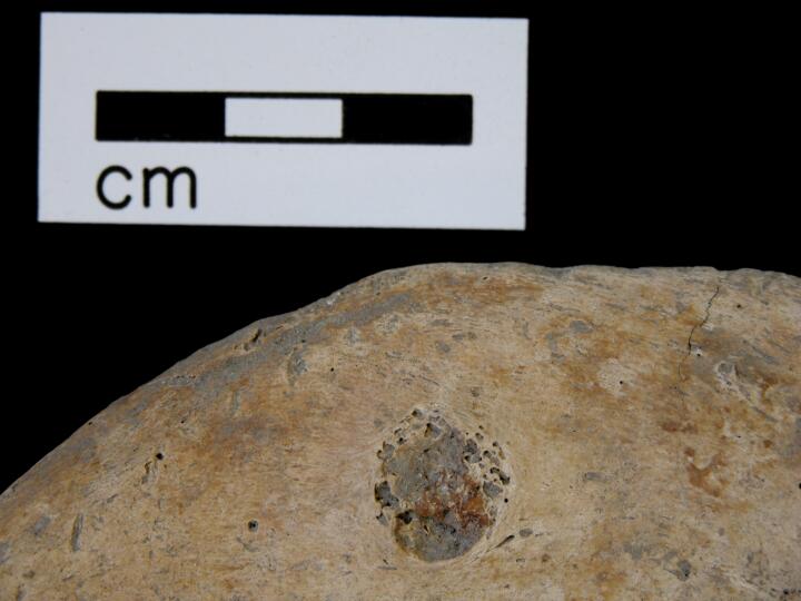
MPY86_3829_10.jpg
|
Left femoral head gross secondary osteoarthritic changes from trauma (medial view/close up)
|
| MPY86
|
3829
|
11
|
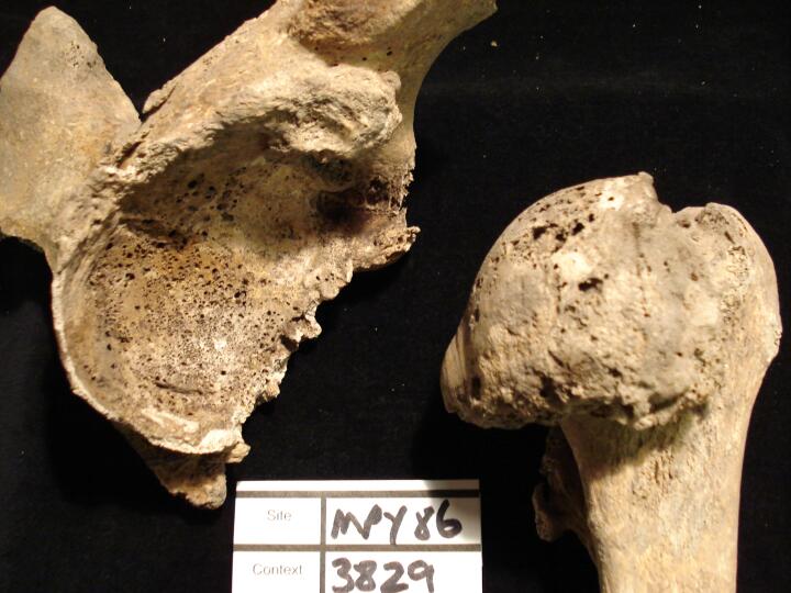
MPY86_3829_11.jpg
|
Left acetabulum & left femoral head gross secondary osteoarthritic changes from trauma
|
| MPY86
|
3829
|
12
|
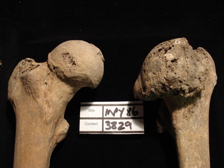
MPY86_3829_12.jpg
|
Left acetabulum & left femoral head gross secondary osteoarthritic changes from trauma (anterior view)
|
| MPY86
|
3829
|
13
|
MPY86_3829_13.jpg
|
Left and right femoral heads to compare pathological change of left & right femoral head (anterior view)
|
| MPY86
|
3878
|
1
|
MPY86_3878_1.jpg
|
Right Os coxa, (ventral aspect) rouded lesion
|
| MPY86
|
3878
|
2
|
MPY86_3878_2.jpg
|
Scapulae, bilateral exostoses, symmetrical
|
| MPY86
|
3878
|
3
|
MPY86_3878_3.jpg
|
Humeri, bilateral exostoses,symmetrical
|
| MPY86
|
3878
|
4
|
MPY86_3878_4.jpg
|
Humeri, bilateral exostoses,symmetrical
|
| MPY86
|
3878
|
5
|
MPY86_3878_5.jpg
|
Thoacic vertebra, Th8, (left lateral aspect)
|
| MPY86
|
3878
|
6
|
MPY86_3878_6.jpg
|
Femora, (anterior aspect) bilateral changes
|
| MPY86
|
3878
|
7
|
MPY86_3878_7.jpg
|
Left femur (anterior aspect)
|
| MPY86
|
3878
|
8
|
MPY86_3878_8.jpg
|
Right ulna, proximal end (posterior aspect)
|
| MPY86
|
3878
|
9
|
MPY86_3878_9.jpg
|
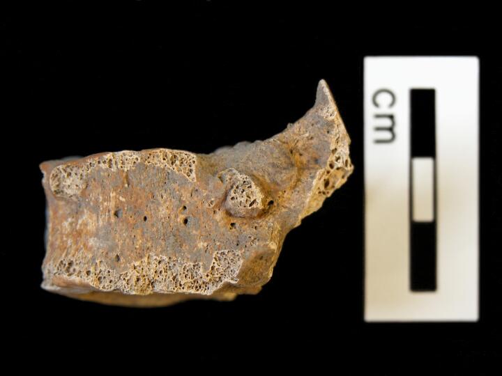
Right rib (inferior aspect) exostosis
|
| MPY86
|
3878
|
10
|
MPY86_3878_10.jpg
|
Left rib (superior aspect) exostosis
|
| MPY86
|
3878
|
11
|
MPY86_3878_11.jpg
|
Femora, (anterior aspect) bilateral exostosis, symmetrical
|
| MPY86
|
3885
|
1
|
MPY86_3885_1.jpg
|
Ankylosis of pubic symphyses
|
| MPY86
|
3885
|
2
|
MPY86_3885_2.jpg
|
Compression fracture of T12-L1
|
| MPY86
|
3885
|
3
|
MPY86_3885_3.jpg
|
Compression fracture of T12-L2
|
| MPY86
|
3885
|
4
|
MPY86_3885_4.jpg
|
Ankylosis of pubic symphyses
|
| MPY86
|
3891
|
1
|
MPY86_3891_1.jpg
|
Healed fracture on central shaft of L tibia
|
| MPY86
|
3904
|
1
|
MPY86_3904_1.jpg
|
Patalla biparte
|
| MPY86
|
3904
|
2
|
MPY86_3904_2.jpg
|
Patalla biparte
|
| MPY86
|
3968
|
1
|
MPY86_3968_1.jpg
|
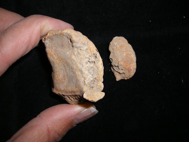
Two small button osteomas to the frontal bone.
|
| MPY86
|
4063
|
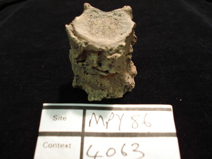
1
|
MPY86_4063_1.jpg
|
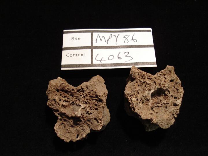
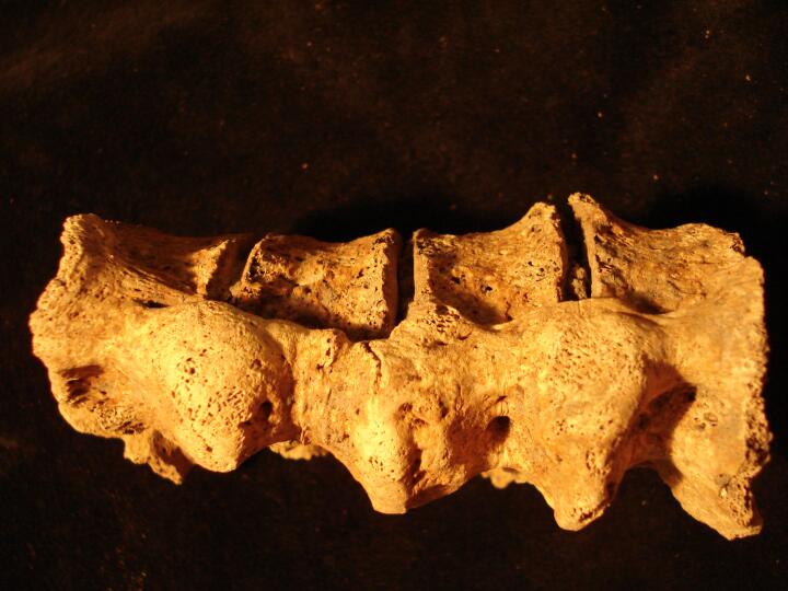
Thoracic vertebrae Th5 (inferior surface) & Th6 (superior surface) possible changes indicative of Tuberculosis
|
| MPY86
|
4063
|
2
|
MPY86_4063_2.jpg
|
Thoracic vertebrae Th5 & Th6 (left side view) changes possibly indicative of Tuberculosis
|
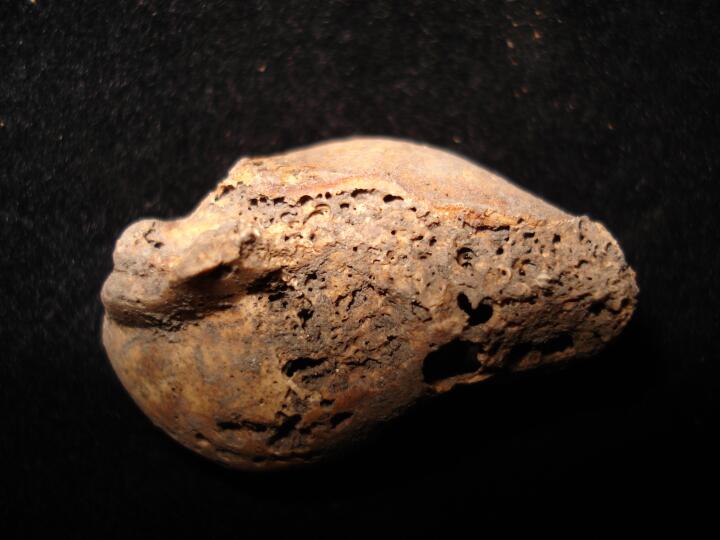
| MPY86
|
4063
|
3
|
MPY86_4063_3.jpg
|
Thoracic vertebrae Th5 & Th6 (anterior view) changes possibly indicative of s Tuberculosis
|
| MPY86
|
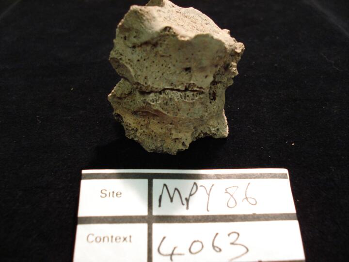
4063
|
4
|
MPY86_4063_4.jpg
|
Thoracic vertebrae Th5 & Th6 (right side view) changes possibly indicative of Tuberculosis
|
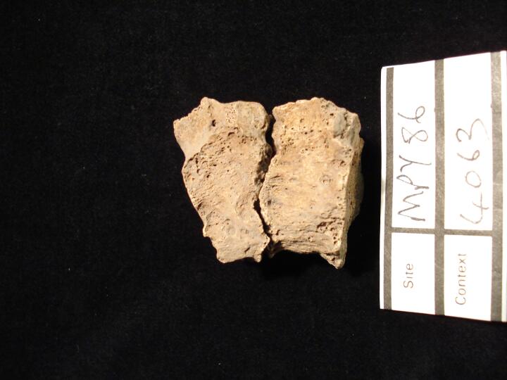
| MPY86
|
4063
|
5
|
MPY86_4063_5.jpg
|
Thoracic vertebrae Th5 & Th6 (left side/ lyingon side) changes possibly indicative Tuberculosis
|
| MPY86
|
4069
|
1
|
MPY86_4069_1.jpg
|
Osteophytic lipping and subchondral cysts on the left scaphoid. (View from the radius)
|
| MPY86
|
4069
|
2
|
MPY86_4069_2.jpg
|
Osteophytic lipping, subchondral cysts and eburnation on the left scaphoid. (View from the radius)
|
| MPY86
|
4069
|
3
|
MPY86_4069_3.jpg
|
Osteoarthritis to the right acetabulum. Pronounced destruction of the joint with osteophytic lipping and subchondral cysts visible.
|
| MPY86
|
4069
|
4
|
MPY86_4069_4.jpg
|
Osteoarthritis to the left acetabulum. Pronounced destruction of the joint with osteophytic lipping and subchondral cysts visible.
|
| MPY86
|
4069
|
5
|
MPY86_4069_5.jpg
|
DISH. 'Dripping candle wax' like fusion of two lower thoracic vertebrae down the anterior longitudinal ligament. Disc spaces have been retained. (Anterior view)
|
| MPY86
|
4069
|
6
|
MPY86_4069_6.jpg
|
DISH. 'Dripping candle wax' like fusion of two lower thoracic vertebrae down the anterior longitudinal ligament. Disc spaces have been retained. (Lateral view)
|
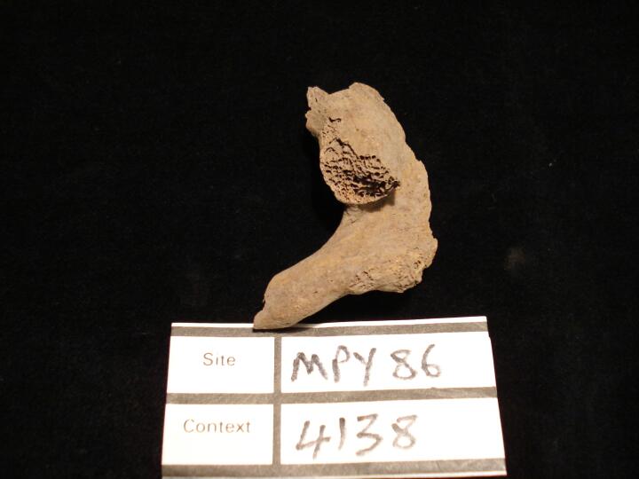
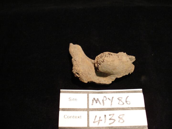
| MPY86
|
4138
|
1
|
MPY86_4138_1.jpg
|
Fusion of right Ist & 2nd rib (superior view)
|
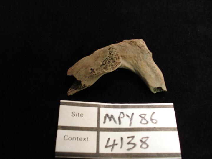
| MPY86
|
4138
|
2
|
MPY86_4138_2.jpg
|
Fusion of right Ist & 2nd rib (posterior view)
|
| MPY86
|
4138
|
3
|
MPY86_4138_3.jpg
|
Fusion of right Ist & 2nd rib (superior view) anterior aspect
|
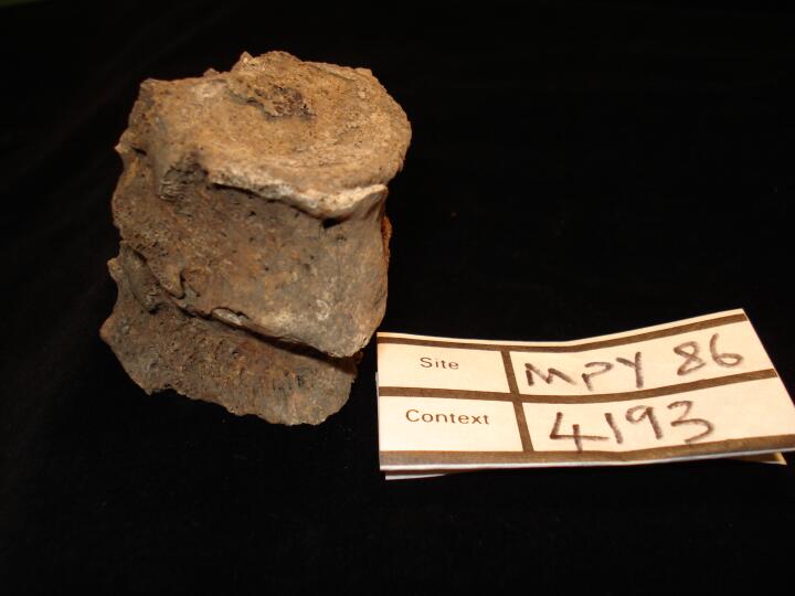
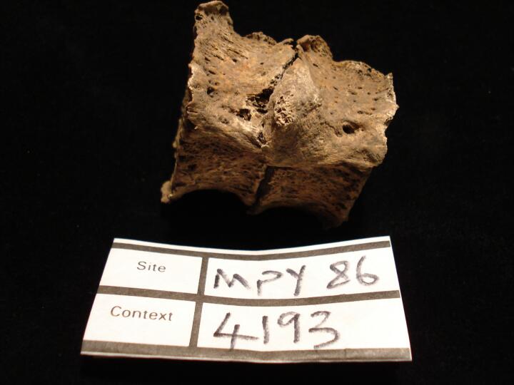
| MPY86
|
4193
|
1
|
MPY86_4193_1.jpg
|
Thoracic vertebrae, Th8 & Th9, fusion (right side view)
|
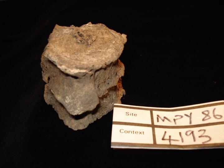
| MPY86
|
4193
|
2
|
MPY86_4193_2.jpg
|
Thoracic vertebrae, Th8 & Th9, fusion (anterior view)
|
| MPY86
|
4193
|
3
|
MPY86_4193_3.jpg
|
Thoracic vertebrae, Th8 & Th9, fusion (anterior view) from superior aspect
|
| MPY86
|
4454
|
1
|
MPY86_4454_1.jpg
|
Gout, scalloped lesions along margins of first MTPJ
|
| MPY86
|
4543
|
1
|
MPY86_4543_1.jpg
|
Gout, scalloped lesions along margins of first MTPJ
|
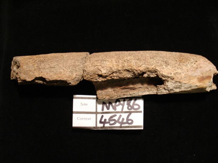
| MPY86
|
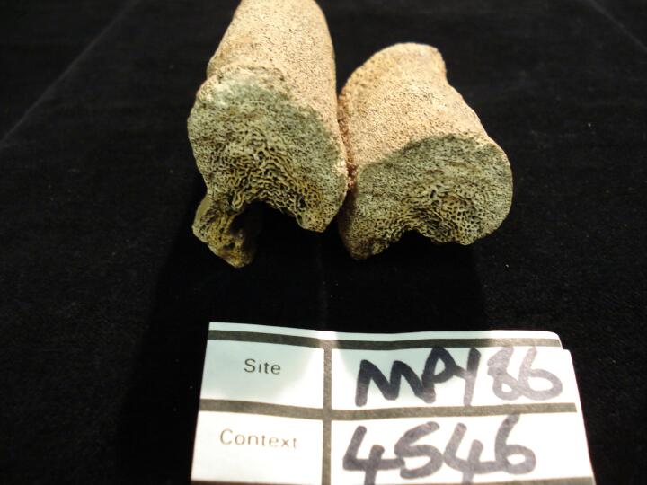
4546
|
1
|
MPY86_4546_1.jpg
|
Left tibia (anterior view) post mortem damage bone thickened & enlarged, possibly Paget's disease
|
| MPY86
|
4546
|
2
|
MPY86_4546_2.jpg
|
Left tibia (medial view) post mortem damage, two fragments, bone thickened & enlarged, possibly Paget's disease
|
| MPY86
|
4546
|
3
|
MPY86_4546_3.jpg
|
Left tibia post mortem damage, two fragments,cross section of deposited bone causing thickeneing to the bone, possibly Paget's disease
|
| MPY86
|
4546
|
4
|
MPY86_4546_4.jpg
|
Left tibia post mortem damage, two fragments,cross section/close up of deposited bone causing thickening, possibly Paget's disease
|
| MPY86
|
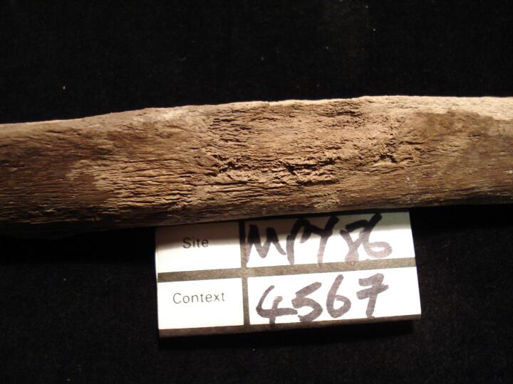
4567
|
1
|
MPY86_4567_1.jpg
|
Left tibia lateral aspect (anterior view) demarcated area of periosteal reaction possibly indicating an ulcer
|
| MPY86
|
4567
|
2
|
MPY86_4567_2.jpg
|
Left tibia lateral aspect (medial view) demarcated area of periosteal reaction possibly indicating an ulcer
|
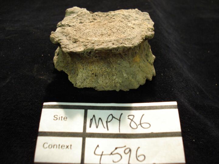
| MPY86
|
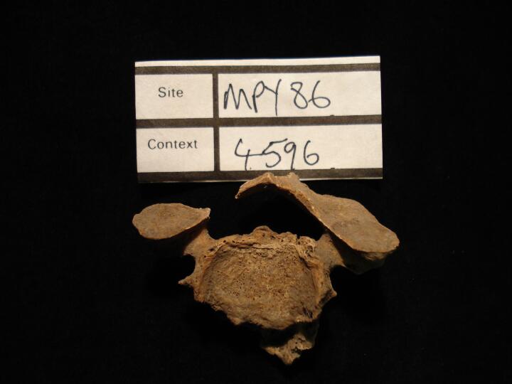
4596
|
1
|
MPY86_4596_1.jpg
|
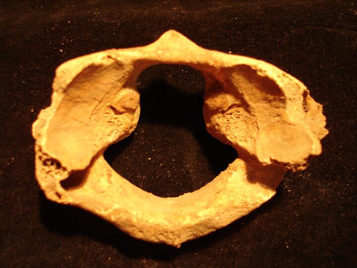
Cervical vertebral degenerative change,?C7 (superior surface) osteophytosis
|
| MPY86
|
4596
|
2
|
MPY86_4596_2.jpg
|
Cervical vertebral degenerative change,?C7 (inferior surface) osteophytosis
|
| MPY86
|
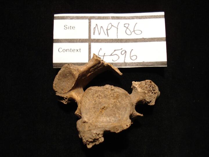
4596
|
3
|
MPY86_4596_3.jpg
|
Thoracic vertebral degenerative change, ?Th12 (anterior view)
|
| MPY86
|
4596
|
4
|
MPY86_4596_4.jpg
|
Thoracic vertebral degenerative change,?Th12 (posterior view)
|
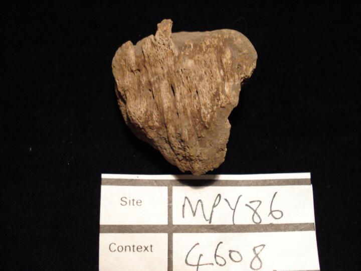
| MPY86
|
4608
|
1
|
MPY86_4608_1.jpg
|
Right talus (superior view) & right tibia posterior articular surface joint change possibly secondary to trauma (post mortem damage)
|
| MPY86
|
4608
|
2
|
MPY86_4608_2.jpg
|
Right patella (anterior view) enthseophytic development
|
| MPY86
|
4676
|
1
|
MPY86_4676_1.jpg
|
Healed sharp force trauma to the left parietal bone.
|
| MPY86
|
4731
|
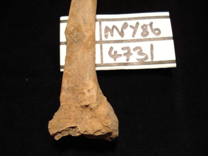
1
|
MPY86_4731_1.jpg
|
Left radius 'Colles' fracture, healed (anterior view)
|
| MPY86
|
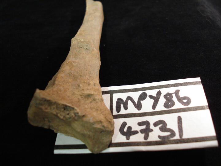
4731
|
2
|
MPY86_4731_2.jpg
|
Left radius 'Colles' fracture (anterior view) close up of fracture site
|
| MPY86
|
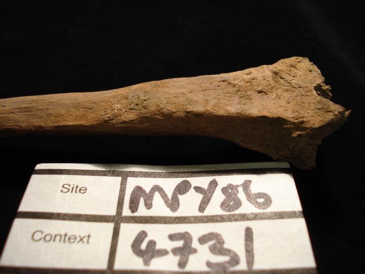
4731
|
3
|
MPY86_4731_3.jpg
|
Left radius 'Colles' fracture, healed (medial view)
|
| MPY86
|
4731
|
4
|
MPY86_4731_4.jpg
|
Left radius 'Colles' fracture, healed (from inferior aspect)
|
| MPY86
|
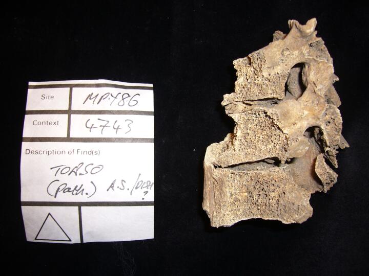
4743
|
1
|
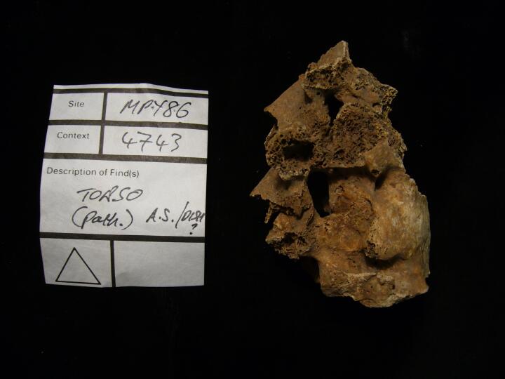
MPY86_4743_1.jpg
|
Vertebrae with osteophytic fusion, DISH or Ankylosing Spondylitis (left side view)
|
| MPY86
|
4743
|
2
|
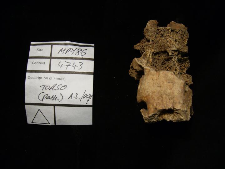
MPY86_4743_2.jpg
|
Vertebrae with osteophytic fusion, DISH or Ankylosing Spondylitis (right side view)
|
| MPY86
|
4743
|
3
|
MPY86_4743_3.jpg
|
Vertebrae with osteophytic fusion, DISH or Ankylosing Spondylitis (anterior view)
|
| MPY86
|
4743
|
4
|
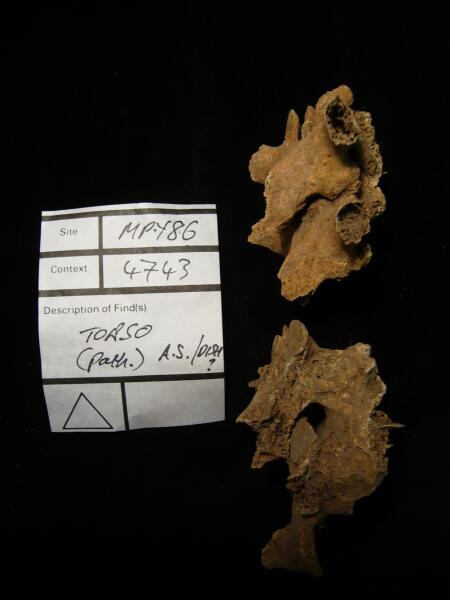
MPY86_4743_4.jpg
|
Vertebral arches with osteophytic fusion, DISH or Ankylosing Spondylitis
|
| MPY86
|
4743
|
5
|
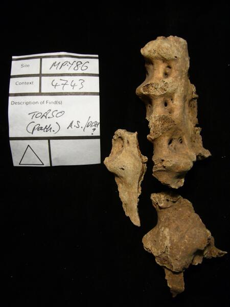
MPY86_4743_5.jpg
|
Vertebrae with osteophytic fusion, DISH or Ankylosing Spondylitis
|
| MPY86
|
4753
|
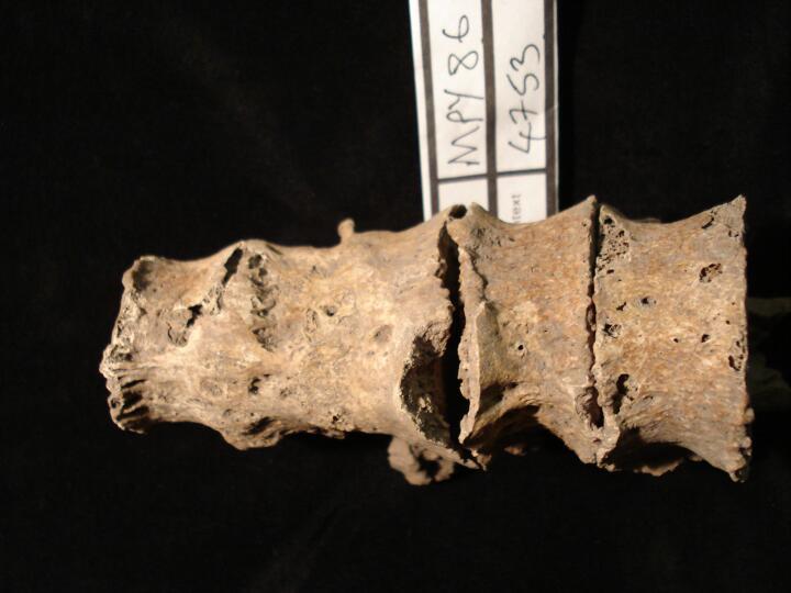
1
|
MPY86_4753_1.jpg
|
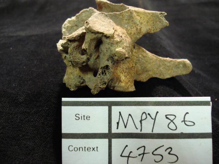
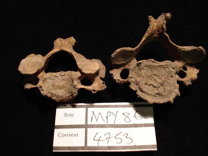
Thoracic vertebrae, Th 8 to Th10 anterior fusion of the vertebral bodies, not DISH,(anterior view)
|
| MPY86
|
4753
|
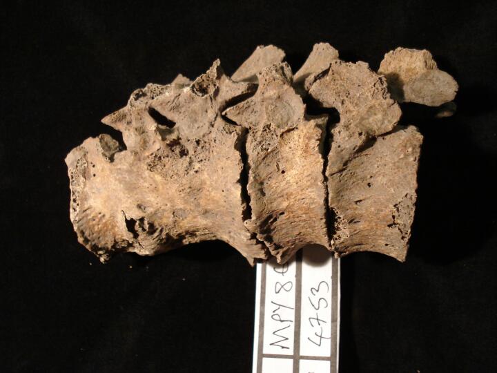
2
|
MPY86_4753_2.jpg
|
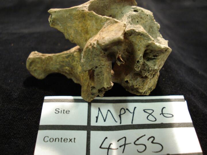
Thoracic vertebrae, Th 8 to Th10 anterior fusion of the vertebral bodies, not DISH,(right side view)
|
| MPY86
|
4753
|
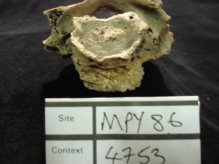
3
|
MPY86_4753_3.jpg
|
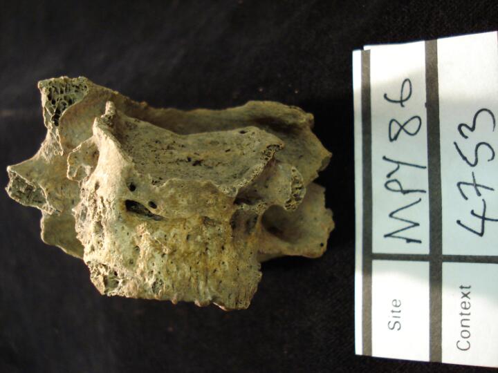
Thoracic vertebrae, Th 8 to Th10 anterior fusion of the vertebral bodies, not DISH,(left side view)
|
| MPY86
|
4753
|
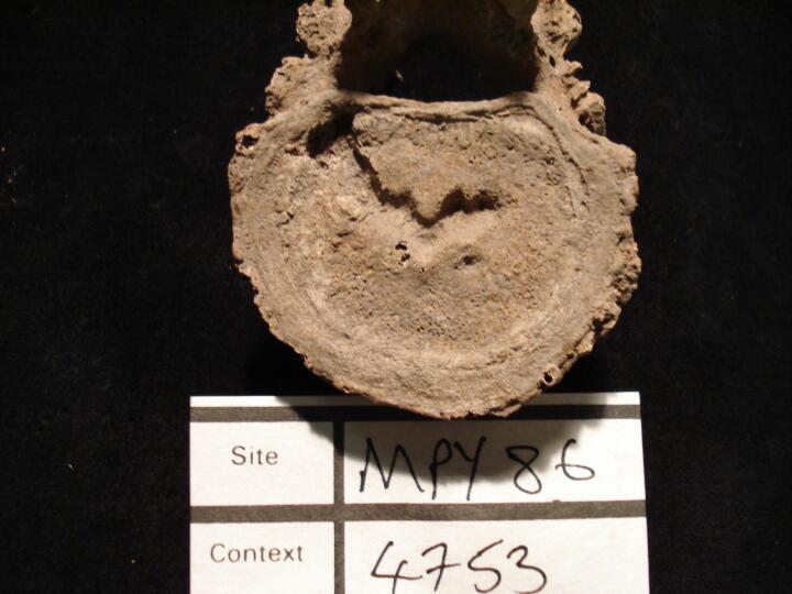
4
|
MPY86_4753_4.jpg
|
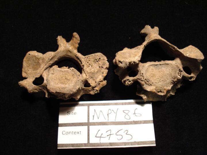
Thoracic vertebra, Th12 (inferior surface) Schmorl's Node
|
| MPY86
|
4753
|
5
|
MPY86_4753_5.jpg
|
Cervical vertebrae C6 & C7 (anterior view) dengereative changes
|
| MPY86
|
4753
|
6
|
MPY86_4753_6.jpg
|
Cervical vertebrae C6 & C7 (left side view) dengereative changes
|
| MPY86
|
4753
|
7
|
MPY86_4753_7.jpg
|
Cervical vertebrae C6 & C7 (right side view) dengereative changes
|
| MPY86
|
4753
|
8
|
MPY86_4753_8.jpg
|
Cervical vertebrae C6 & C7 (anterior view) dengereative changes
|
| MPY86
|
4753
|
9
|
MPY86_4753_9.jpg
|
Cervicl vertebra C4 (inferior surface) osteoarthritis, C5 (superior surface) osteophytosis
|
| MPY86
|
4753
|
10
|
MPY86_4753_10.jpg
|
Cervicl vertebra C4 (superior surface) osteoarthritis, C5 (inferior surface) osteophytosis
|
| MPY86
|
4753
|
11
|
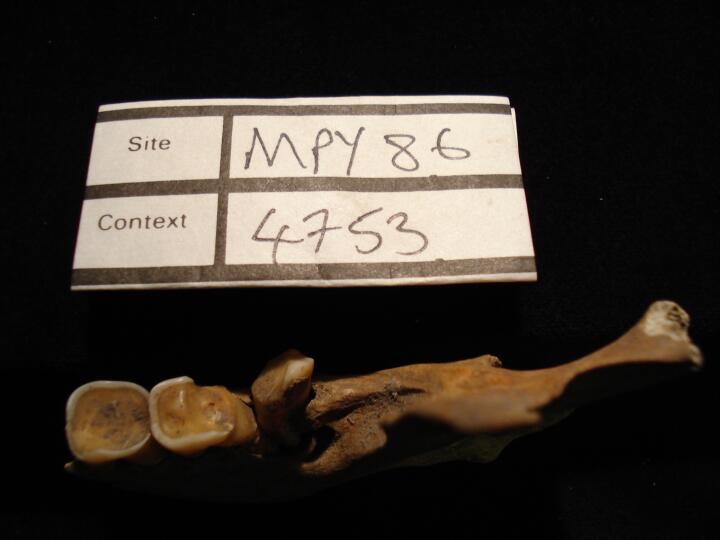
MPY86_4753_11.jpg
|
Left side of mandible (occlusal view) heavy wear of permanent molars
|
| MPY86
|
4753
|
12
|
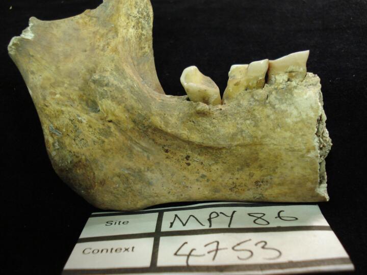
MPY86_4753_12.jpg
|
Left side of mandible (buccal view) heavy wear of permanent molars & external draining periapical lesion, 3rd molar
|
| MPY86
|
4753
|
13
|
MPY86_4753_13.jpg
|
Left side of mandible (lingual view) heavy wear of permanent molars & external draining periapical lesion, 3rd molar
|
| MPY86
|
4848
|
1
|
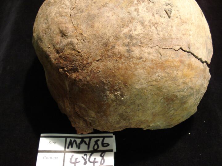
MPY86_4848_1.jpg
|
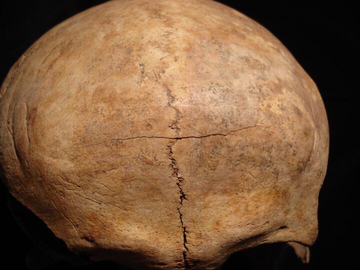
Skull (right side view) protruding & step like effect of occipital bone, possibly Bathrocephaly
|
| MPY86
|
4848
|
2
|
MPY86_4848_2.jpg
|
Skull (right side view/close up) protruding & step like effect of occipital bone, possibly Bathrocephaly
|
| MPY86
|
4848
|
3
|
MPY86_4848_3.jpg
|
Skull (posterior view) protruding & step like effect of occipital bone, possibly Bathrocephaly
|
| MPY86
|
4890
|
1
|
MPY86_4890_1.jpg
|
Large caries of T46. Pulp cavity exposed. Destruction of the mesial surface and partial destruction of the lingual and occlusal surfaces. (occlusal view)
|
| MPY86
|
4890
|
2
|
MPY86_4890_2.jpg
|
Close up of large caries of T46. Pulp cavity exposed. Destruction of the mesial surface and partial destruction of the lingual and occlusal surfaces. (occlusal view)
|
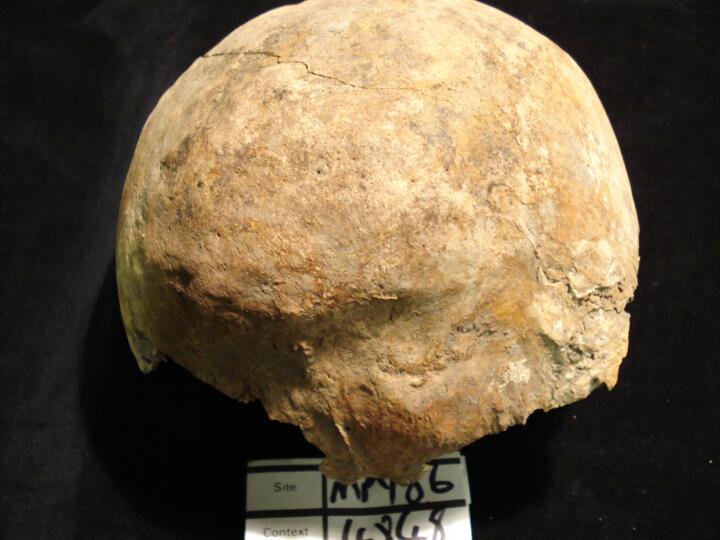
| MPY86
|
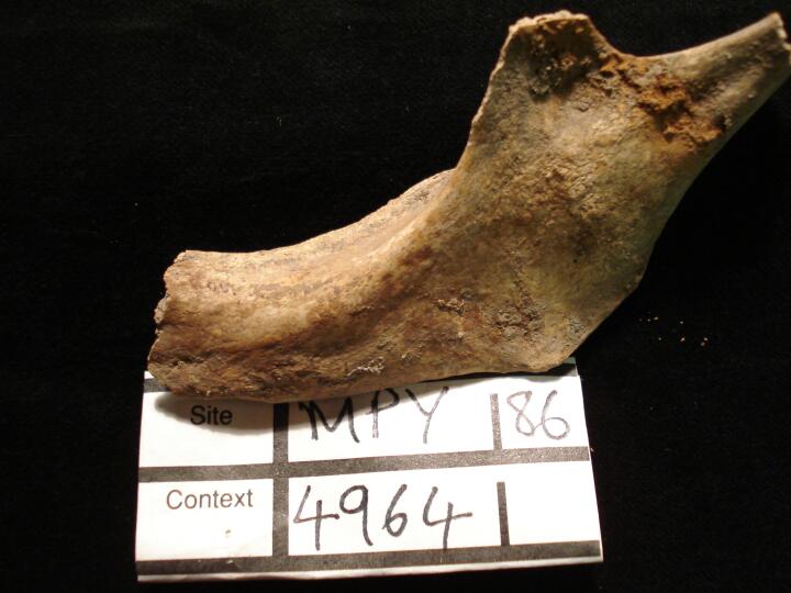
4964
|
1
|
MPY86_4964_1.jpg
|
Skull left parietal button osteoma (anterior view)
|
| MPY86
|
4964
|
2
|
MPY86_4964_2.jpg
|
Skull left parietal button osteoma (left side view)
|
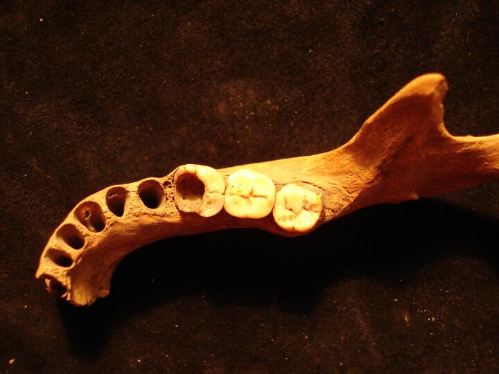
| MPY86
|
4964
|
3
|
MPY86_4964_3.jpg
|
Mandible left side ante mortem tooth loss
|
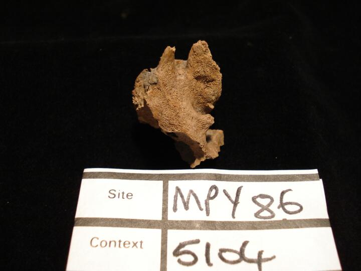
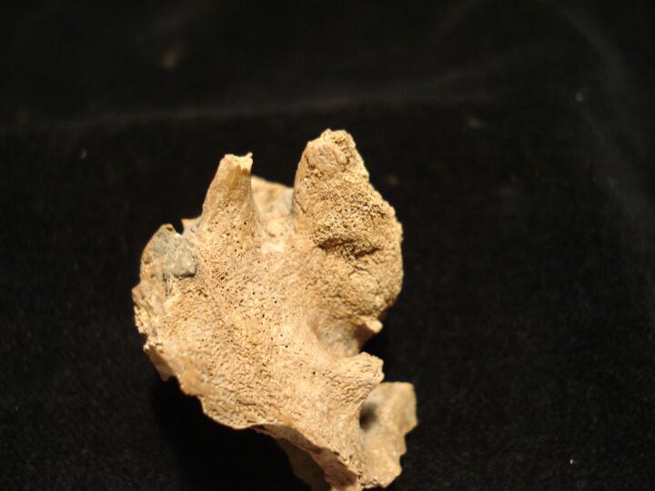
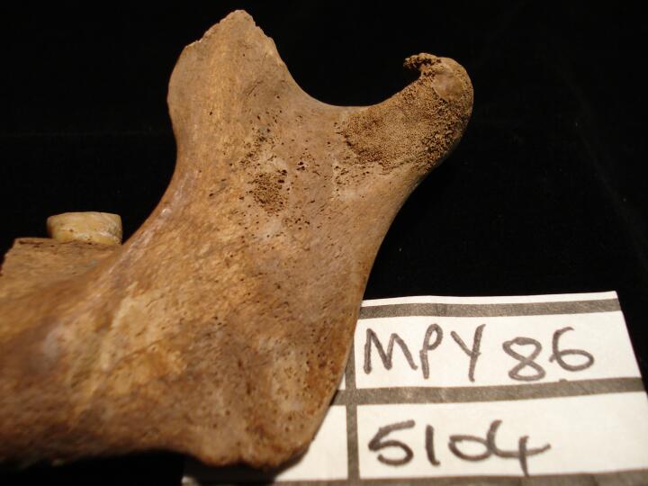
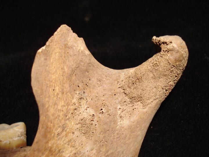
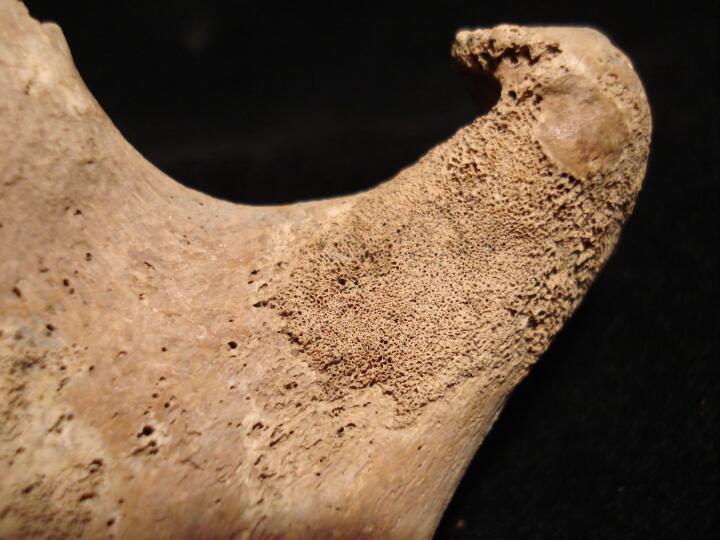
| MPY86
|
5104
|
1
|
MPY86_5104_1.jpg
|
Skull right orbit, layer of fine active new bone growth (anterior view) possibly indicator of scurvy
|
| MPY86
|
5104
|
2
|
MPY86_5104_2.jpg
|
Sphenoid body,active new bone growth (posterior view) possibly an indicator of scurvy
|
| MPY86
|
5104
|
3
|
MPY86_5104_3.jpg
|
Skull occipital bone (endocranial surface) layer of active new bone growth possibly an indicator of scurvy
|
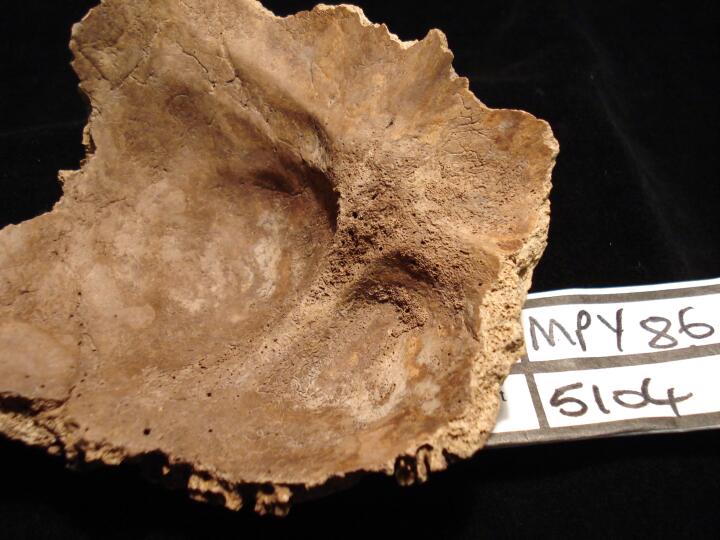
| MPY86
|
5104
|
4
|
MPY86_5104_4.jpg
|
Skull occipital bone (endocranial surface/close up) layer of active new bone growth possibly an indicator of scurvy
|
| MPY86
|
5104
|
5
|
MPY86_5104_5.jpg
|
Mandible left ramus area of fine active new bone growth possibly an indicator of scurvy
|
| MPY86
|
5104
|
6
|
MPY86_5104_6.jpg
|
Mandible left ramus (buccal view) area of fine active new bone growth possibly an indicator of scurvy
|
| MPY86
|
5104
|
7
|
MPY86_5104_7.jpg
|
Mandible left ramus (buccal view/close up) area of fine active new bone growth possibly an indicator of scurvy
|
| MPY86
|
5104
|
8
|
MPY86_5104_8.jpg
|
Mandible left ramus (lingual view) area of fine active new bone growth possibly an indicator of scurvy
|
| MPY86
|
5104
|
9
|
MPY86_5104_9.jpg
|
Mandible left ramus (lingual view/close up) area of fine active new bone growth possibly an indicator of scurvy
|
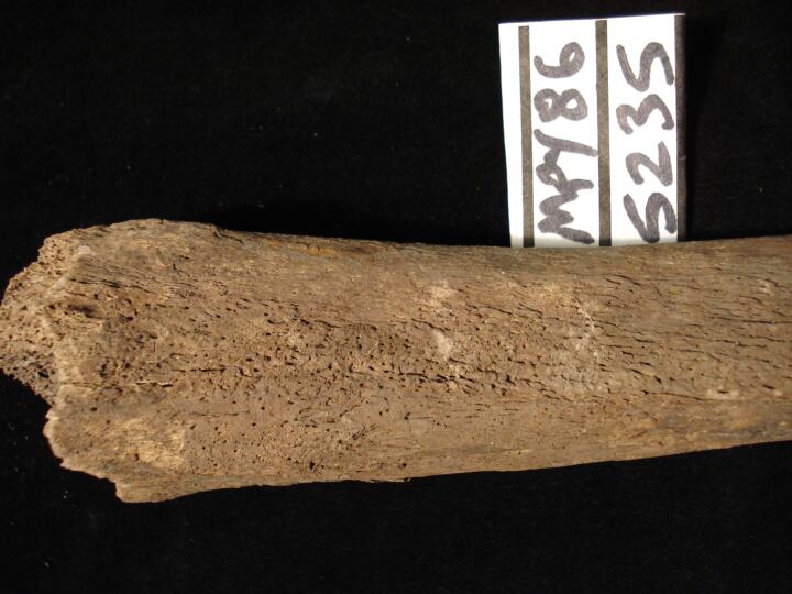
| MPY86
|
5235
|
1
|
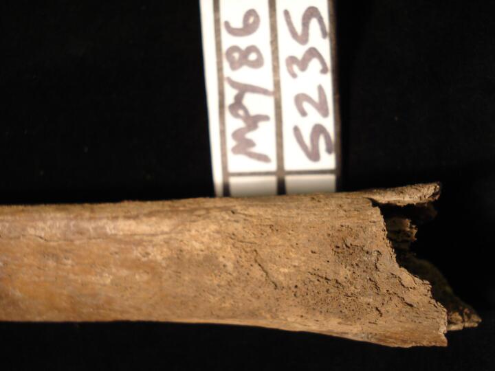
MPY86_5235_1.jpg
|
Right tibia, proximal to mid shaft (lateral view) non specific periosteal changes
|
| MPY86
|
5235
|
2
|
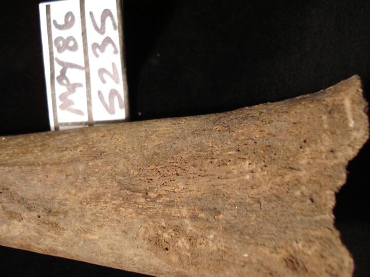
MPY86_5235_2.jpg
|
Right tibia, distal end of shaft (medial view) non specific periosteal changes
|
| MPY86
|
5235
|
3
|
MPY86_5235_3.jpg
|
Right femur distal end (posterior view) non specific periosteal changes
|
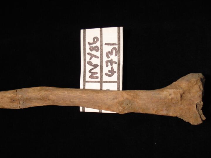
| MPY86
|
5247
|
1
|
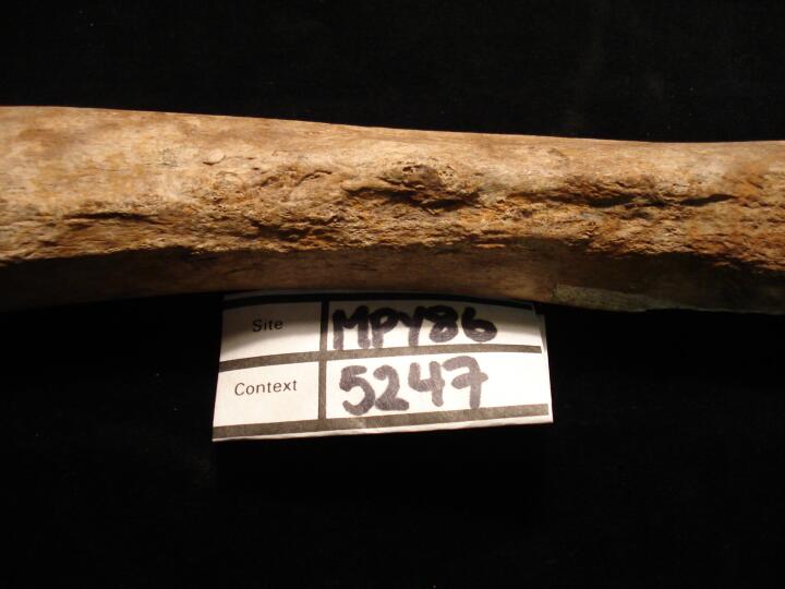
MPY86_5247_1.jpg
|
Right femur proximal/mid shaft (medial view) swollen area possibly a very well healed fracture
|
| MPY86
|
5247
|
2
|
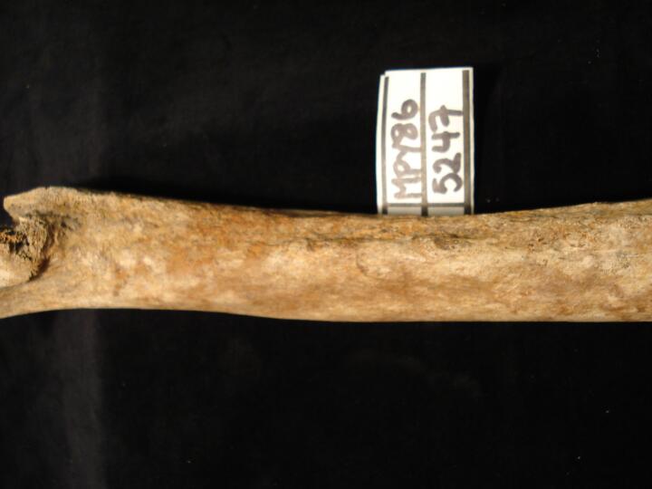
MPY86_5247_2.jpg
|
Right femur proximal/mid shaft (posterior view) swollen area possibly a very well healed fracture
|
| MPY86
|
5277
|
1
|
MPY86_5277_1.jpg
|
Well healed sharp force trauma to the right frontal bone.
|
| MPY86
|
5277
|
2
|
MPY86_5277_2.jpg
|
Close up of a well healed sharp force trauma to the right frontal bone. Showing displacement of the bone. Wound is positioned superior-inferior.
|
| MPY86
|
5277
|
3
|
MPY86_5277_3.jpg
|
Close up of a well healed sharp force trauma to the right frontal bone. Wound is positioned superior-inferior.
|
| MPY86
|
5277
|
4
|
MPY86_5277_4.jpg
|
Osteoid Osteoma? Thickened ovular bony protrusion of bone on the posterior surface of the right femur, medial to the linea aspera.
|
| MPY86
|
5277
|
5
|
MPY86_5277_5.jpg
|
DISH. Dripping wax like osteophytes causing fusion to the right anterior bodies of T6-9. Disc spaces are retained.
|
| MPY86
|
5321
|
1
|
MPY86_5321_1.jpg
|
Large button osteoma to the left frontal bone and a metopic suture.
|
| MPY86
|
7159
|
1
|
MPY86_7159_1.jpg
|
DISH? 'Dripping candle wax' like osteophytes down the right vertebral bodies causing fusion of T9 and T10.
|
| MPY86
|
7401
|
1
|
MPY86_7401_1.jpg
|
Pronounced osteophytic lipping of a lumbar vertebra.
|
| MPY86
|
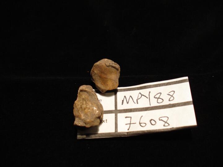
7608
|
1
|
MPY86_7608_1.jpg
|
Right trapezium & 1st metacarpal, osteoarthritis of the joint with eburnation of articulating surfaces (superior view)
|
| MPY86
|
7608
|
2
|
MPY86_7608_2.jpg
|
Right trapezium & 1st metacarpal, osteoarthritis of the joint with eburnation of articulating surfaces
|
| MPY86
|
7655
|
1
|
MPY86_7655_1.jpg
|
Malaligned healed fracture of L radius
|
| MPY86
|
7689
|
1
|
MPY86_7689_1.jpg
|
Tuberculosis, pott's spine
|
| MPY86
|
7689
|
2
|
MPY86_7689_2.jpg
|
Tuberculosis, pott's spine
|
| MPY86
|
7689
|
3
|
MPY86_7689_3.jpg
|
Tuberculosis, pott's spine
|
| MPY86
|
7689
|
4
|
MPY86_7689_4.jpg
|
Tuberculosis, pott's spine
|
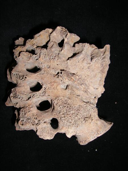
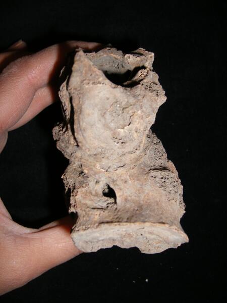
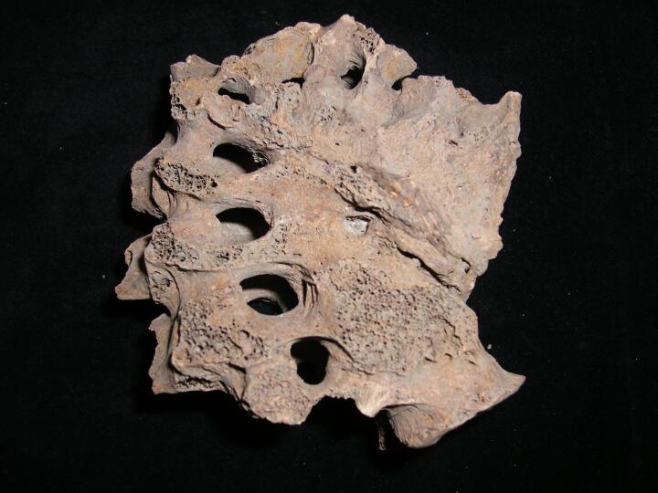
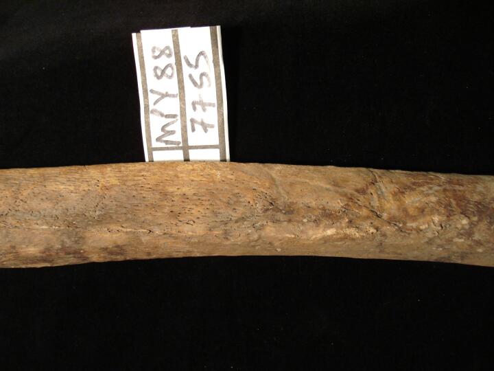
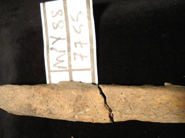
| MPY86
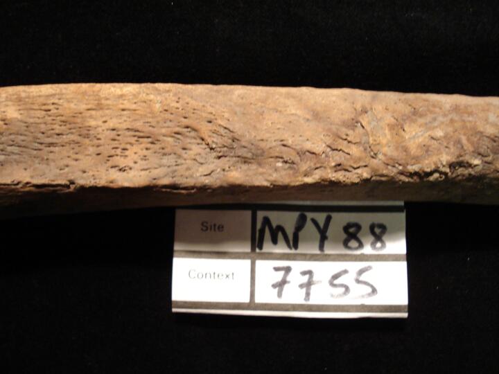
|
7755
|
1
|
MPY86_7755_1.jpg
|
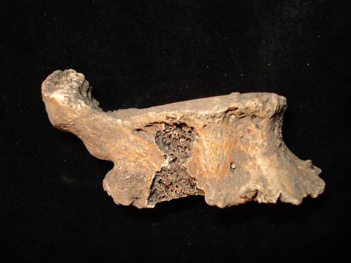
Right tibia lateral aspect (anterior view) pronounced periosteal changes possiby indication of osteomyelitis or syphilis
|
| MPY86
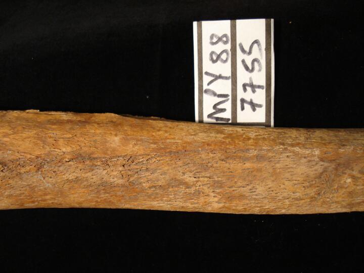
|
7755
|
2
|
MPY86_7755_2.jpg
|
Right tibia lateral aspect (anterior view) pronounced periosteal changes possiby indication of osteomyelitis or syphilis
|
| MPY86
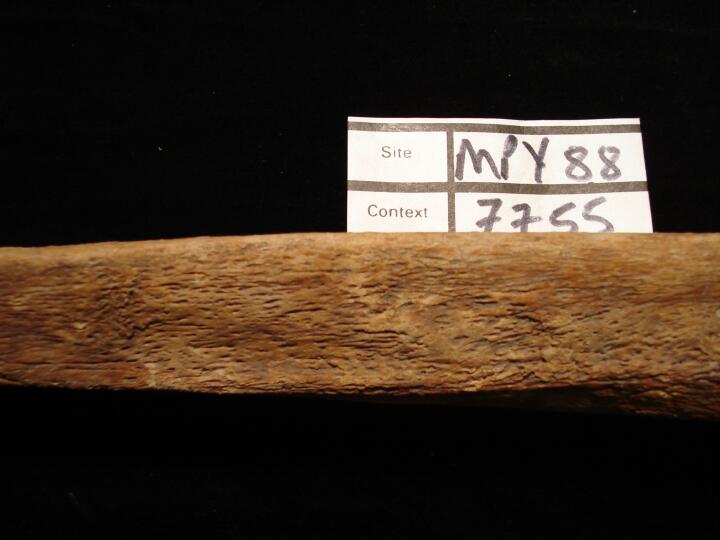
|
7755
|
3
|
MPY86_7755_3.jpg
|
Left tibia lateral aspect (superior view) pronounced periosteal changes possiby indication of osteomyelitis or syphilis
|
| MPY86
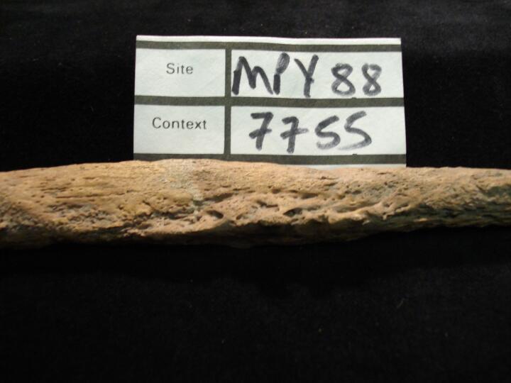
|
7755
|
4
|
MPY86_7755_4.jpg
|
Left tibia lateral aspect (superior view) pronounced periosteal changes possiby indication of osteomyelitis or syphilis
|
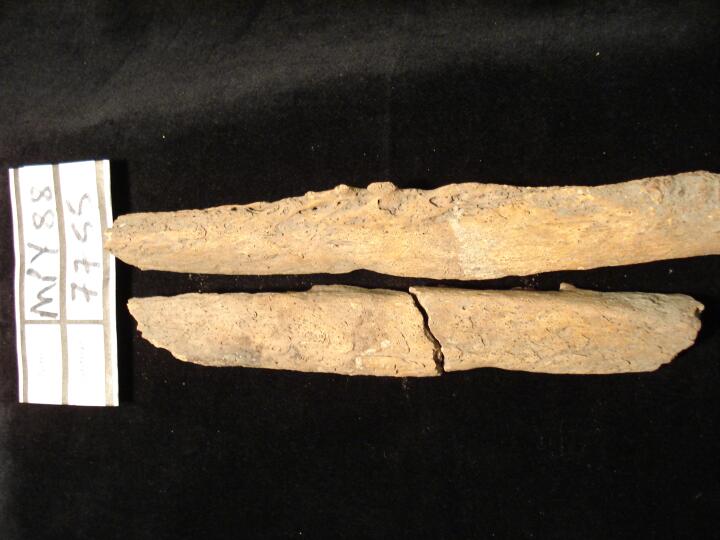
| MPY86
|
7755
|
5
|
MPY86_7755_5.jpg
|
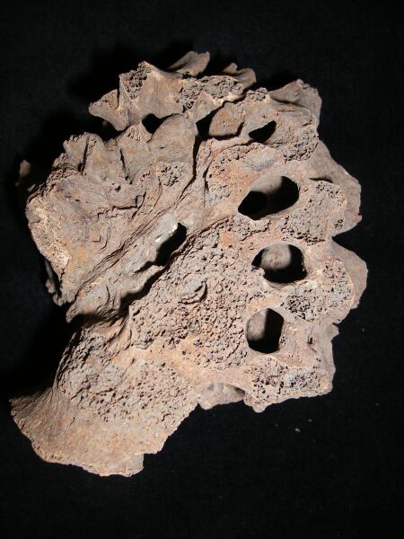
Left fibula mid shaft (superior view) pronounced periosteal changes possiby indication of osteomyelitus or syphilis
|
| MPY86
|
7755
|
6
|
MPY86_7755_6.jpg
|
Left fibula mid shaft (superior view) pronounced periosteal changes possiby indication of osteomyelitis or syphilis
|
| MPY86
|
7755
|
7
|
MPY86_7755_7.jpg
|
Left fibula mid shaft (superior view) pronounced periosteal changes possiby indication of osteomyelitis or syphilis
|
| MPY86
|
7755
|
8
|
MPY86_7755_8.jpg
|
Left fibula mid shaft (superior view) pronounced periosteal changes possiby indication of osteomyelitis or syphilis
|
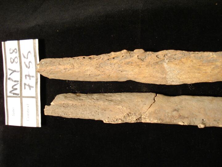
| MPY86
|
7755
|
9
|
MPY86_7755_9.jpg
|
Left & right fibula mid shaft (superior view) florid periosteal changes & bony projections, possiby indication of osteomyelitis or syphilis,
|
| MPY86
|
7755
|
10
|
MPY86_7755_10.jpg
|
Left & right fibula mid shaft (superior view) pronounced periosteal changes possiby indication of osteomyelitis or syphilis
|
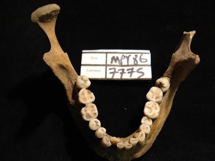
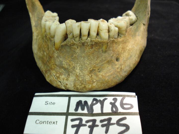
| MPY86
|
7775
|
1
|
MPY86_7775_1.jpg
|
Mandible (occlusal view) over crowding of teeth & impaction of left 3rd molar
|
| MPY86
|
7775
|
2
|
MPY86_7775_2.jpg
|
Mandible, anterior teeth over crowding (buccal view)
|
| MPY86
|
7775
|
3
|
MPY86_7775_3.jpg
|
Mandible left side (buccal view) impaction of 3rd molar
|
| MPY86
|
7775
|
4
|
MPY86_7775_4.jpg
|
Mandible left canine linear hypoplastic defect
|
| MPY86
|
7775
|
5
|
MPY86_7775_5.jpg
|
Mandible left canine linear hypoplastic defect (lingual view)
|
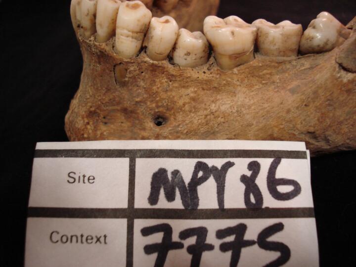
| MPY86
|
7775
|
6
|
MPY86_7775_6.jpg
|
Mandible overcrowding & calculus (lingual view/close up)
|


