| Site code
|
Context
|
Frame number
|
Photo
|
Description
|
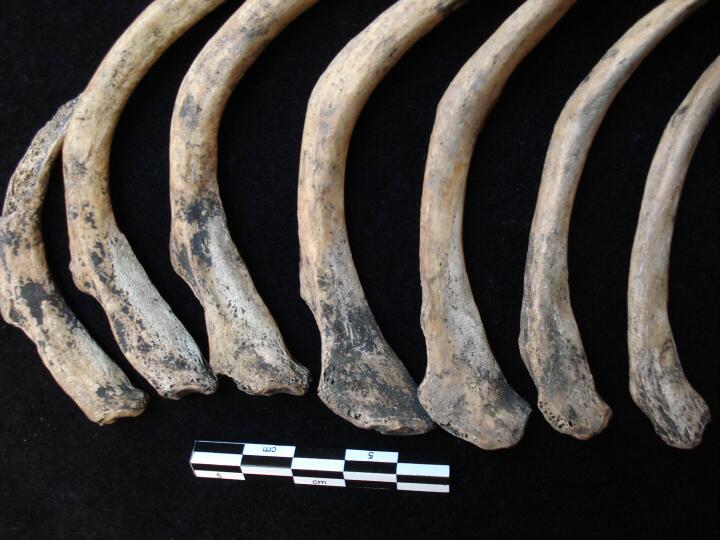
| NRT85
|
385
|
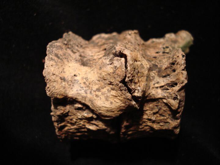
1
|
NRT85_385_1.jpg
|
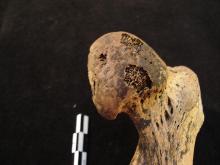
Left ribs active non-specific infection (visceral surface) localised to head/angle of ribs
|
| NRT85
|
385
|
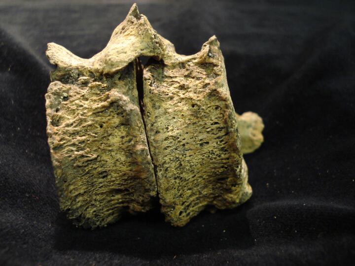
2
|
NRT85_385_2.jpg
|
Two left ribs active non-specific infection (visceral surface) localised to head/angle of ribs
|
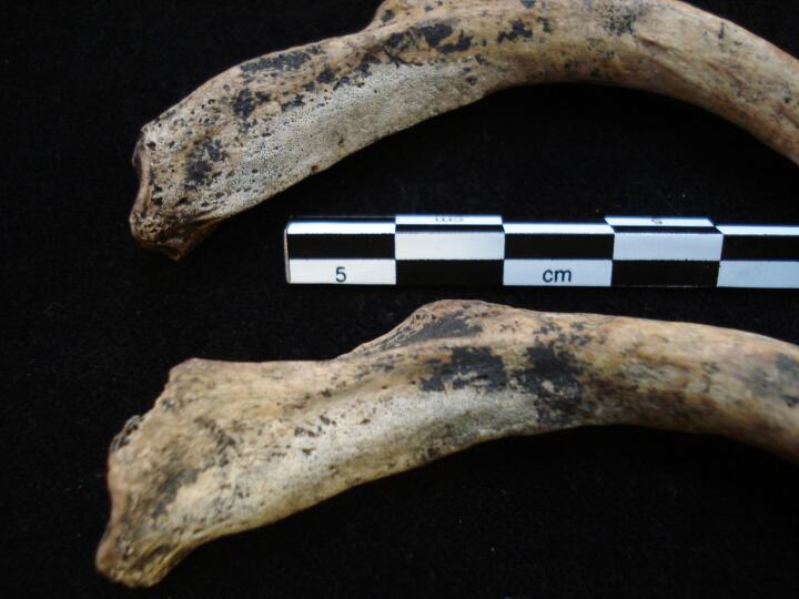
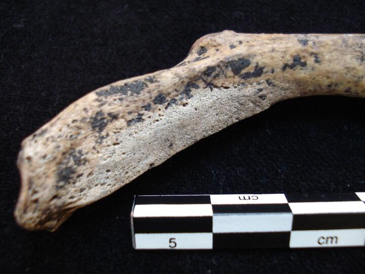
| NRT85
|
385
|
3
|
NRT85_385_3.jpg
|
One left rib head/angle (visceral surface/close up) active non-specific infection
|
| NRT85
|
385
|
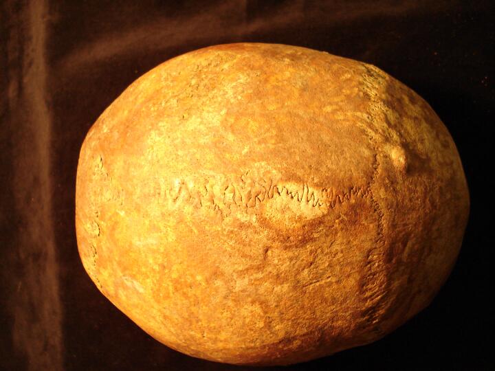
4
|
NRT85_385_4.jpg
|
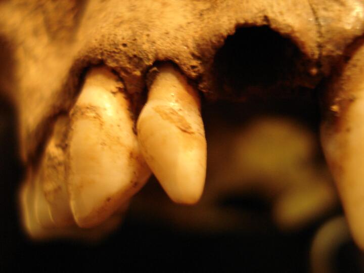
Mandible (occlusal view/close up) 'peg' like 3rd mandibular molars
|
| NRT85
|
385
|
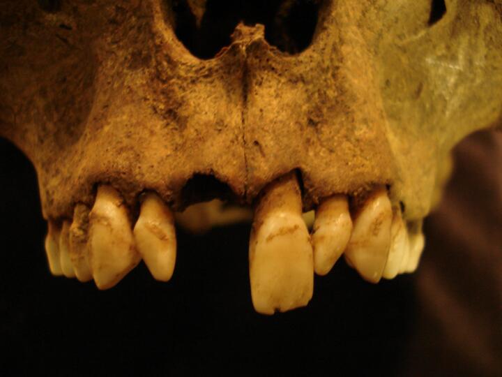
5
|
NRT85_385_5.jpg
|
Mandible (anterior view) showing all dentition & 'peg' like 3rd mandibular molars
|
| NRT85
|
385
|
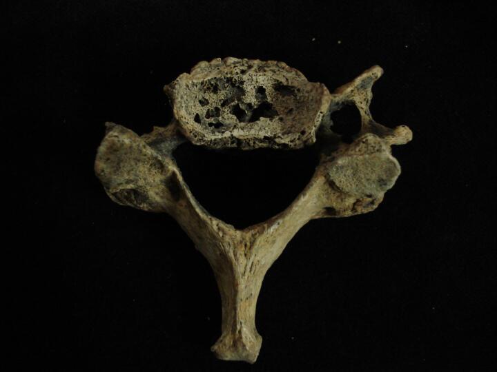
6
|
NRT85_385_6.jpg
|
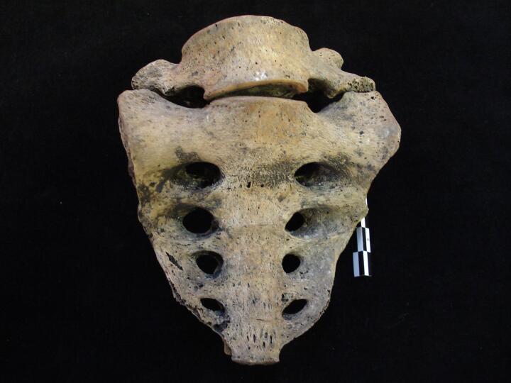
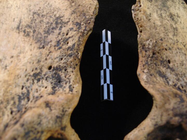
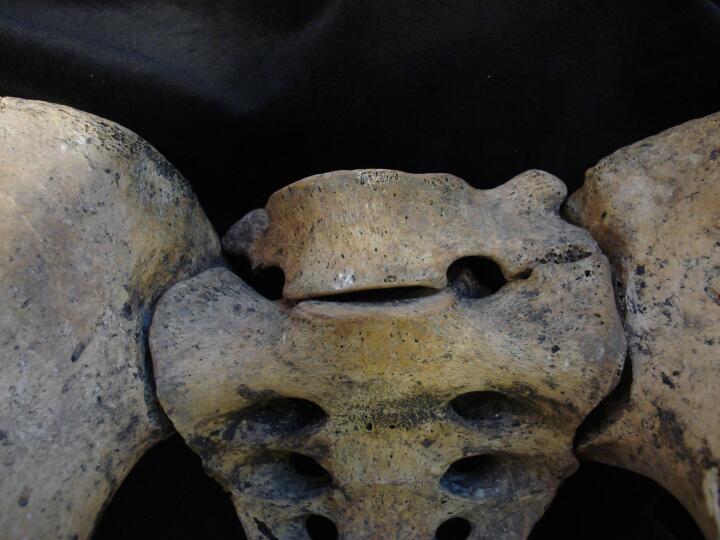
Sacrum & Lumbar L5 with articulating left side ala like wing (anterior view)
|
| NRT85
|
385
|
7
|
NRT85_385_7.jpg
|
Sacrum & Lumbar L5 with articulating left side ala like wing (anterior view)
|
| NRT85
|
385
|
8
|
NRT85_385_8.jpg
|
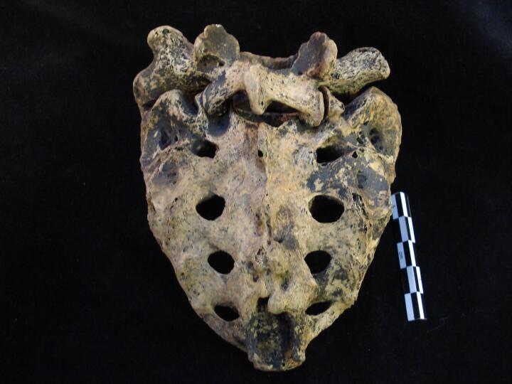
Sacrum & Lumbar L5 with articulating left side ala like wing (posterior view)
|
| NRT85
|
385
|
9
|
NRT85_385_9.jpg
|
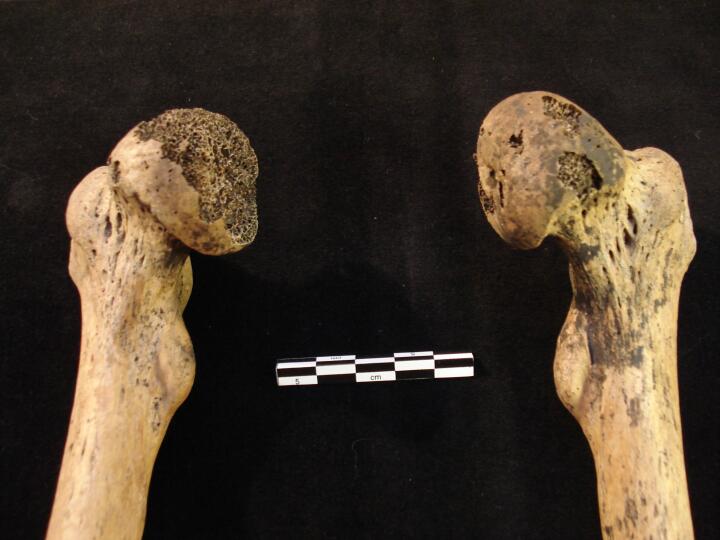
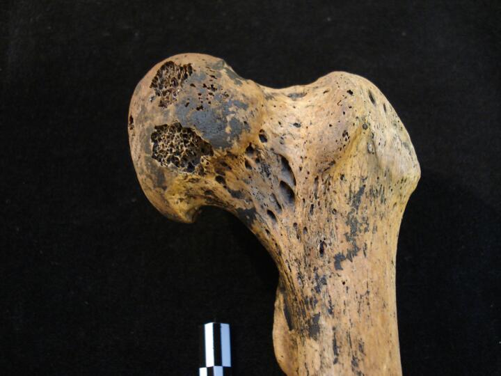
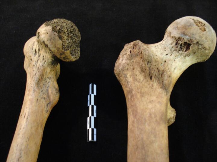
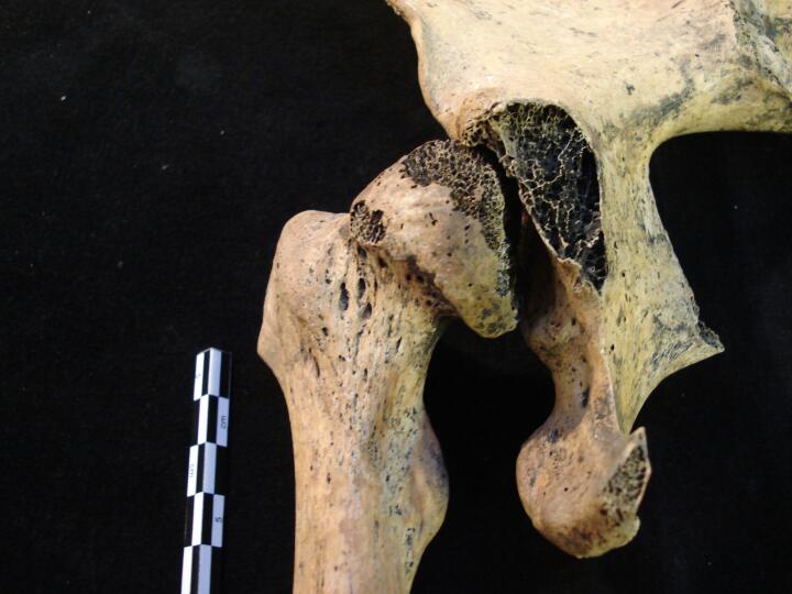
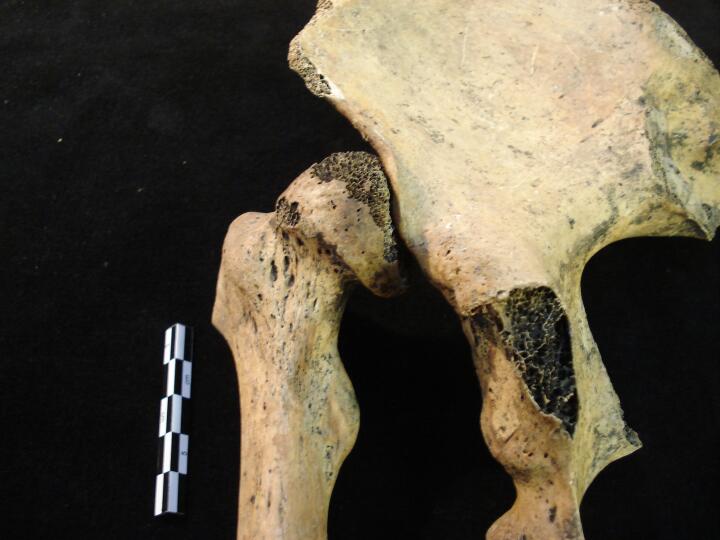
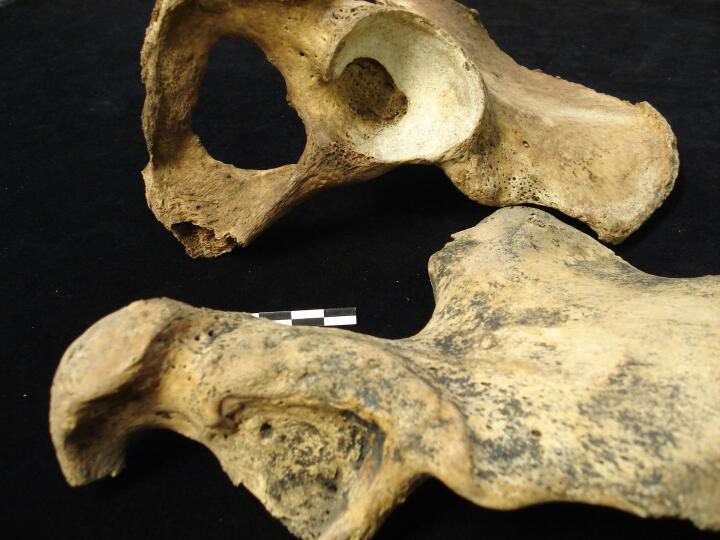
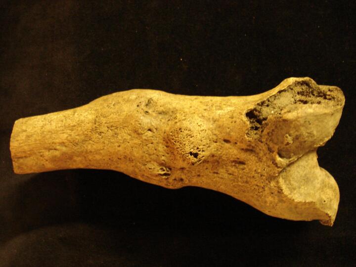
Right and Left femora (anterior view) showing proximal 1/3 & distorted femoral heads from Congenital Hip Displacement
|
| NRT85
|
385
|
10
|
NRT85_385_10.jpg
|
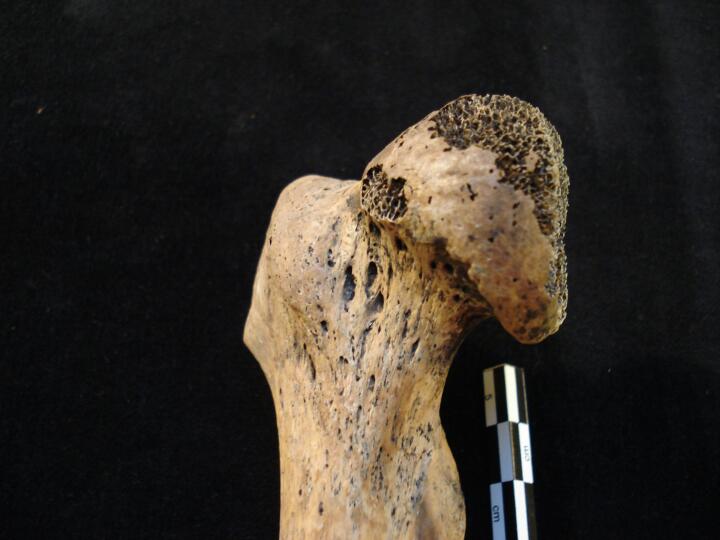
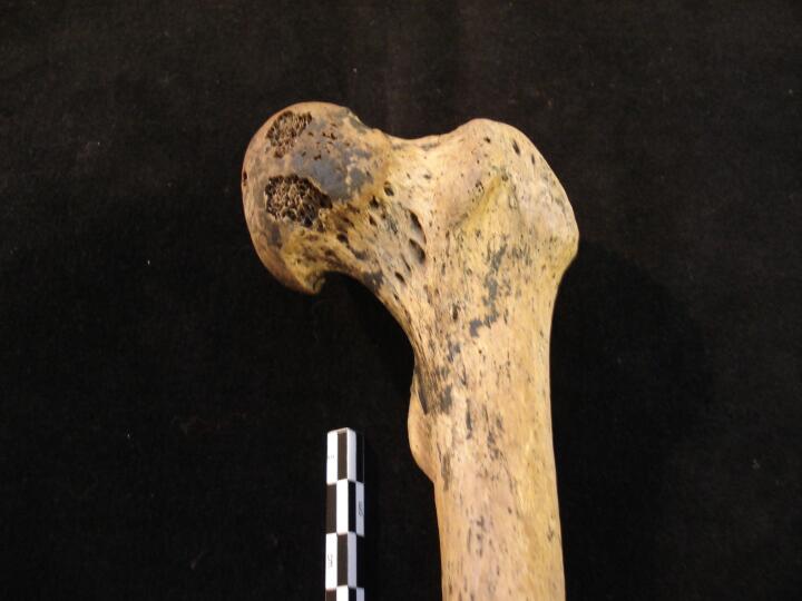
Right femur (anterior view) showing proximal 1/3 & distorted femoral head from Congenital Hip Displacement
|
| NRT85
|
385
|
11
|
NRT85_385_11.jpg
|
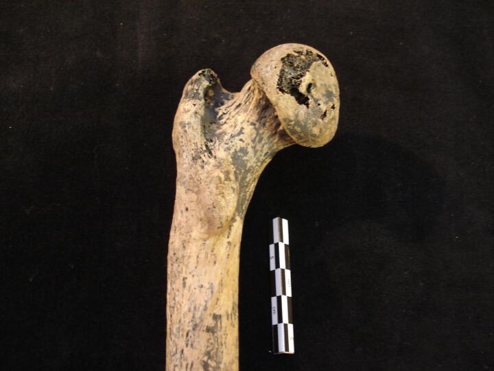
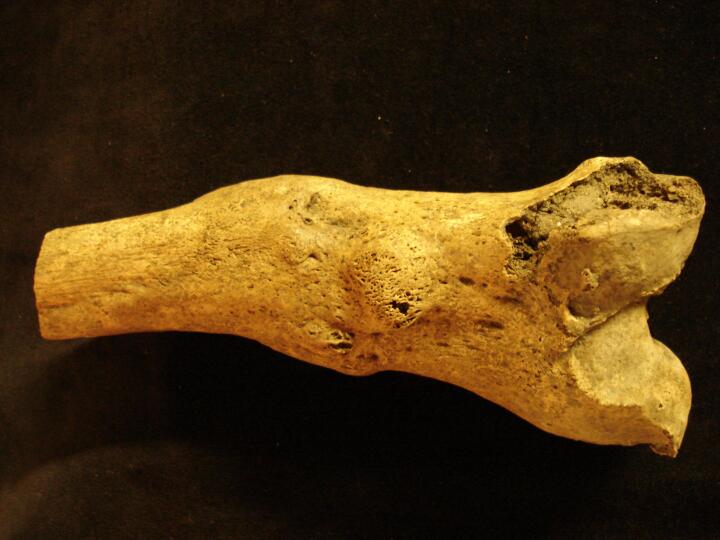
Right femur (posterior) showing proximal 1/3 & distorted femoral heads from Congenital Hip Displacement
|
| NRT85
|
385
|
12
|
NRT85_385_12.jpg
|
Right femur (anterior view/close up) showing proximal 1/3 & distorted femoral head from Congenital Hip Displacement
|
| NRT85
|
385
|
13
|
NRT85_385_13.jpg
|
Left femur (anterior view) showing proximal 1/3 & distorted femoral head from Congenital Hip Displacement
|
| NRT85
|
385
|
14
|
NRT85_385_14.jpg
|
Left femur (posterior view) showing proximal 1/3 & distorted femoral head from Congenital Hip Displacement
|
| NRT85
|
385
|
15
|
NRT85_385_15.jpg
|
Left femur (anterior view/close up) showing proximal 1/3 & distorted femoral head from Congenital Hip Displacement
|
| NRT85
|
385
|
16
|
NRT85_385_16.jpg
|
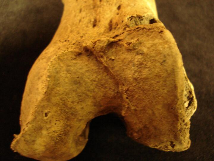
Left femur (medial view/close up) showing medially flattening of the femoral head
|
| NRT85
|
385
|
17
|
NRT85_385_17.jpg
|
Comparison between normal femoral head (female) and right femoral head from individual with congenital hip displacement (anterior view)
|
| NRT85
|
385
|
18
|
NRT85_385_18.jpg
|
Comparison between normal femoral head (female) and left femoral head from individual with congenital hip displacement (anterior view)
|
| NRT85
|
385
|
19
|
NRT85_385_19.jpg
|
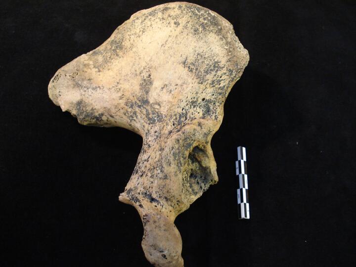
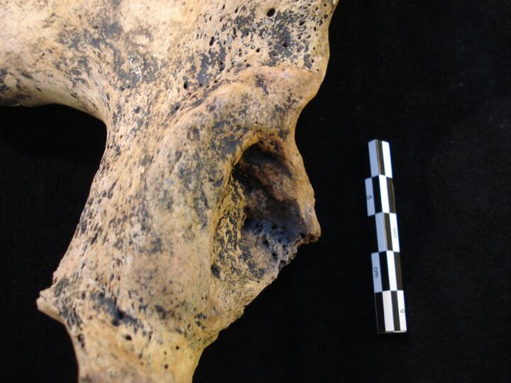
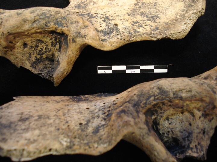
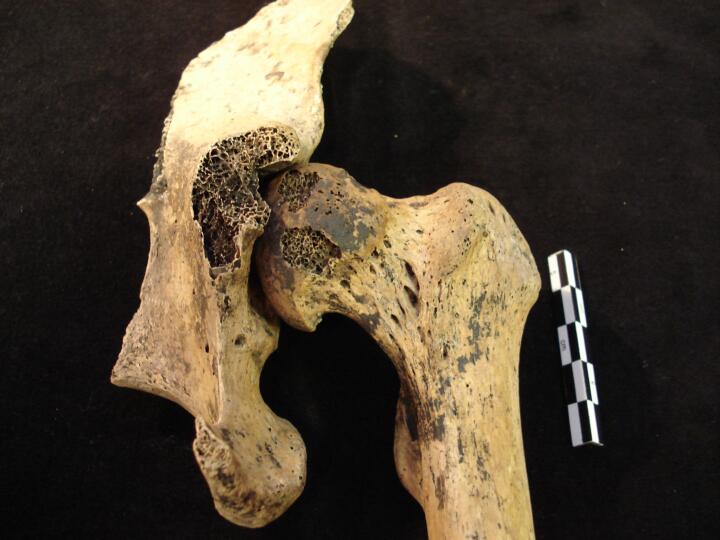
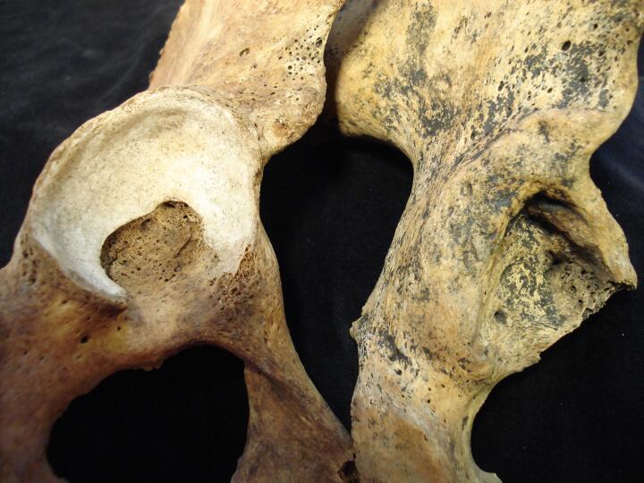
Right pelvis (posterior/medial view) showing malformed acetabulum from congenital hip displacement
|
| NRT85
|
385
|
20
|
NRT85_385_20.jpg
|
Right pelvis (posterior/medial view) close up showing malformed acetabulum from congenital hip displacement
|
| NRT85
|
385
|
21
|
NRT85_385_21.jpg
|
Right pelvis (medial view) close up showing malformed acetabulum from congenital hip displacement
|
| NRT85
|
385
|
22
|
NRT85_385_22.jpg
|
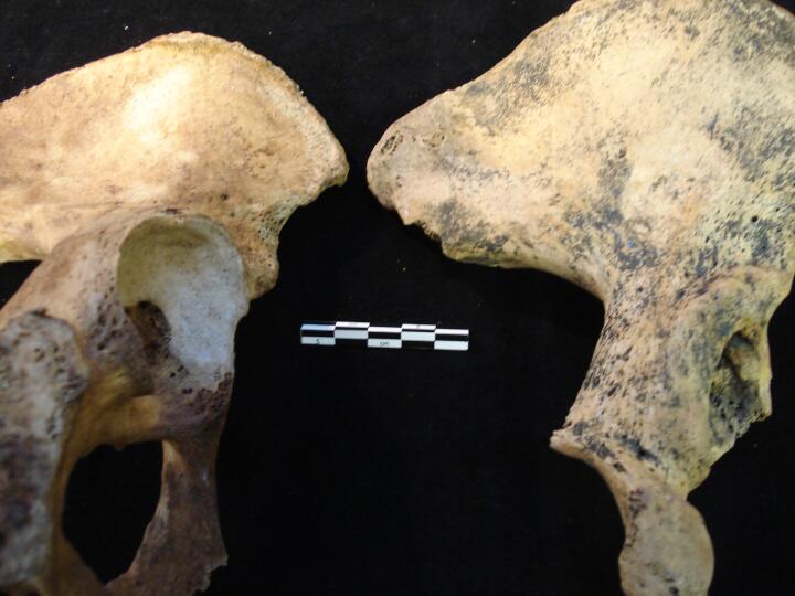
Left and Right pelves (medial view) showing acetabulae with bilateral congenital hip displacement
|
| NRT85
|
385
|
23
|
NRT85_385_23.jpg
|
Left and Right pelvis showing superior aspect of malformed acetabulae from bilateral congenital hip displacement (posterior view)
|
| NRT85
|
385
|
24
|
NRT85_385_24.jpg
|
Left and Right pelvis showing superior aspect of malformed acetabulae from bilateral congenital hip displacement (medial view)
|
| NRT85
|
385
|
25
|
NRT85_385_25.jpg
|
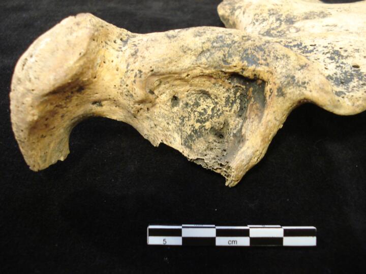
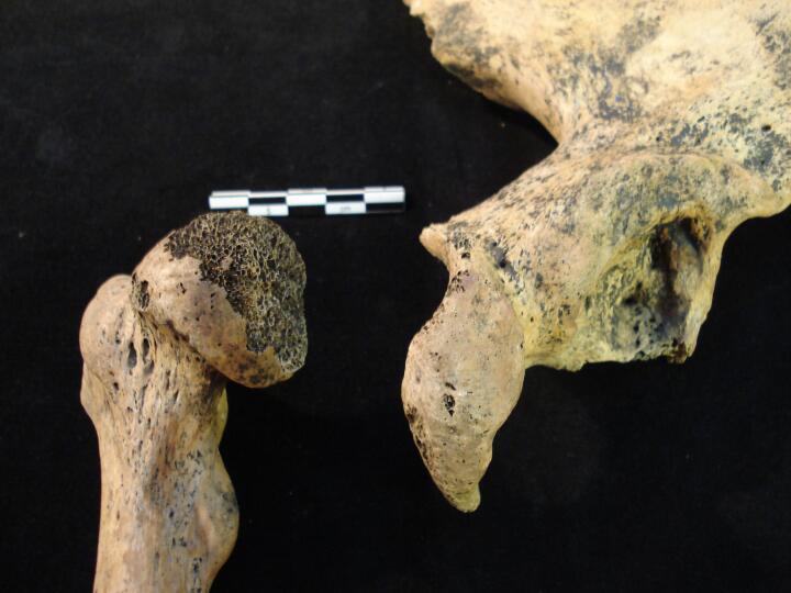
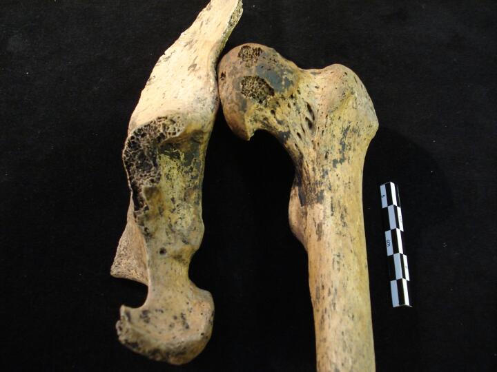
Right femoral head & right pelvis showing congenital hip displacement (anterior view)
|
| NRT85
|
385
|
26
|
NRT85_385_26.jpg
|
Normal articulated right femoral head & right pelvis (acetabulum) in individual with congenital hip displacement (anterior view)
|
| NRT85
|
385
|
27
|
NRT85_385_27.jpg
|
Abnormal articulated right femoral head & right pelvis, showing displacement of the femoral head to the superior aspect of acetabulum in individual with congenital hip displacement (anterior view)
|
| NRT85
|
385
|
28
|
NRT85_385_28.jpg
|
Normal articulated left femoral head & left pelvis (acetabulum) in individual with congenital hip displacement (anterior view)
|
| NRT85
|
385
|
29
|
NRT85_385_29.jpg
|
Abnormal articulated left femoral head & left pelvis, showing displacement of the femoral head to the superior aspect of acetabulum in individual with congenital hip displacement (anterior view)
|
| NRT85
|
385
|
30
|
NRT85_385_30.jpg
|
Comparison of normal right (female) pelvis-acetabulum and abnormal pelvis-acetabulum form congenital hip displacement (posterior view)
|
| NRT85
|
385
|
31
|
NRT85_385_31.jpg
|
Comparison of normal right (female) pelvis-acetabulum and abnormal pelvis-acetabulum form congenital hip displacement (medial view)
|
| NRT85
|
385
|
32
|
NRT85_385_32.jpg
|
Comparison (close up) of normal right (female) pelvis-acetabulum and abnormal pelvis-acetabulum form congenital hip displacement (posterior view)
|
| NRT85
|
385
|
33
|
NRT85_385_33.jpg
|
Articulation of pelves, sacrum & lumbar L5 with articulating left ala like wing (anterior view)
|
| NRF88
|
1304
|
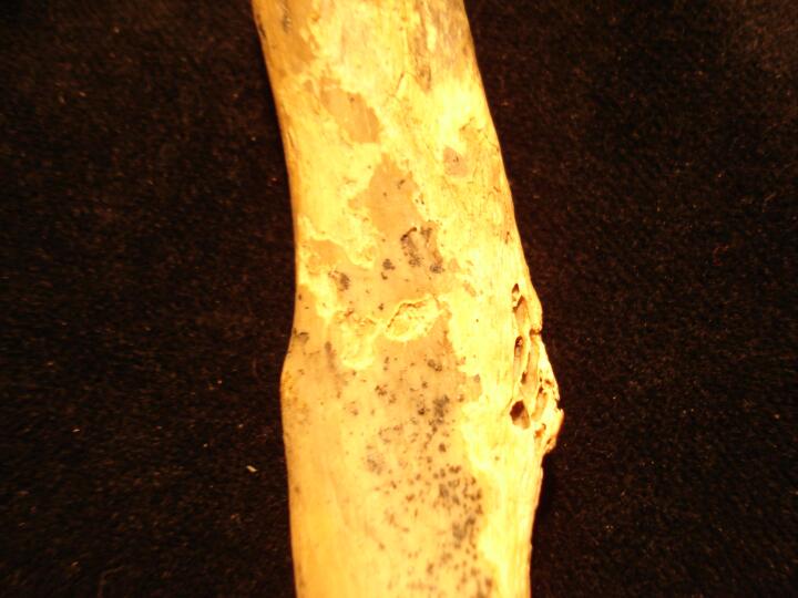
2
|
NRF88_1304_2.jpg
|
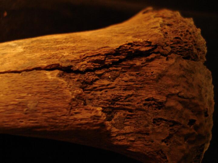
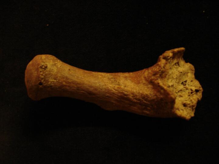
Osgood Schlatters disease? Proximal tibia anterior view (close up of the tibial tuberosity)
|
| NRF88
|
1456
|
1
|
NRF88_1456_1.jpg
|
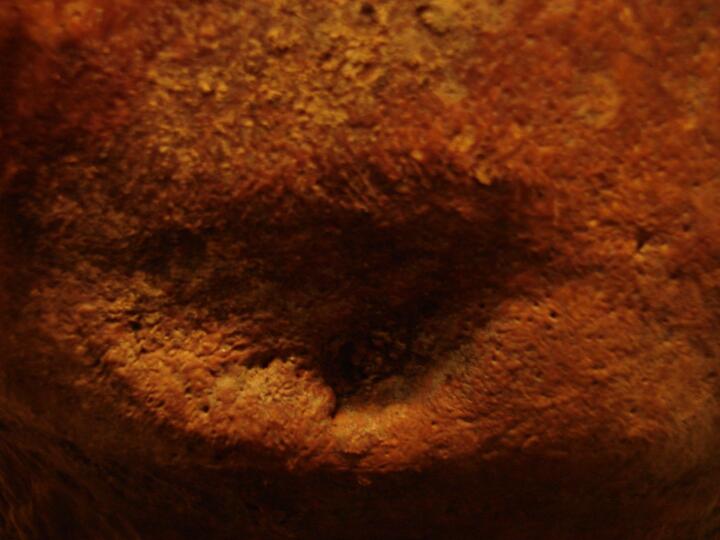
Button osteoma on the frontal bone (Superior view of the skull)
|
| NRF88
|
1456
|
2
|
NRF88_1456_2.jpg
|
Button osteoma on the frontal bone (Close up)
|
| NRF88
|
1456
|
3
|
NRF88_1456_3.jpg
|
Well healed blunt force trauma wound to the occipital bone
|
| NRF88
|
1456
|
4
|
NRF88_1456_4.jpg
|
Well healed blunt force trauma wound to the occipital bone (Close up)
|
| NRF88
|
1569
|
1
|
NRF88_1569_1.jpg
|
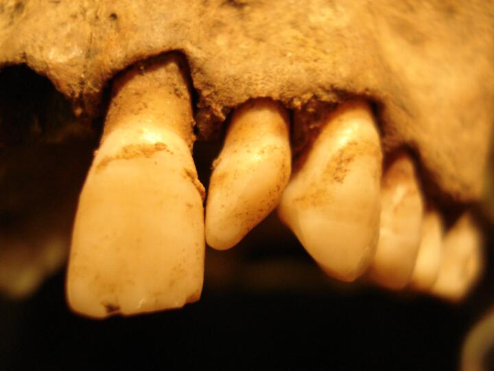
Peg shaped lateral maxillary incisors
|
| NRF88
|
1569
|
2
|
NRF88_1569_2.jpg
|
Peg shaped lateral maxillary incisors (Close up left lateral incisor)
|
| NRF88
|
1569
|
3
|
NRF88_1569_3.jpg
|
Peg shaped lateral maxillary incisors (Close up right lateral incisor)
|
| NRT85
|
339
|
1
|
NRT85_339_1.jpg
|
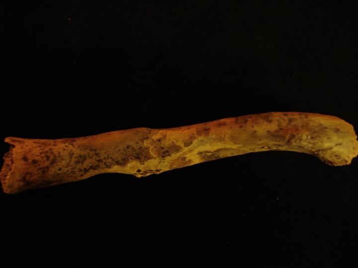
Left femur distal end, healed malaligned fracture (anterior view)
|
| NRT85
|
339
|
2
|
NRT85_339_2.jpg
|
Left femur distal end, healed malaligned fracture (anterior view)
|
| NRT85
|
339
|
3
|
NRT85_339_3.jpg
|
Left femur distal end, articular surfaces (anterior view)
|
| NRT85
|
339
|
4
|
NRT85_339_4.jpg
|
Left femur distal end, healed malaligned fracture (posterior view)
|
| NRT89
|
269
|
1
|
NRT89_269_1.jpg
|
Tuberculosis/trauma to thoracic and lumbar vertebrae
|
| SPQ88
|
123
|
1
|
SPQ88_123_1.jpg
|
DISH 'Dripping candle wax' like osteophytes down the right anterior ligament of the vertebral bodies of T9 and T10 (anterior view)
|
| SPQ88
|
123
|
2
|
SPQ88_123_2.jpg
|
DISH 'Dripping candle wax' like osteophytes down the right anterior ligament of the vertebral bodies of T9 and T10
|
| SPQ88
|
123
|
3
|
SPQ88_123_3.jpg
|
IVD on the superior vertebral body of C7
|
| SPQ88
|
277
|
1
|
SPQ88_277_1.jpg
|
Misalignment of a well healed right rib fracture
|
| SPQ88
|
277
|
2
|
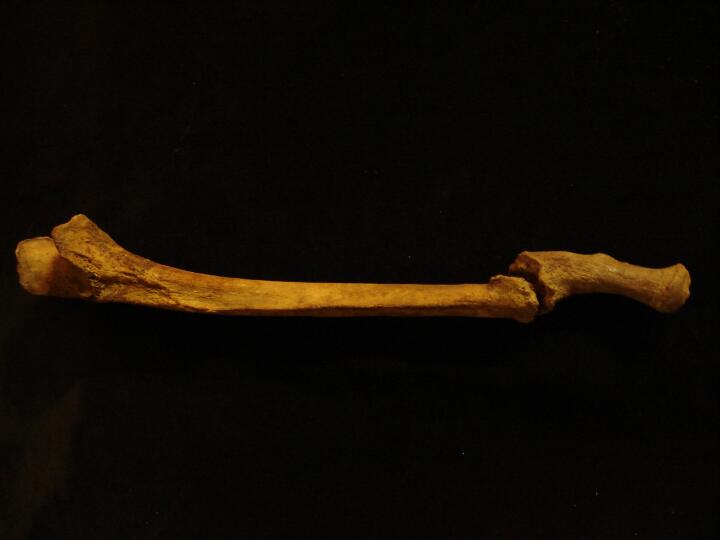
SPQ88_277_2.jpg
|
Well healed non union fracture of the right ulna
|
| SPQ88
|
277
|
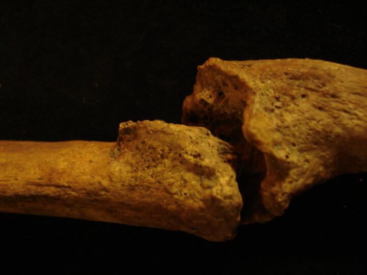
3
|
SPQ88_277_3.jpg
|
Well healed non union fracture of the right ulna (Close up)
|
| SPQ88
|
277
|
4
|
SPQ88_277_4.jpg
|
Well healed non union fracture of the right ulna. Close up of the proximal fracture surface)
|
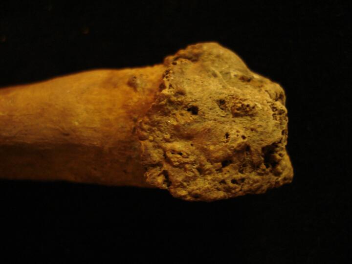
| SPQ88
|
277
|
5
|
SPQ88_277_5.jpg
|
Well healed non union fracture of the right ulna (Close up of the distal fracture surface)
|
| SSQ88
|
405
|
1
|
SSQ88_405_1.jpg
|
Well healed fracture ot the midshaft of the right clavicle
|


