| Site code
|
Context
|
Frame number
|
Photo
|
Description
|
| REW92
|
6
|
2
|
REW92_6_2.jpg
|
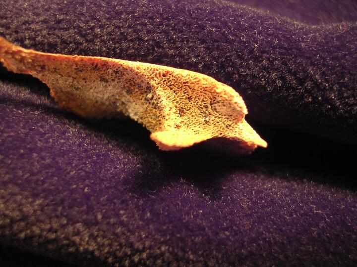
Left scapula (medial view) close up of bone changes, prostate cancer
|
| REW92
|
6
|
3
|
REW92_6_3.jpg
|
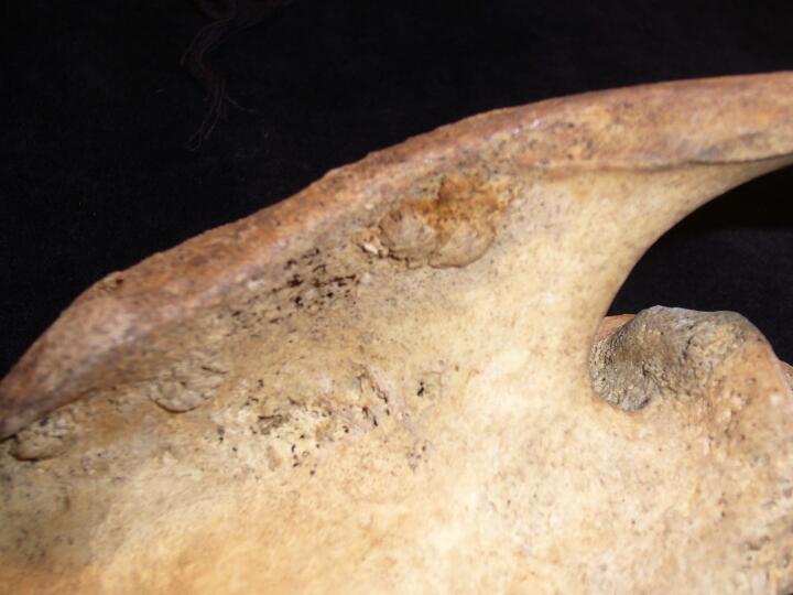
Scapula acromion lytic & osteoblastic changes
|
| REW92
|
6
|
4
|
REW92_6_4.jpg
|
Manubrium (anterior view) lytic lesions
|
| REW92
|
6
|
5
|
REW92_6_5.jpg
|
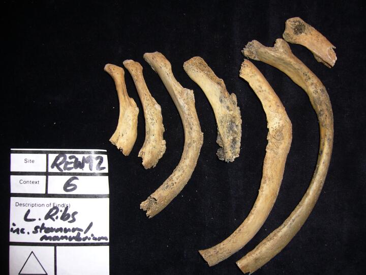
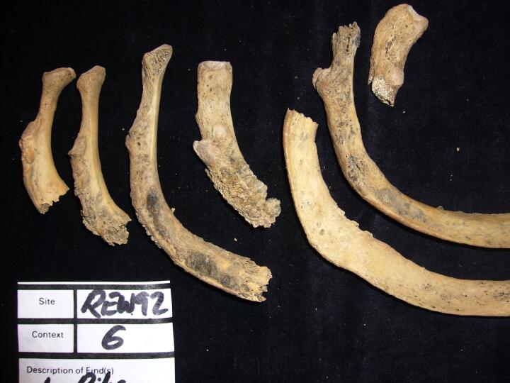
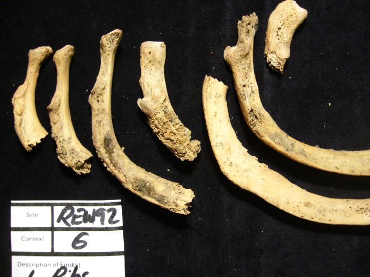
Left ribs osteoblastic changes
|
| REW92
|
6
|
6
|
REW92_6_6.jpg
|
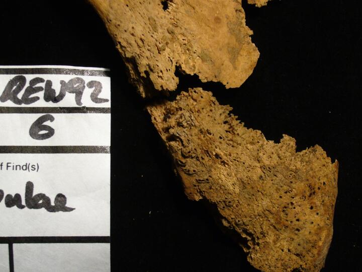
Left ribs osteoblastic changes (visceral surface)
|
| REW92
|
6
|
7
|
REW92_6_7.jpg
|
Left ribs osteoblastic changes (visceral surface)
|
| REW92
|
6
|
8
|
REW92_6_8.jpg
|
Infection on viseceral surface of ribs
|
| REW92
|
6
|
9
|
REW92_6_9.jpg
|
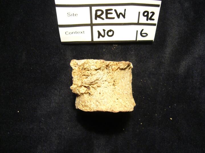
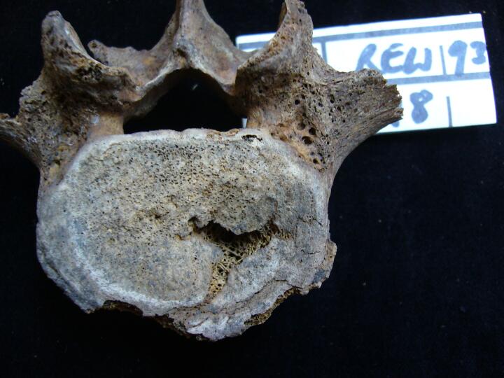
Osteoblastic bone on lateral aspect on lower vertebra - metastatic carcinoma?
|
| REW92
|
6
|
10
|
REW92_6_10.jpg
|
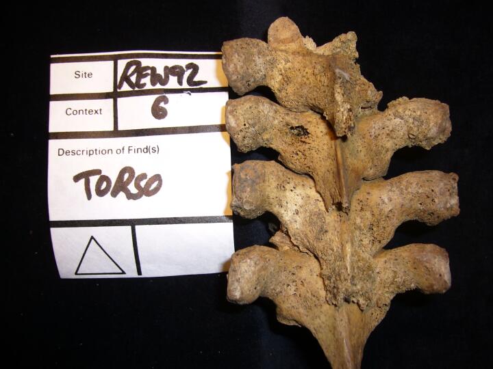
Thoracic vertebrae, spinous processes (posterior view) lytic lesions
|
| REW92
|
6
|
11
|
REW92_6_11.jpg
|
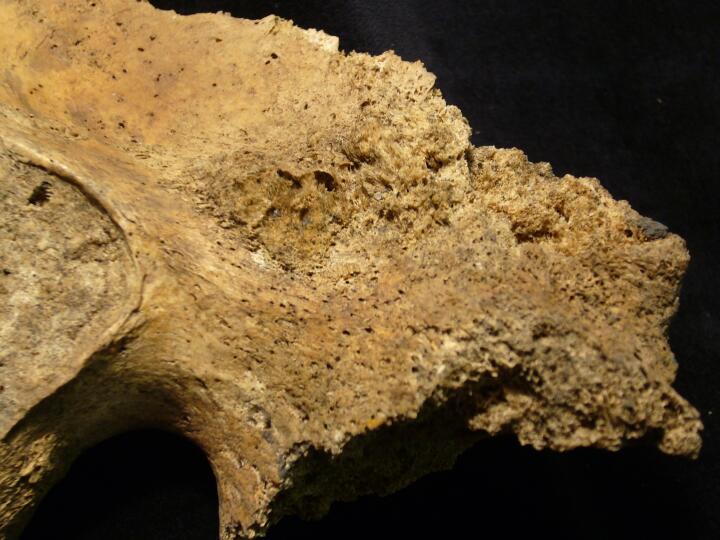
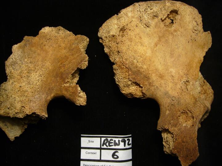
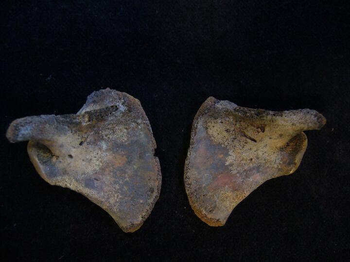
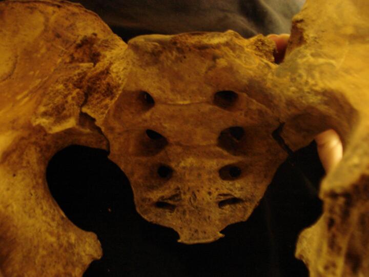
Pelves (ventral view) florid osteoblastic changes, ?prostate cancer
|
| REW92
|
6
|
12
|
REW92_6_12.jpg
|
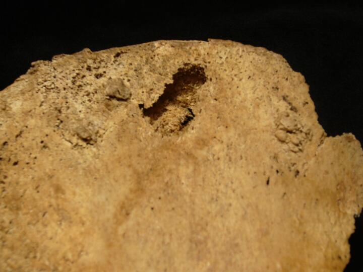
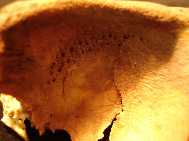
Left pelvis, iliac fossa, osteoblastic changes,?prostate cancer
|
| REW92
|
6
|
13
|
REW92_6_13.jpg
|
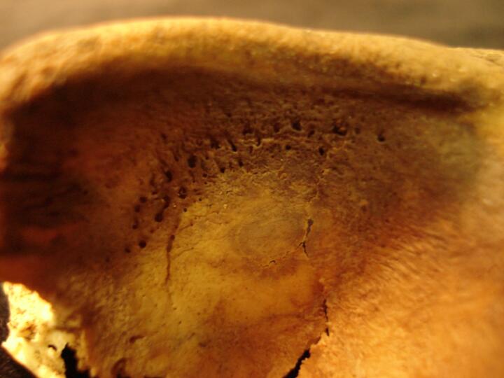
Left pelvis, iliac fossa, osteoblastic changes,?prostate cancer
|
| REW92
|
6
|
14
|
REW92_6_14.jpg
|
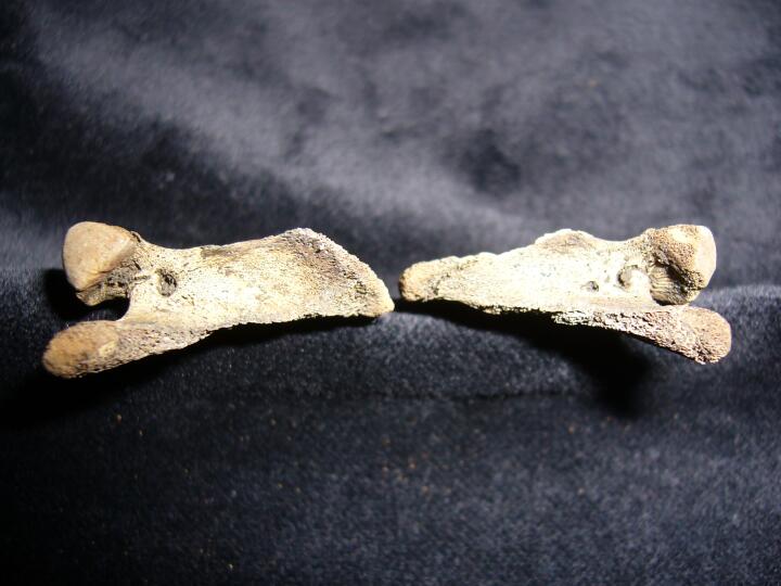
Pelves (dorsal view)
|
| REW92
|
6
|
15
|
REW92_6_15.jpg
|
Pelvic rim & fossa with lesions
|
| REW92
|
58.2
|
1
|
REW92_58.2_1.jpg
|
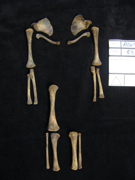
Sub adult long bones anatomically positioned
|
| REW92
|
58.2
|
2
|
REW92_58.2_2.jpg
|
Scapulae & clavicles
|
| REW92
|
58.2
|
3
|
REW92_58.2_3.jpg
|
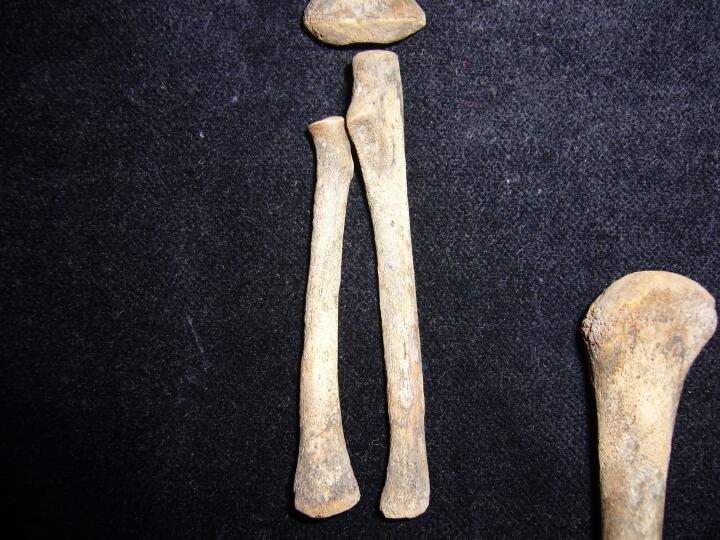
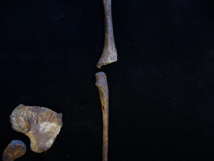
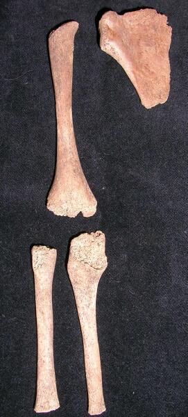
Right ulna & radius porous new bone on shafts,?scurvy
|
| REW92
|
58.2
|
4
|
REW92_58.2_4.jpg
|
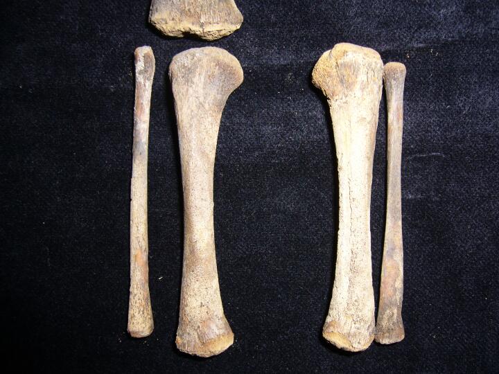
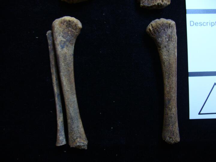
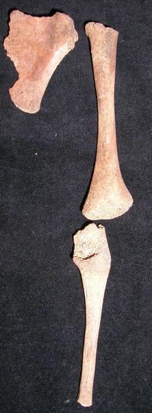
Tibiae & fibulae porous new bone on shafts, ?scurvy
|
| REW92
|
58.2
|
5
|
REW92_58.2_5.jpg
|
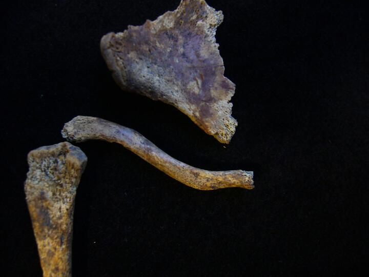
Scapulae, new bone growth
|
| REW92
|
58.2
|
6
|
REW92_58.2_6.jpg
|
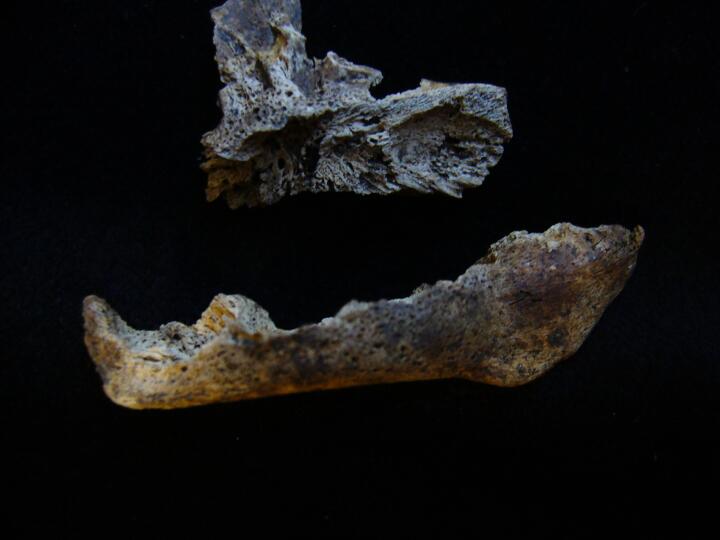
Scapulae, supraspinous fossa new bone growth
|
| REW92
|
62
|
1
|
REW92_62_1.jpg
|
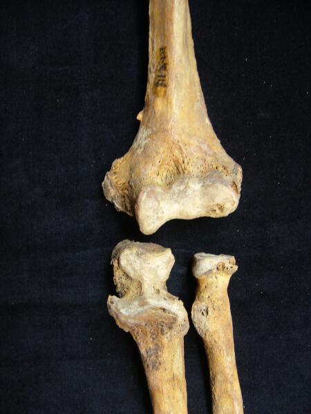
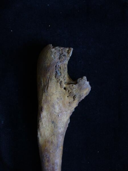
Right ulna & radius, distal end, fracture with infection & secondary joint changes
|
| REW92
|
62
|
2
|
REW92_62_2.jpg
|
Right radius
|
| REW92
|
62
|
3
|
REW92_62_3.jpg
|
Right ulna
|
| REW92
|
62
|
4
|
REW92_62_4.jpg
|
Right 2nd & 3rd metacarpals with ankylosed carpals (dorsal view)
|
| REW92
|
62
|
5
|
REW92_62_5.jpg
|
Right 2nd & 3rd metacarpals with ankylosed carpals (palmar view)
|
| REW92
|
62
|
6
|
REW92_62_6.jpg
|
Right 2nd & 3rd metacarpals with ankylosed carpals (medial view)
|
| REW92
|
62
|
7
|
REW92_62_7.jpg
|
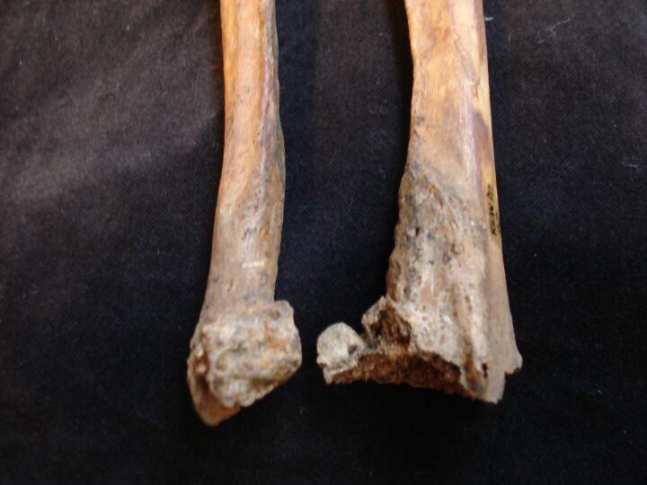
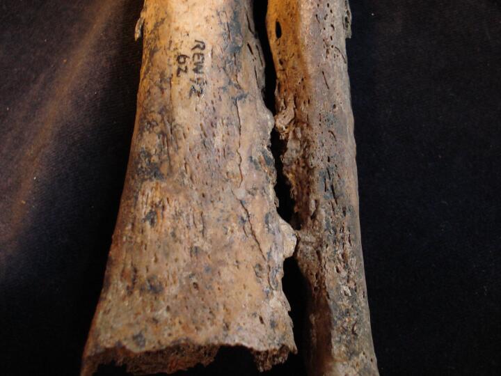
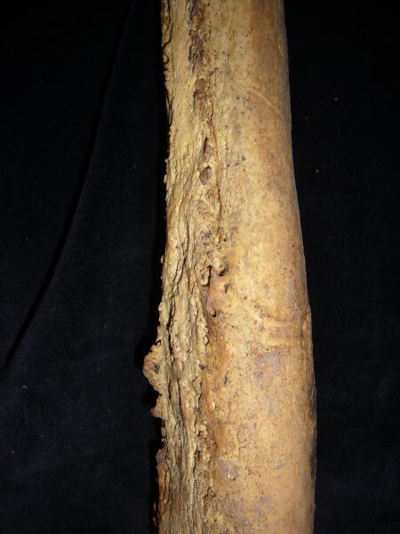
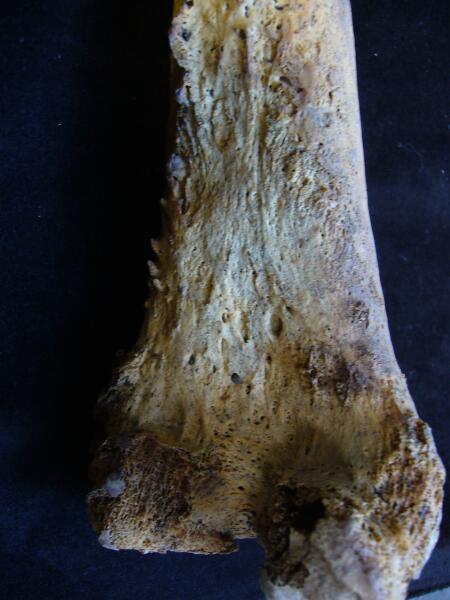
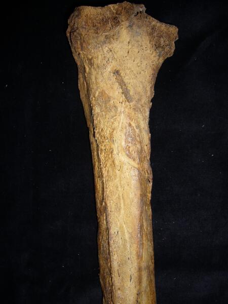
Left tibia gross changes, ?osteomyelitis (medial view)
|
| REW92
|
62
|
8
|
REW92_62_8.jpg
|
Left tibia gross changes, ?osteomyelitis (posterior view)
|
| REW92
|
62
|
9
|
REW92_62_9.jpg
|
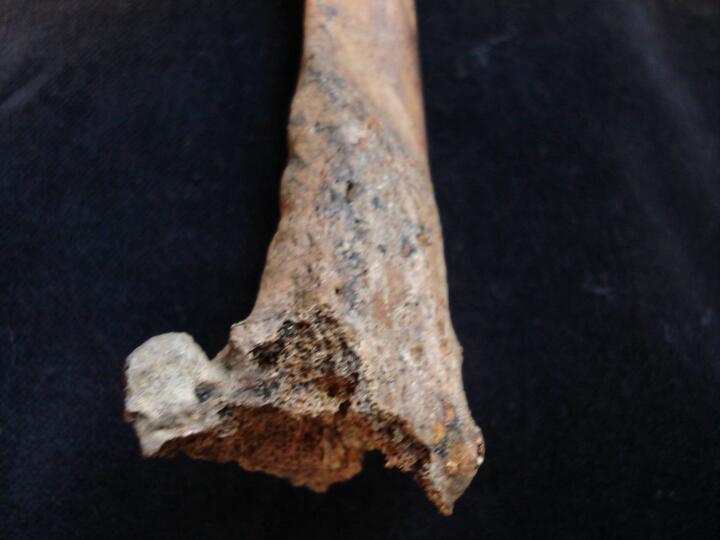
Left tibia gross changes, ?osteomyelitis (medial view/close up)
|
| REW92
|
62
|
10
|
REW92_62_10.jpg
|
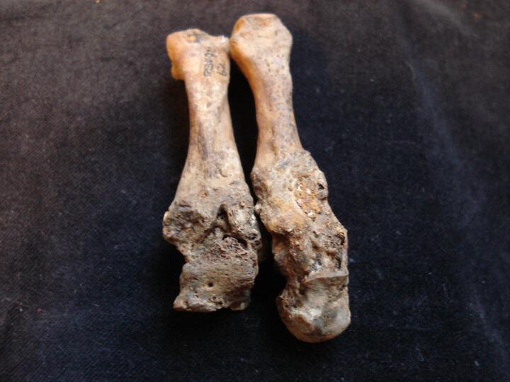
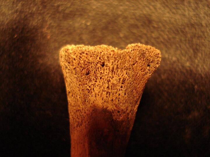
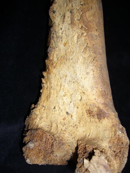
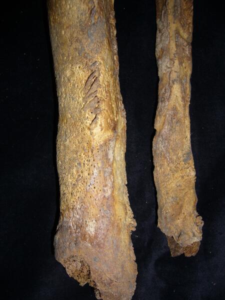
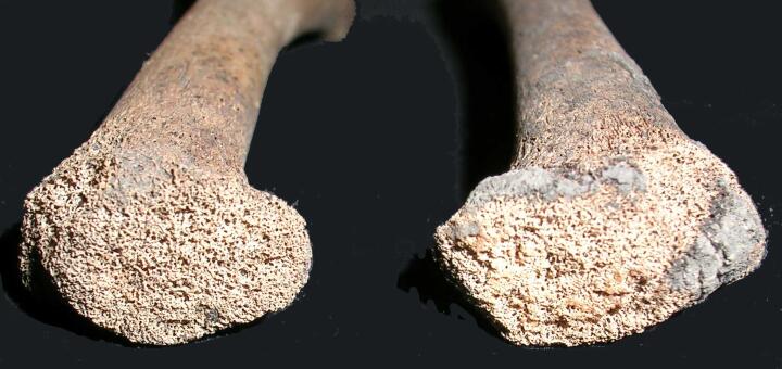
Left fibula gross & florid periosteal changes, ?osteomyelitis
|
| REW92
|
62
|
11
|
REW92_62_11.jpg
|
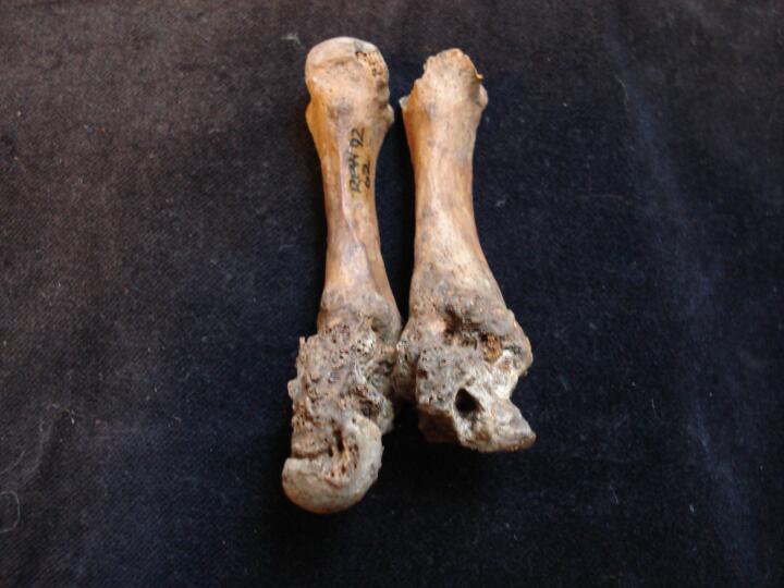
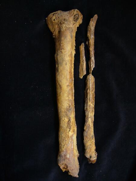
Left tibia & fibula gross & florid periosteal changes with fusion in distal 1/3
|
| REW92
|
78
|
1
|
REW92_78_1.jpg
|
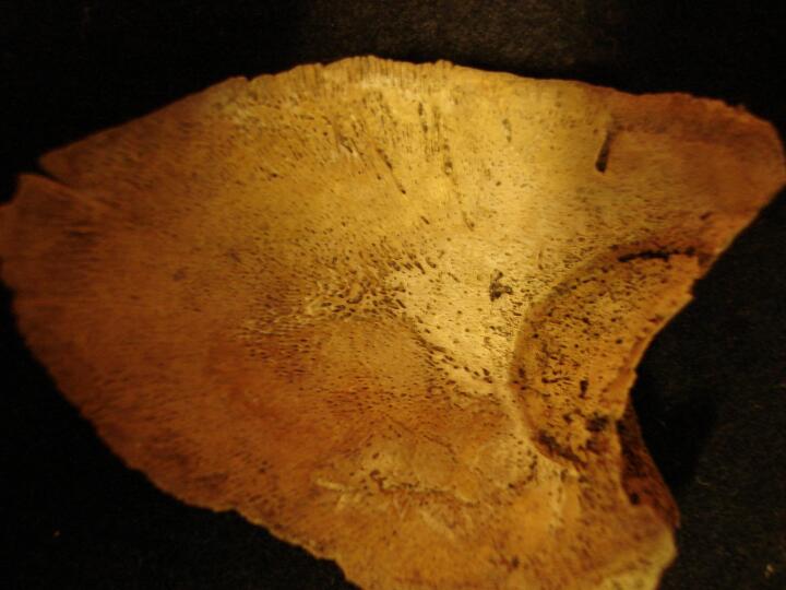
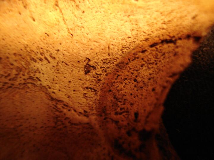
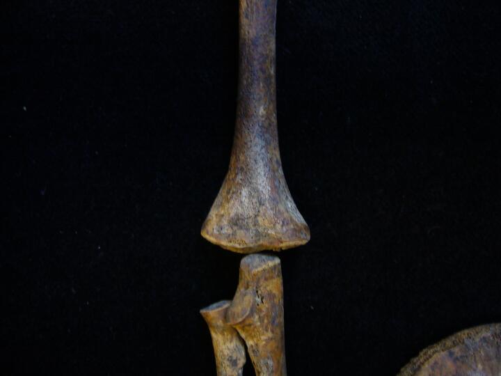
Right radius distal end flaring (posterior view/close up) ?rickets
|
| REW92
|
78
|
2
|
REW92_78_2.jpg
|
Right radius distal end flaring (posterior view) ?rickets
|
| REW92
|
78
|
3
|
REW92_78_3.jpg
|
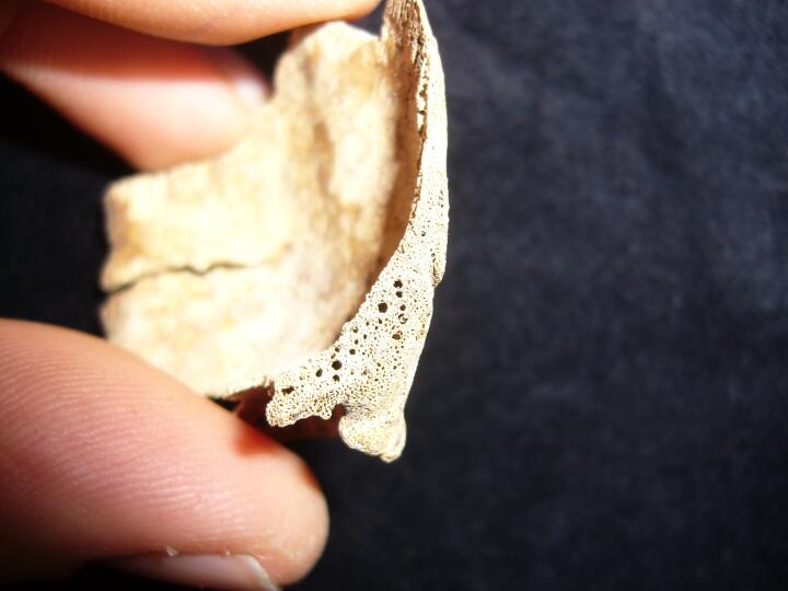
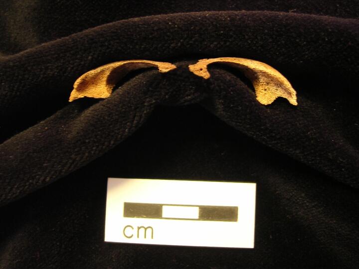
Right ribs sternal end flaring, ?rickets
|
| REW92
|
82
|
1
|
REW92_82_1.jpg
|
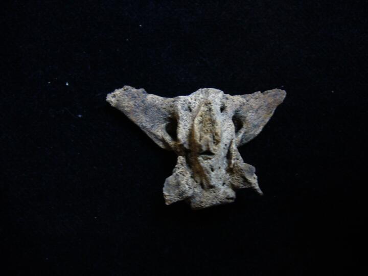
Left parietal (endocranial surface) circular ?lesion eminating from left orbit
|
| REW92
|
82
|
2
|
REW92_82_2.jpg
|
Left parietal (endocranial surface/close up) circular ?lesion eminating from left orbit
|
| REW92
|
84
|
1
|
REW92_84_1.jpg
|
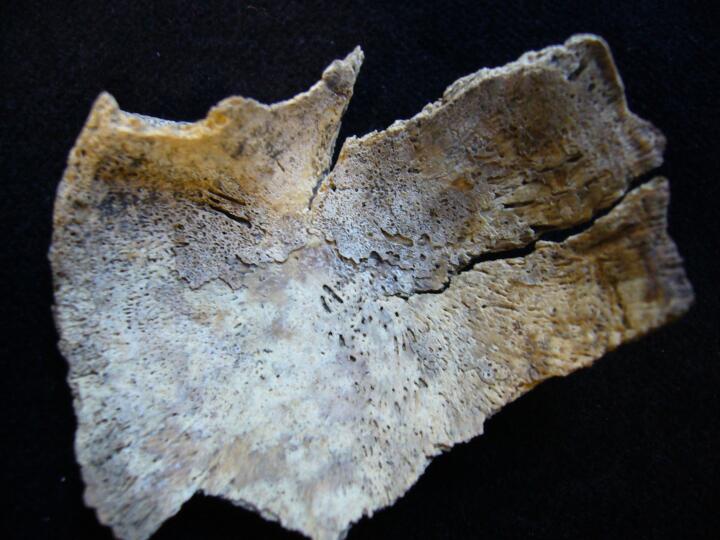
Left scapula (dorsal view) fine new bone plaque
|
| REW92
|
84
|
2
|
REW92_84_2.jpg
|
Left scapula (dorsal view/close up) fine new bone plaque
|
| REW92
|
84
|
3
|
REW92_84_3.jpg
|
Right scapula (dorsal view) fine new bone plaque
|
| REW92
|
93
|
1
|
REW92_93_1.jpg
|
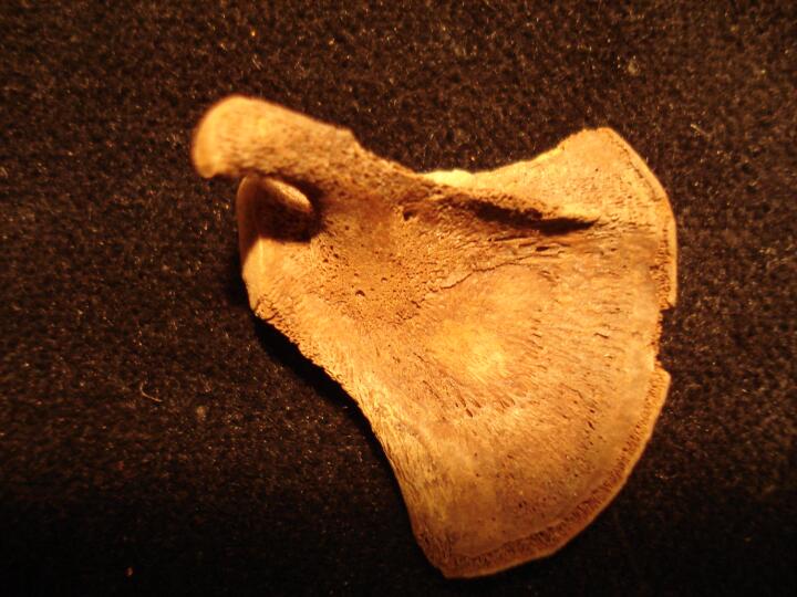
Right zygomatic bone (orbital view) increased porosity & plaque like deposit
|
| REW92
|
96
|
1
|
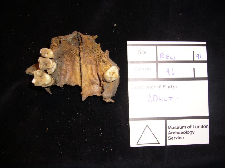
REW92_96_1.jpg
|
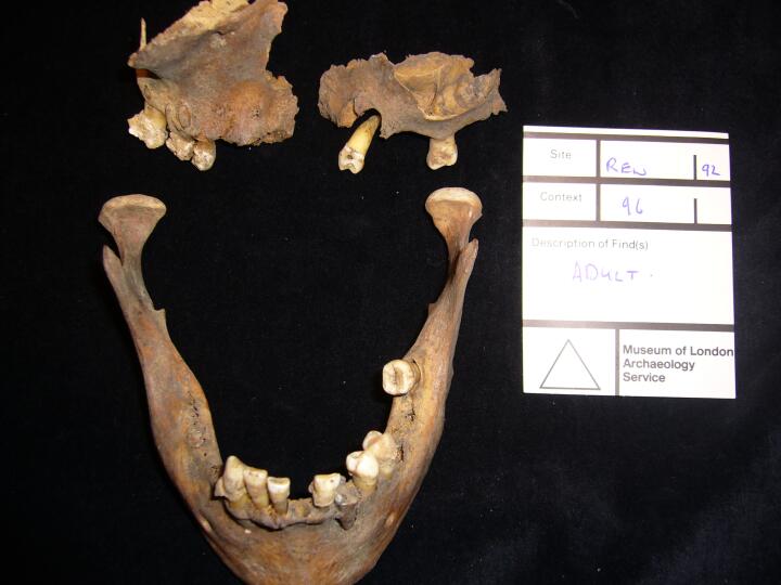
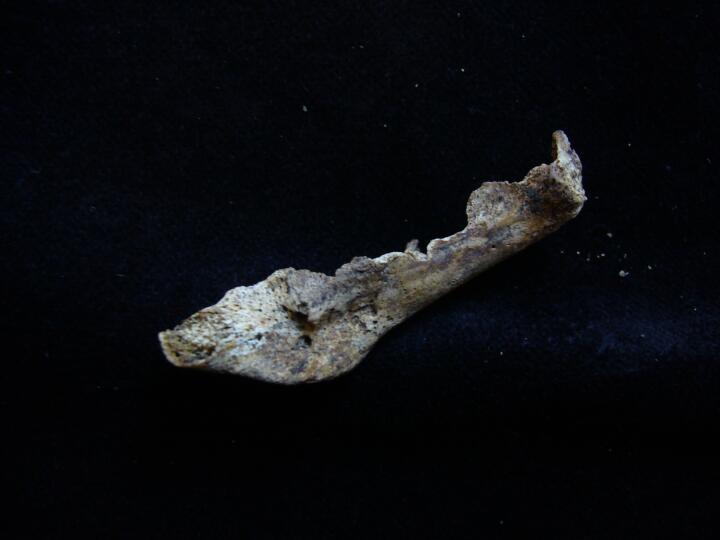
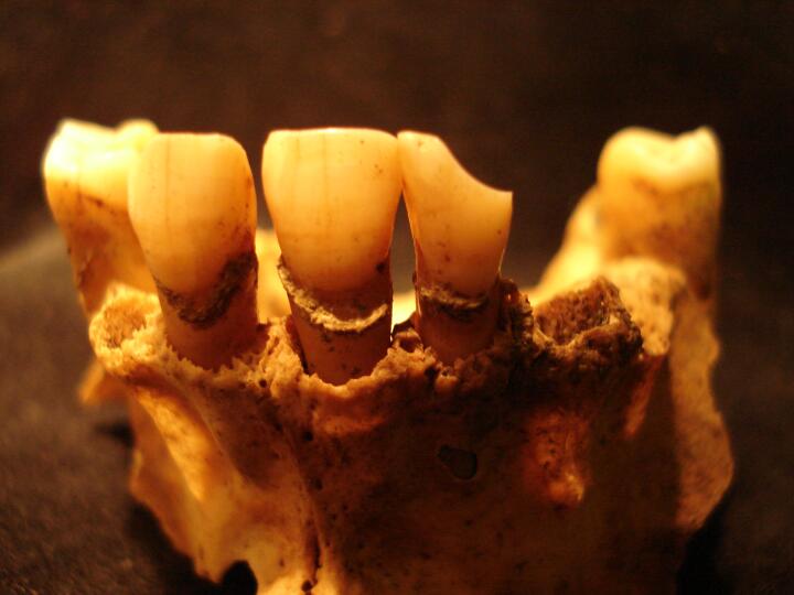
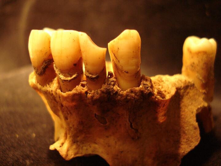
Maxilla & mandible
|
| REW92
|
96
|
2
|
REW92_96_2.jpg
|
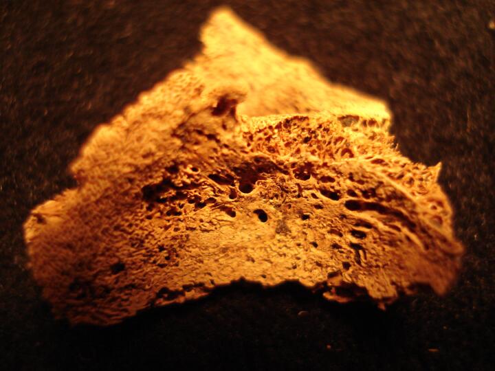
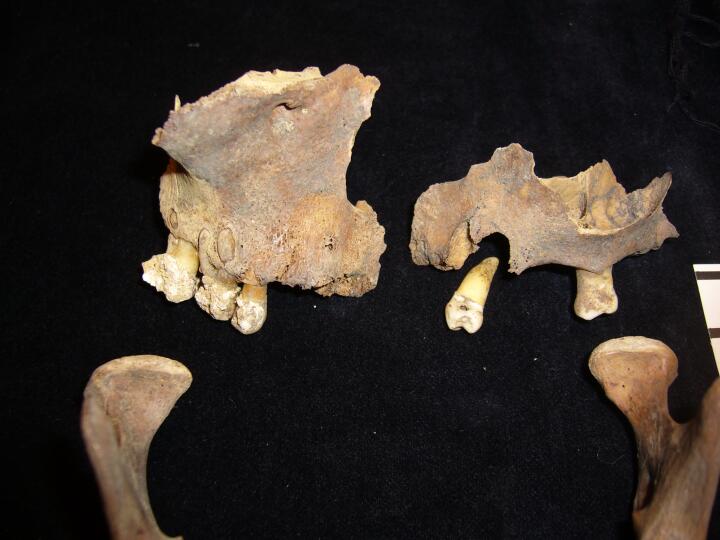
Maxilla
|
| REW92
|
96
|
3
|
REW92_96_3.jpg
|
Maxilla (palatal view)
|
| REW92
|
96
|
4
|
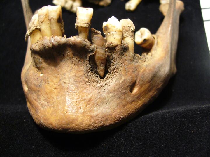
REW92_96_4.jpg
|
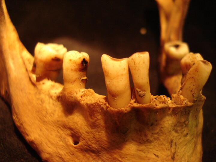
Mandible (buccal surface)
|
| REW92
|
96
|
5
|
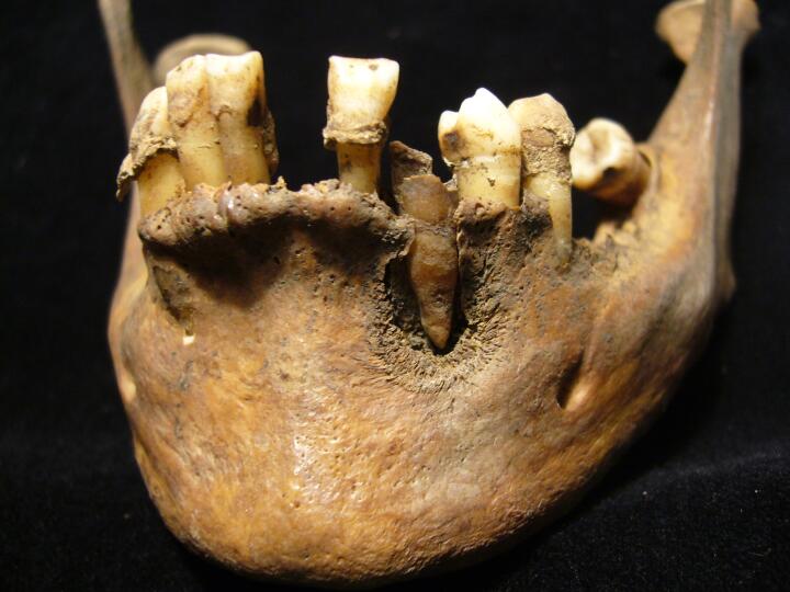
REW92_96_5.jpg
|
Mandible (buccal surface)
|
| REW92
|
96
|
6
|
REW92_96_6.jpg
|
Mandible (buccal surface)
|
| REW92
|
96
|
7
|
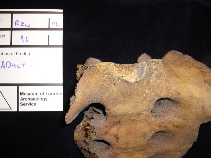
REW92_96_7.jpg
|
Sacrum (anterior view)
|
| REW92
|
99
|
1
|
REW92_99_1.jpg
|
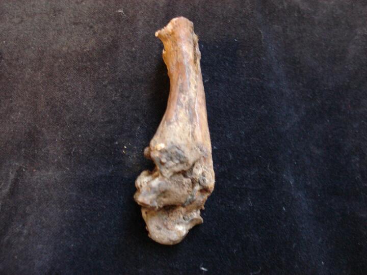
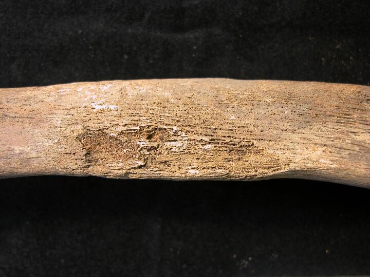
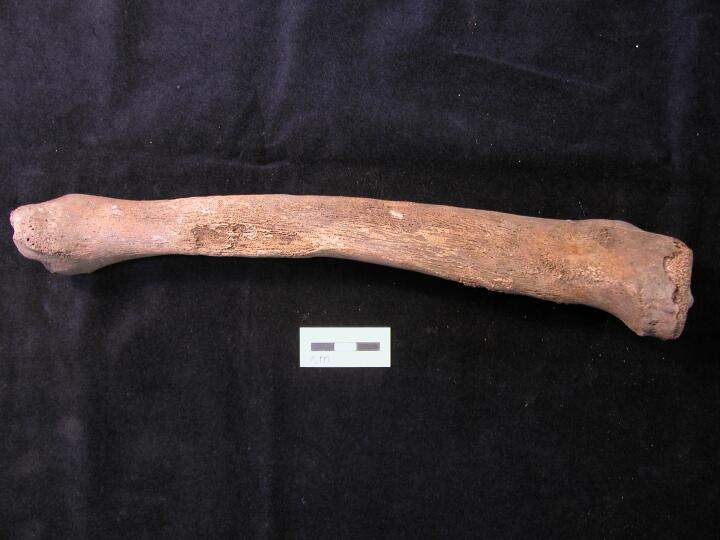
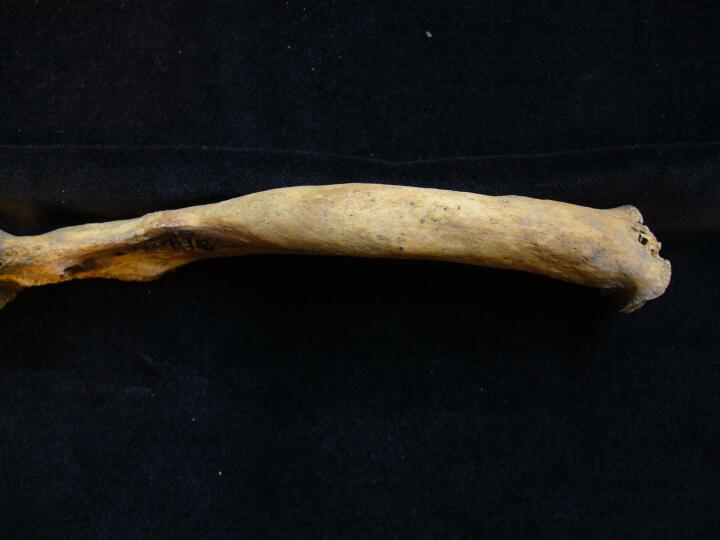
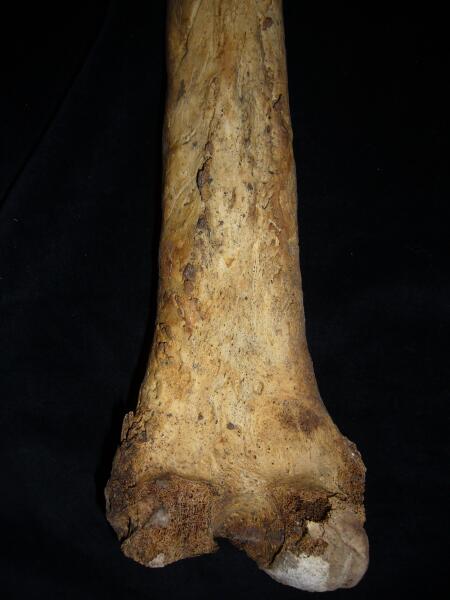
Right tibia periosteal changes associated with syphilis
|
| REW92
|
99
|
2
|
REW92_99_2.jpg
|
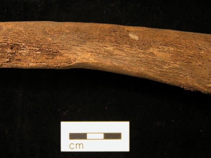
Cranium frontal bone/glabella gross changes, sypilis
|
| REW92
|
99
|
3
|
REW92_99_3.jpg
|
Left fibula, periosteal changes associated with syphilis
|
| REW92
|
99
|
4
|
REW92_99_4.jpg
|
Left leg periosteal changes associated with syphilis
|
| REW92
|
99
|
5
|
REW92_99_5.jpg
|
Left leg periosteal changes associated with syphilis
|
| REW92
|
99
|
6
|
REW92_99_6.jpg
|
Left tibia periosteal changes associated with syphilis
|
| REW92
|
99
|
7
|
REW92_99_7.jpg
|
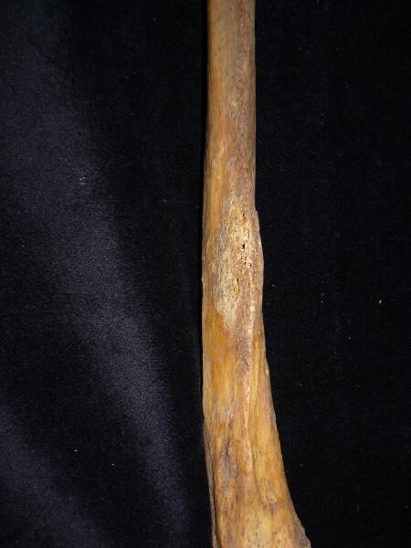
Right tibia periosteal changes associated with syphilis
|
| REW92
|
99
|
8
|
REW92_99_8.jpg
|
Right tibia periosteal changes associated with syphilis
|
| REW92
|
103
|
1
|
REW92_103_1.jpg
|
Left humerus (anterior view) distal 1/3 porosity & new bone, ?scurvy
|
| REW92
|
103
|
2
|
REW92_103_2.jpg
|
Left scapula, new bone plaque on surface (dorsal view/close up) ?scurvy
|
| REW92
|
112
|
1
|
REW92_112_1.jpg
|
Left petrous (endocranial view) growth& development
|
| REW92
|
112
|
2
|
REW92_112_2.jpg
|
Left petrous (endocranial view) growth& development
|
| REW92
|
112
|
3
|
REW92_112_3.jpg
|
Left petrous (endocranial view) growth& development
|
| REW92
|
118
|
1
|
REW92_118_1.jpg
|
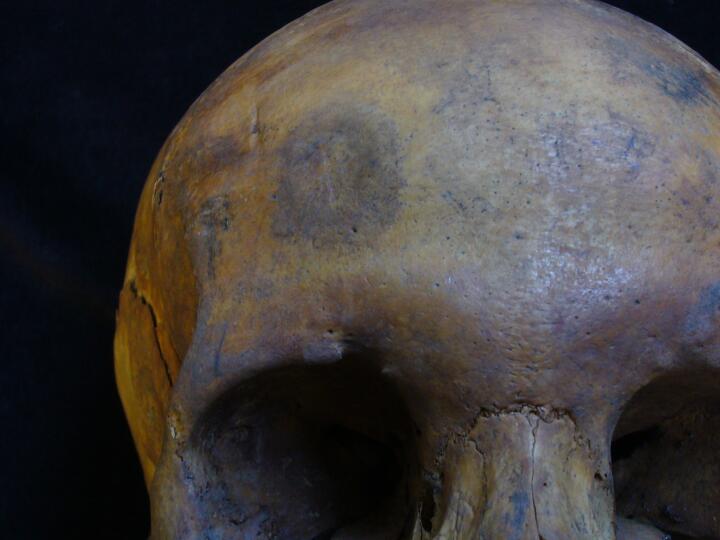
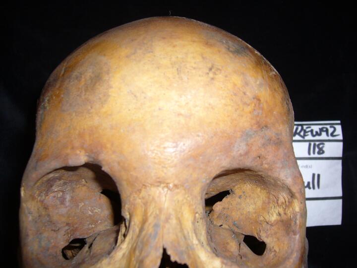
Skull, right frontal bone, ?stellate lesion/syphilis
|
| REW92
|
118
|
2
|
REW92_118_2.jpg
|
Skull, right frontal bone, ?stellate lesion/syphilis
|
| REW92
|
118
|
3
|
REW92_118_3.jpg
|
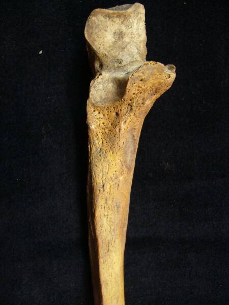
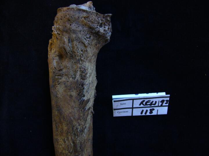
Right ulna, proximal end (medial view) increased porosity, ?syphilis
|
| REW92
|
118
|
4
|
REW92_118_4.jpg
|
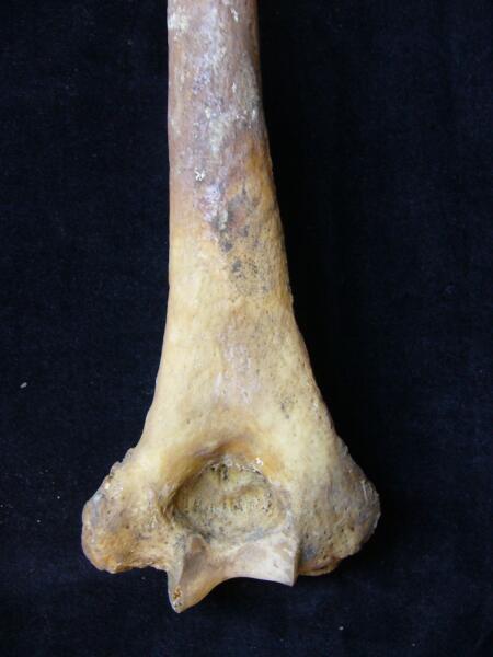
Right humerus, distal end (posteior view) increased porosity, ?syphilis
|
| REW92
|
118
|
5
|
REW92_118_5.jpg
|
Left clavicle, lesions sternal end
|
| REW92
|
118
|
6
|
REW92_118_6.jpg
|
Right clavicle, increased porosity mid shaft
|
| REW92
|
118
|
7
|
REW92_118_7.jpg
|
Right elbow joint (anterior view)
|
| REW92
|
118
|
8
|
REW92_118_8.jpg
|
Ulnae shaft surfaces
|
| REW92
|
118
|
9
|
REW92_118_9.jpg
|
Ulna shaft surface periosteal changes
|
| REW92
|
118
|
10
|
REW92_118_10.jpg
|
Right ulna (lateral view)
|
| REW92
|
118
|
11
|
REW92_118_11.jpg
|
Left humerus (anterior view) distal end increased porosity, ?syphilis
|
| REW92
|
118
|
12
|
REW92_118_12.jpg
|
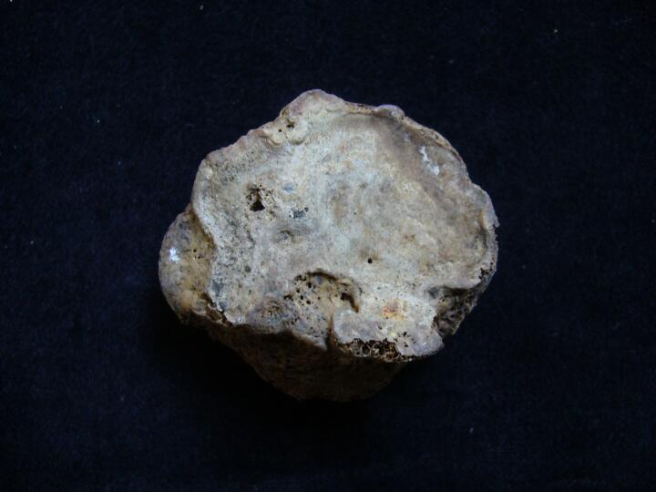
Left 1st metatarsal, distal articular surface, DJD
|
| REW92
|
118
|
13
|
REW92_118_13.jpg
|
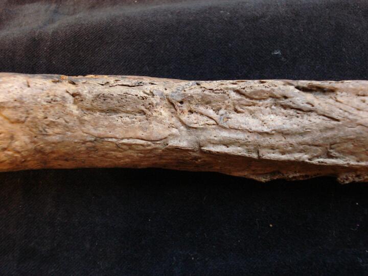
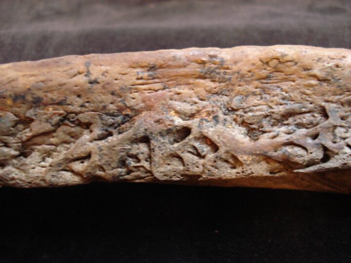
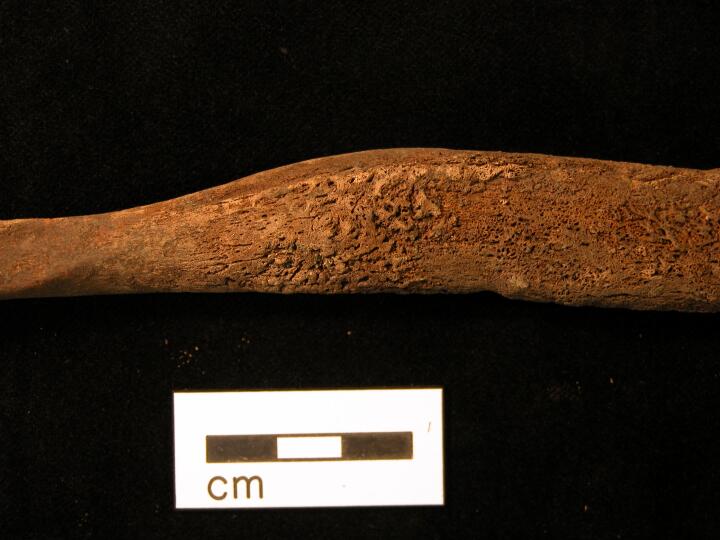
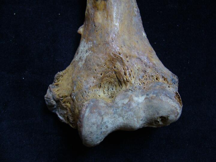
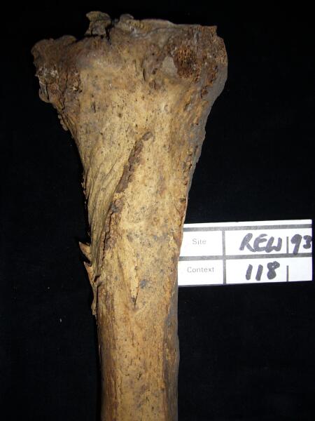
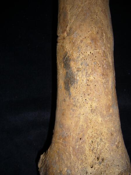
Right tibia florid periosteal changes associated with syphilis (anterior view)
|
| REW92
|
118
|
14
|
REW92_118_14.jpg
|
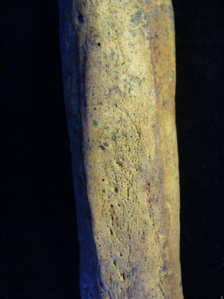
Right tibia periosteal changes associated with syphilis (medial view)
|
| REW92
|
118
|
15
|
REW92_118_15.jpg
|
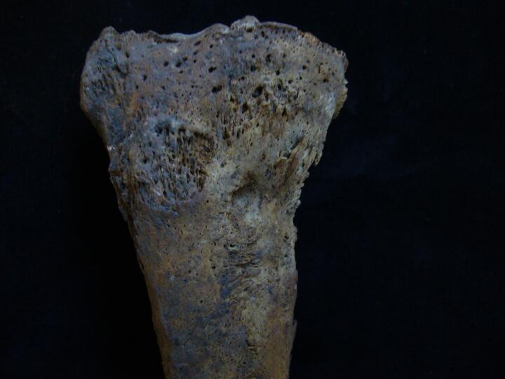
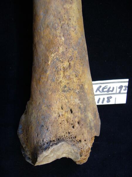
Right tibia periosteal changes associated with syphilis (posterior view)
|
| REW92
|
118
|
16
|
REW92_118_16.jpg
|
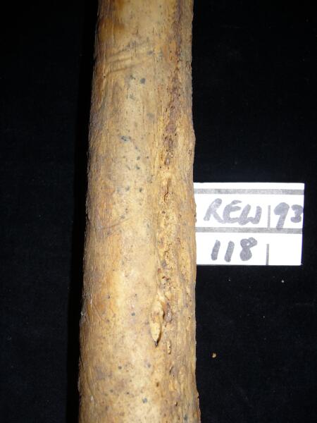
Right femur mid shaft (anterior view) increased porosity
|
| REW92
|
118
|
17
|
REW92_118_17.jpg
|
Right femur mid shaft (anterior view) periosteal changes
|
| REW92
|
118
|
18
|
REW92_118_18.jpg
|
Right femur mid shaft (anterior view) periosteal changes,?syphilis (posterior view)
|
| REW92
|
118
|
19
|
REW92_118_19.jpg
|
Femur mid shaft periosteal changes,?syphilis
|
| REW92
|
118
|
20
|
REW92_118_20.jpg
|
Right femur distal articular joint surface changes, ?septic arthropathy
|
| REW92
|
118
|
21
|
REW92_118_21.jpg
|
Left femur distal 1/3 of shaft (anterior view) increased porosity & enlargement of bone
|
| REW92
|
118
|
22
|
REW92_118_22.jpg
|
Left femur (posterior view) periosteal changes
|
| REW92
|
118
|
23
|
REW92_118_23.jpg
|
Left femur mid shaft (posterior view) florid periosteal changes,?syphilis
|
| REW92
|
118
|
24
|
REW92_118_24.jpg
|
Left femur (posterior view) periosteal changes,?syphilis
|
| REW92
|
118
|
25
|
REW92_118_25.jpg
|
Left femur (posterior view) periosteal changes,?syphilis
|
| REW92
|
118
|
26
|
REW92_118_26.jpg
|
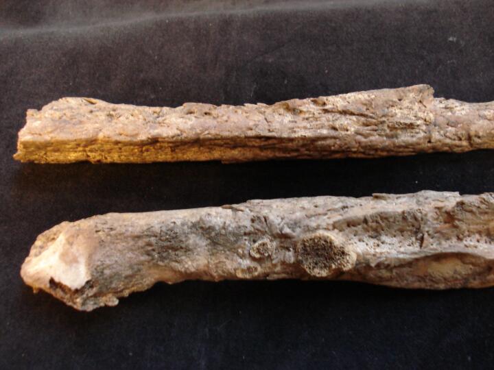
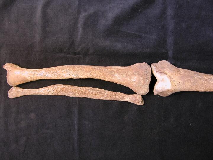
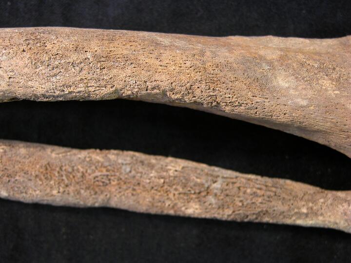
Left tibia & fibula florid periosteal changes, ?syphilis
|
| REW92
|
118
|
27
|
REW92_118_27.jpg
|
Left tibia & fibula (distal 1/2) florid periosteal changes, ?syphilis
|
| REW92
|
118
|
28
|
REW92_118_28.jpg
|
Left tibia proximal end (posterior view) periosteal changes, ?syphilis
|
| REW92
|
118
|
29
|
REW92_118_29.jpg
|
Left tibia proximal articular surfaces (superior view)
|
| REW92
|
118
|
30
|
REW92_118_30.jpg
|
Left tibia distal articular surface (superior view)
|
| REW92
|
118
|
31
|
REW92_118_31.jpg
|
Lumbar vertebra (L5)
|
| REW92
|
119.1
|
1
|
REW92_119.1_1.jpg
|
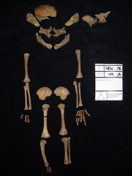
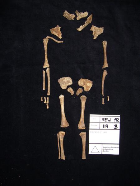
Sub adult anatomically positioned
|
| REW92
|
119.1
|
2
|
REW92_119.1_2.jpg
|
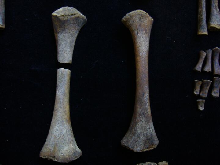
Left tibia/fibula &right tibia. Left tibia new bne growth
|
| REW92
|
119.1
|
3
|
REW92_119.1_3.jpg
|
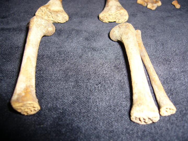
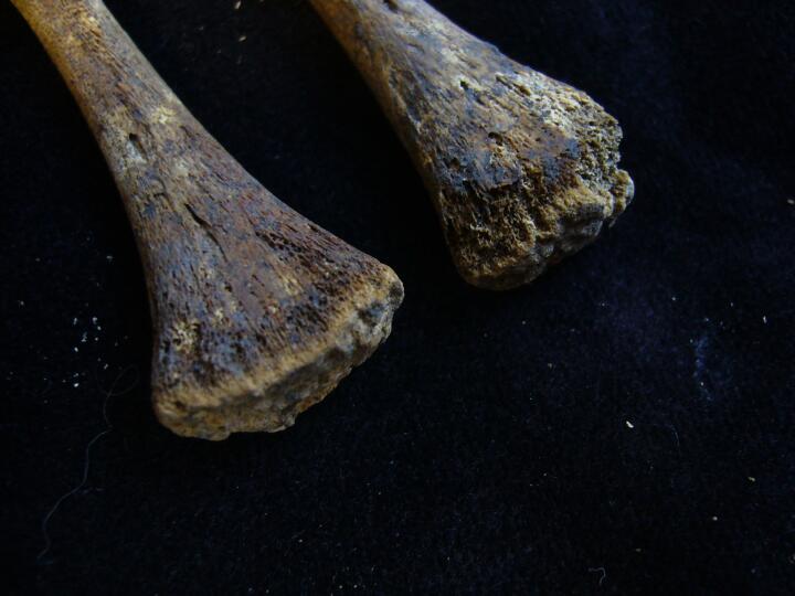
Femora
|
| REW92
|
119.1
|
4
|
REW92_119.1_4.jpg
|
Right ulna & radius distal end (anterior view) increased porosity/post mortem damage
|
| REW92
|
119.1
|
5
|
REW92_119.1_5.jpg
|
Left elbow joint (anterior view) increased porosity new bone/post mortem damage
|
| REW92
|
119.1
|
6
|
REW92_119.1_6.jpg
|
Left ulna & radius increased porosity new bone/post mortem damage
|
| REW92
|
119.1
|
7
|
REW92_119.1_7.jpg
|
Right elbow joint (anterior view) increased porosity new bone/post mortem damage
|
| REW92
|
119.1
|
8
|
REW92_119.1_8.jpg
|
Sphenoid increased porosity/new bone growth
|
| REW92
|
119.1
|
9
|
REW92_119.1_9.jpg
|
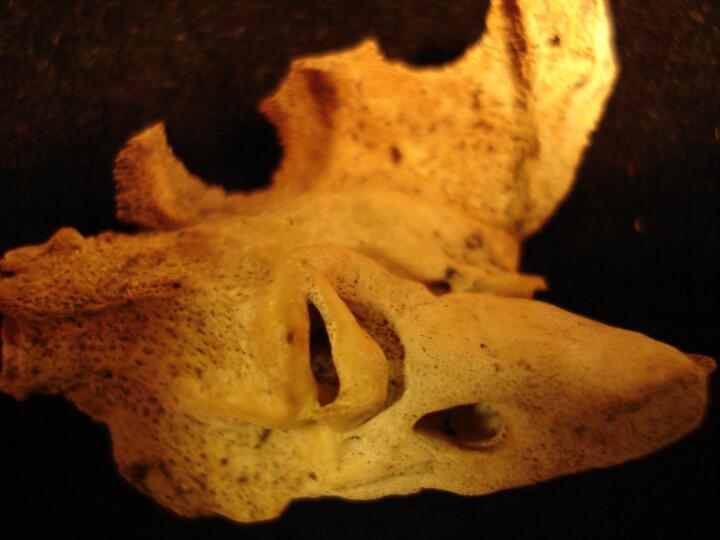
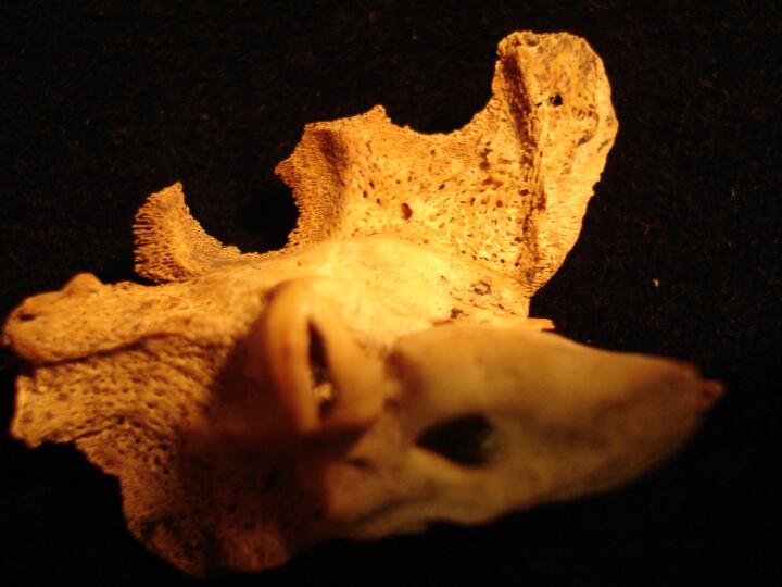
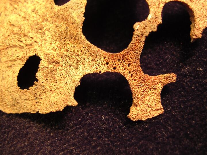
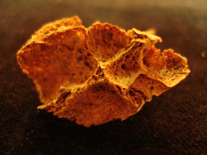
Right orbit (cross section) cribra orbitalia (Type 5)
|
| REW92
|
119.1
|
10
|
REW92_119.1_10.jpg
|
Parietal (endocranial surface) new bone growth
|
| REW92
|
119.1
|
11
|
REW92_119.1_11.jpg
|
Petrous bones increased porosity
|
| REW92
|
119.1
|
12
|
REW92_119.1_12.jpg
|
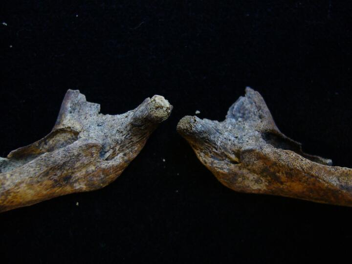
Mandible (left & right side) buccal surface
|
| REW92
|
119.1
|
13
|
REW92_119.1_13.jpg
|
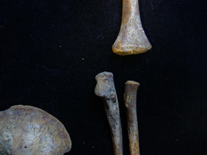
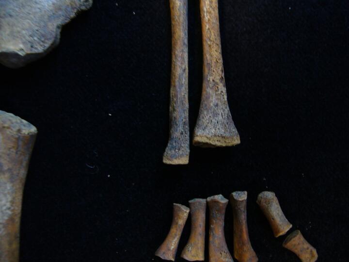
Left & right leg
|
| REW92
|
119.2
|
1
|
REW92_119.2_1.jpg
|
Sub adult anatomically positioned
|
| REW92
|
119.2
|
2
|
REW92_119.2_2.jpg
|
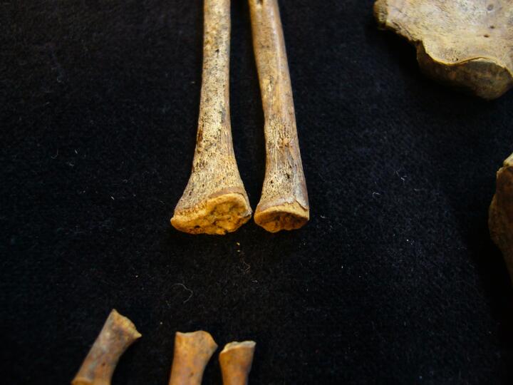
Left tibia increased porosity & right tibia/fibula, ?congenital syphilis
|
| REW92
|
119.2
|
3
|
REW92_119.2_3.jpg
|
Left humerus (distal end) & left ulna
|
| REW92
|
119.2
|
4
|
REW92_119.2_4.jpg
|
Right shoulder joint (anterior view)
|
| REW92
|
119.2
|
5
|
REW92_119.2_5.jpg
|
Left maxilla & left mandible
|
| REW92
|
119.2
|
6
|
REW92_119.2_6.jpg
|
Maxillary processes
|
| REW92
|
119.2
|
7
|
REW92_119.2_7.jpg
|
Left side of mandible (buccal view) area of new bone
|
| REW92
|
119.2
|
8
|
REW92_119.2_8.jpg
|
Femora
|
| REW92
|
125
|
1
|
REW92_125_1.jpg
|
Orbit showing smaller rounded lesion,?Histiocytosis-X
|
| REW92
|
125
|
2
|
REW92_125_2.jpg
|
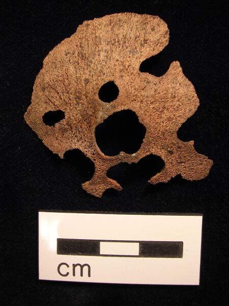
Parietal (ectocranial surface) multiple circular lesions, possibly Histiosyctosis-X
|
| REW92
|
125
|
3
|
REW92_125_3.jpg
|
Parietal (endocranial surface) multiple circular lesions ?bevelled edged, possibly Histiosyctosis-X
|
| REW92
|
125
|
4
|
REW92_125_4.jpg
|
Parietal (endocranial surface) multiple circular lesions ?bevelled edged, possibly Histiosyctosis-x
|
| REW92
|
125
|
5
|
REW92_125_5.jpg
|
Greater Wings of sphenoid, ?lesions, possibly Histiocytois-X
|
| REW92
|
125
|
6
|
REW92_125_6.jpg
|
Occipital with bevelled edged lesions, possibly Histiocytosis-X
|
| REW92
|
125
|
7
|
REW92_125_7.jpg
|
Occipital (close up) with lesions, possibly Histiocytosis-X
|
| REW92
|
125
|
8
|
REW92_125_8.jpg
|
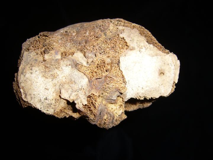
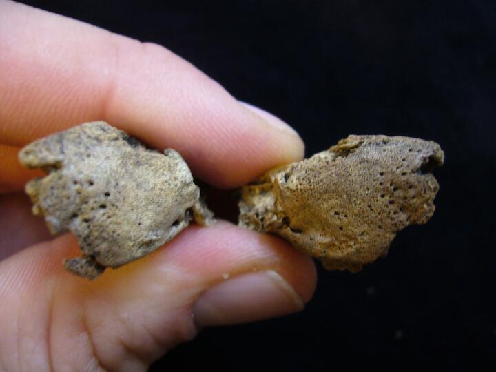
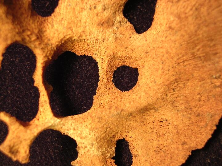
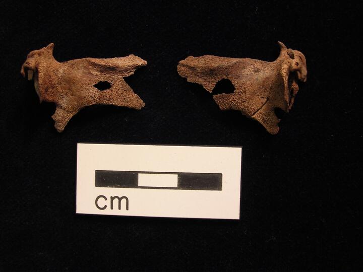
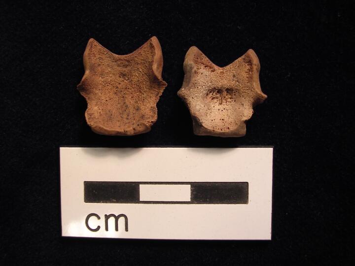
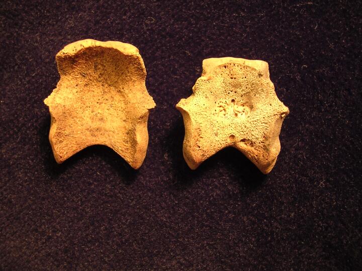
Comparison of pars basilaris, left appears flattened & enlarged
|
| REW92
|
125
|
9
|
REW92_125_9.jpg
|
Comparison of pars basilaris, left appears flattened & enlarged
|
| REW92
|
125
|
10
|
REW92_125_10.jpg
|
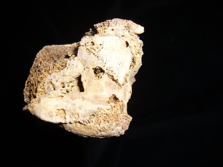
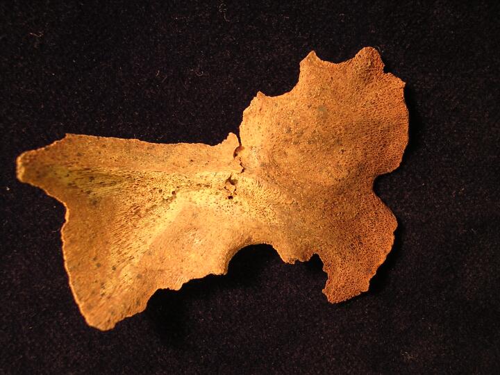
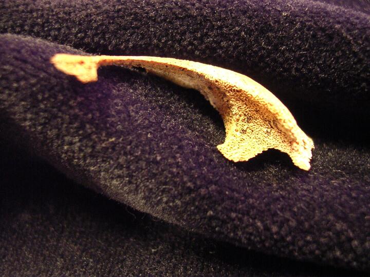
Pars bassilaris (medial view) to show lesion
|
| REW92
|
125
|
11
|
REW92_125_11.jpg
|
Parietal (ectocranial surface) group of lesions & single circular lesion, ?Histiocytosis-X
|
| REW92
|
125
|
12
|
REW92_125_12.jpg
|
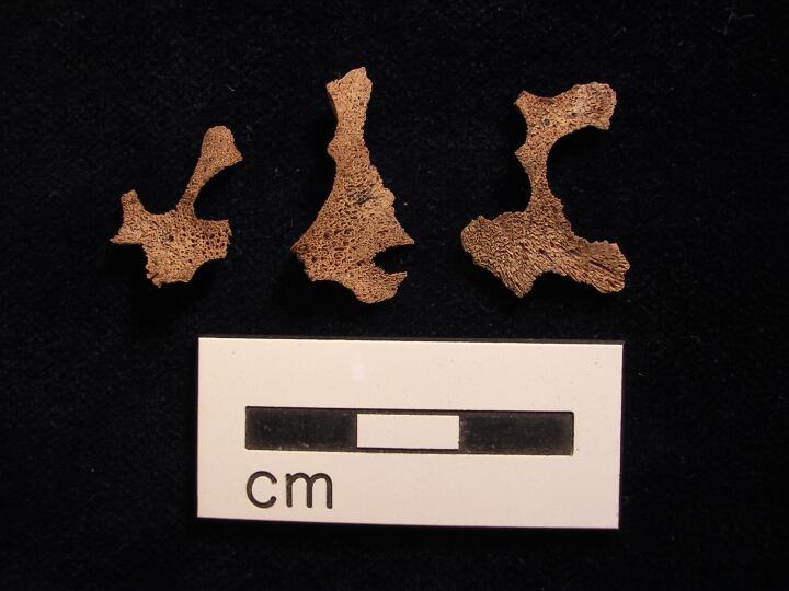
Skull fragments, with lesions, ?Histiocytosis-X
|
| REW92
|
125
|
13
|
REW92_125_13.jpg
|
Orbit showing smaller rounded lesion,?Histiocytosis-X
|
| REW92
|
125
|
14
|
REW92_125_14.jpg
|
Left orbit (close up) of lesion
|
| REW92
|
125
|
15
|
REW92_125_15.jpg
|
Orbits Bilateral large rounded lesions on the medial and central aspect of the orbital roof,?Histiocytosis_X
|
| REW92
|
131
|
1
|
REW92_131_1.jpg
|
Right maxilla increased porosity & developing tooth sockets (palatal view)
|
| REW92
|
131
|
2
|
REW92_131_2.jpg
|
Right maxilla increased porosity & developing tooth sockets (palatal view)
|
| REW92
|
133
|
1
|
REW92_133_1.jpg
|
Right elbow - destructive lesions - possible smallpox
|
| REW92
|
133
|
2
|
REW92_133_2.jpg
|
Right elbow - destructive lesions - possible smallpox
|
| REW92
|
133
|
3
|
REW92_133_3.jpg
|
Left elbow - destructive lesions - possible smallpox
|
| REW92
|
133
|
4
|
REW92_133_4.jpg
|
Left ulna - destructive lesions - possible smallpox
|
| REW92
|
133
|
5
|
REW92_133_5.jpg
|
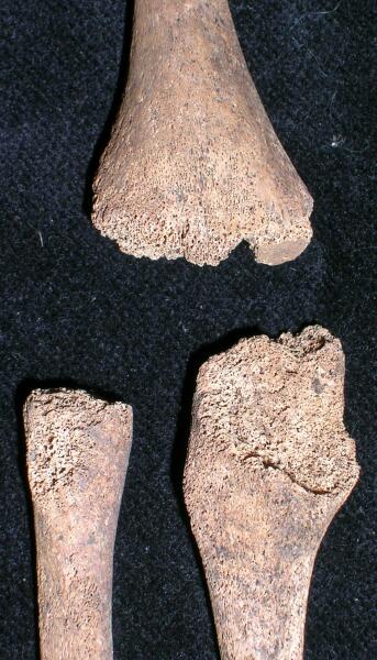
Femora - flattening of the femoral heads
|
| REW92
|
165
|
1
|
REW92_165_1.jpg
|
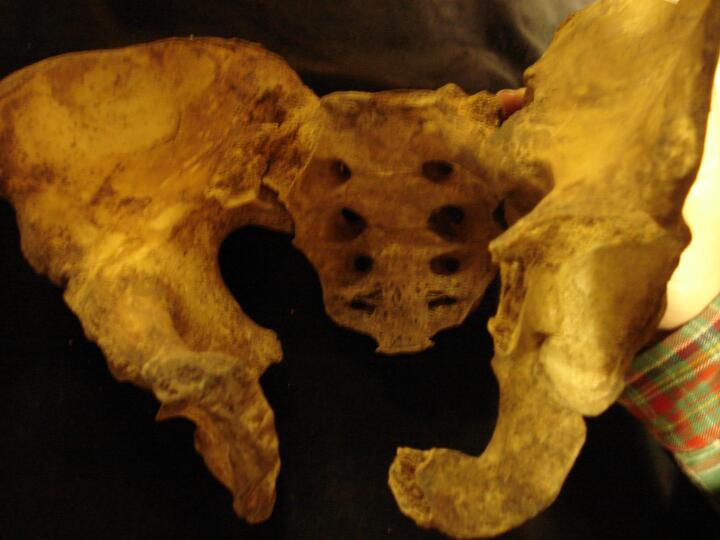
Pelves & sacrum bilateral sacroiliac fusion
|
| REW92
|
165
|
2
|
REW92_165_2.jpg
|
Pelves & sacrum bilateral sacroiliac fusion
|
| REW92
|
167
|
1
|
REW92_167_1.jpg
|
Right orbit cribra orbitalia (Type 3)
|
| REW92
|
167
|
2
|
REW92_167_2.jpg
|
Right orbit cribra orbitalia (Type 3)
|
| REW92
|
167
|
3
|
REW92_167_3.jpg
|
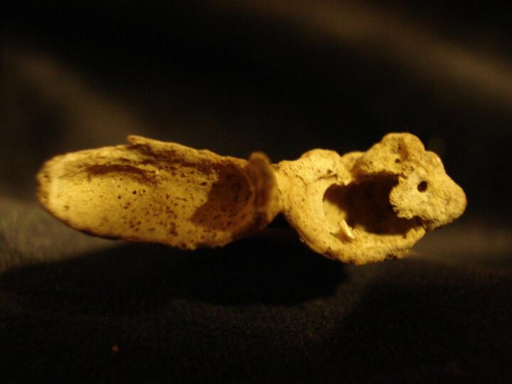
Maxillary process pipe facet (right lateral incisor)
|
| REW92
|
167
|
4
|
REW92_167_4.jpg
|
Maxillary process pipe facets (right lateral incisor & canine)
|
| REW92
|
167
|
5
|
REW92_167_5.jpg
|
Mandible pipe facets (right lateral incisor & canine)
|
| REW92
|
167
|
6
|
REW92_167_6.jpg
|
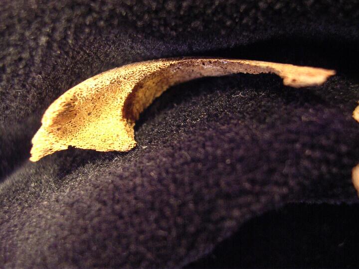
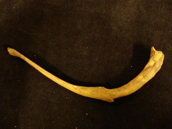
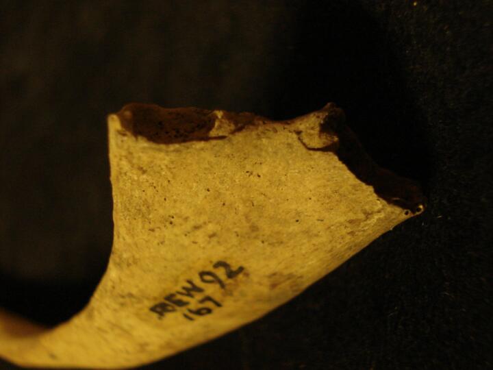
Right 11th rib mid shaft healed fracture
|
| REW92
|
167
|
7
|
REW92_167_7.jpg
|
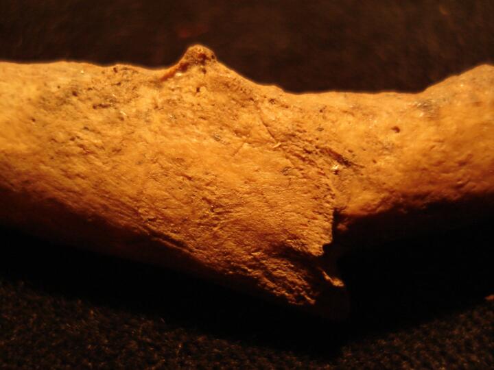
Right 11th rib mid shaft healed fracture (close up of fracture)
|
| REW92
|
167
|
8
|
REW92_167_8.jpg
|
Right 1st rib (sternal end) bifurcate
|
| REW92
|
167
|
9
|
REW92_167_9.jpg
|
Right lower rib (sternal end) bifurcate
|


