| Site code
|
Context
|
Frame number
|
Photo
|
Description
|
| FAO90
|
1052
|
2
|
FAO90_1052_2.jpg
|
Eburnation of 1st CMC joints
|
| FAO90
|
1052
|
3
|
FAO90_1052_3.jpg
|
Eburnation of inter phalangeal joints of the hand
|
| FAO90
|
1052
|
4
|
FAO90_1052_4.jpg
|
Eburnation of interphalangeal joints of the hand
|
| FAO90
|
1052
|
5
|
FAO90_1052_5.jpg
|
Eburnation of distal femoral joint
|
| FAO90
|
1052
|
6
|
FAO90_1052_6.jpg
|
Eburnation of proximal tibial joint
|
| FAO90
|
1052
|
7
|
FAO90_1052_7.jpg
|
Myositis ossificans of the lesser trochanter
|
| FAO90
|
1052
|
8
|
FAO90_1052_8.jpg
|
Healed fracture of left MT5
|
| FAO90
|
1052
|
9
|
FAO90_1052_9.jpg
|
Healed fracture of left MT5
|
| FAO90
|
1055
|
1
|
FAO90_1055_1.jpg
|
New bone on the visceral surface of the ribs
|
| FAO90
|
1055
|
2
|
FAO90_1055_2.jpg
|
Manubrium and sternum, manubrium displaying cut marks on the left side through the ossified cartilage
|
| FAO90
|
1055
|
3
|
FAO90_1055_3.jpg
|
Manubrium with autopsy cut across ossified cartilage, anterior view
|
| FAO90
|
1055
|
4
|
FAO90_1055_4.jpg
|
Manubrium with autopsy cut across ossified cartilage, lateral view
|
| FAO90
|
1055
|
5
|
FAO90_1055_5.jpg
|
Treponematosis? R clavicle with new bone on the surface.
|
| FAO90
|
1055
|
6
|
FAO90_1055_6.jpg
|
Treponematosis? Bony reaction on distal anterior shaft if the R radius and ulna
|
| FAO90
|
1055
|
7
|
FAO90_1055_7.jpg
|
Treponematosis? Bony reaction on proximal anterior shaft if the L radius and ulna
|
| FAO90
|
1055
|
8
|
FAO90_1055_8.jpg
|
New active bone on the shaft of the fibula
|
| FAO90
|
1055
|
9
|
FAO90_1055_9.jpg
|
New active bone on the shaft of the tibia
|
| FAO90
|
1055
|
10
|
FAO90_1055_10.jpg
|
Healed New bone formation of the distal aspect of L femur and tibia
|
| FAO90
|
1055
|
11
|
FAO90_1055_11.jpg
|
Healed New bone formation of the distal aspect of L femur.
|
| FAO90
|
1119
|
1
|
FAO90_1119_1.jpg
|
Inflammation on the visceral surface of the ribs
|
| FAO90
|
1120
|
1
|
FAO90_1120_1.jpg
|
Osteoarthritis of the hip joint
|
| FAO90
|
1120
|
2
|
FAO90_1120_2.jpg
|
Eburnation of femoral head
|
| FAO90
|
1121
|
1
|
FAO90_1121_1.jpg
|
Scurvy: bone formation on orbital roof
|
| FAO90
|
1121
|
2
|
FAO90_1121_2.jpg
|
Active rickets: flaring and destruction of sternal ends note the rounded lesions
|
| FAO90
|
1121
|
3
|
FAO90_1121_3.jpg
|
Scurvy?? Porosity on endocranial aspect of the petrous bone
|
| FAO90
|
1121
|
4
|
FAO90_1121_4.jpg
|
Scurvy?? Marked porosity of Pars lateralis
|
| FAO90
|
1121
|
5
|
FAO90_1121_5.jpg
|
Scurvy: Porotic hyperostosis of the frontal bone
|
| FAO90
|
1121
|
6
|
FAO90_1121_6.jpg
|
Active rickets and scurvy: Femora with flatting of the proximal diaphyses and flaring of distal diaphyses and extra cortical bone growth
|
| FAO90
|
1121
|
7
|
FAO90_1121_7.jpg
|
Flaring and destruction of growth plate of distal femora
|
| FAO90
|
1121
|
8
|
FAO90_1121_8.jpg
|
Active rickets and scurvy of tibiae: with angulation of distal diaphyses and new bone formation formation around the shaft.
|
| FAO90
|
1121
|
9
|
FAO90_1121_9.jpg
|
Active rickets: flaring and destruction of growth plate of distal radii
|
| FAO90
|
1121
|
10
|
FAO90_1121_10.jpg
|
Active rickets: Destruction of growth plates of proximal femora
|
| FAO90
|
1121
|
11
|
FAO90_1121_11.jpg
|
Active rickets and scurvy: Gross new bone formation on shaft, flaring of distal diaphyses and lateral bowing of ulnae
|
| FAO90
|
1121
|
12
|
FAO90_1121_12.jpg
|
Active rickets and scurvy: Gross new bone formation on shaft, flaring of distal diaphyses and lateral bowing of ulnae
|
| FAO90
|
1121
|
13
|
FAO90_1121_13.jpg
|
Active rickets: Distal growth plate destruction of ulnae
|
| FAO90
|
1121
|
14
|
FAO90_1121_14.jpg
|
Active rickets and scurvy: new bone formation on shaft and flaring of distal diaphyses of radii
|
| FAO90
|
1121
|
15
|
FAO90_1121_15.jpg
|
Active rickets with flaring and destruction of growth plate of radii
|
| FAO90
|
1121
|
16
|
FAO90_1121_16.jpg
|
Active rickets and scurvy: new bone formation and flattening of proximal diaphyses of humeri
|
| FAO90
|
1121
|
17
|
FAO90_1121_17.jpg
|
Active rickets: Flaring of sternal rib ends
|
| FAO90
|
1123
|
1
|
FAO90_1123_1.jpg
|
Asymmetry of sacrum
|
| FAO90
|
1123
|
2
|
FAO90_1123_2.jpg
|
Osteoarthritis of sacral facets
|
| FAO90
|
1124
|
1
|
FAO90_1124_1.jpg
|
Residual rickets, marked anterior bowing of R femur
|
| FAO90
|
1124
|
2
|
FAO90_1124_2.jpg
|
Residual rickets, marked bowing of femur and tibia
|
| FAO90
|
1125
|
1
|
FAO90_1125_1.jpg
|
Lesions on visceral surface of rib
|
| FAO90
|
1125
|
2
|
FAO90_1125_2.jpg
|
Destructive lesions on transverse process of thoracic vertebra
|
| FAO90
|
1125
|
3
|
FAO90_1125_3.jpg
|
Destructive lesions on inferior aspect of the spinous process of the vertebra
|
| FAO90
|
1125
|
4
|
FAO90_1125_4.jpg
|
Destructive lesions on spinous process of lumbar vertebra
|
| FAO90
|
1125
|
5
|
FAO90_1125_5.jpg
|
Possible lesion on scapula in area of infraglenoid tubercle
|
| FAO90
|
1125
|
6
|
FAO90_1125_6.jpg
|
Deformation of superior left superior facet of L5
|
| FAO90
|
1125
|
7
|
FAO90_1125_7.jpg
|
Marked curvature of metacarpals (congenital or trauma??)
|
| FAO90
|
1125
|
8
|
FAO90_1125_8.jpg
|
Periosteal bone growth on ischium
|
| FAO90
|
1125
|
9
|
FAO90_1125_9.jpg
|
Marked porosity of the lateral aspect of the greater sphenoid wing
|
| FAO90
|
1126
|
1
|
FAO90_1126_1.jpg
|
Bilateral spondylolisis of L5
|
| FAO90
|
1126
|
2
|
FAO90_1126_2.jpg
|
Healed fracture of left MT5
|
| FAO90
|
1126
|
3
|
FAO90_1126_3.jpg
|
Healed fracture of left MT5
|
| FAO90
|
1126
|
4
|
FAO90_1126_4.jpg
|
Healed fracture of left MT5
|
| FAO90
|
1126
|
5
|
FAO90_1126_5.jpg
|
Coffin nail penetrating illium
|
| FAO90
|
1127
|
1
|
FAO90_1127_1.jpg
|
Paget's disease? Marked irregular bone formation on the endocranial aspect of the L temporal bone
|
| FAO90
|
1127
|
2
|
FAO90_1127_2.jpg
|
Paget's disease? Endocranial view of skull
|
| FAO90
|
1127
|
3
|
FAO90_1127_3.jpg
|
Paget's disease? Irregular bone growth on endocranial frontal bone
|
| FAO90
|
1127
|
4
|
FAO90_1127_4.jpg
|
Paget's disease? Irregular bone growth on endocranial frontal bone
|
| FAO90
|
1127
|
5
|
FAO90_1127_5.jpg
|
Edentulous mandible
|
| FAO90
|
1127
|
6
|
FAO90_1127_6.jpg
|
Osteoarthritis of the CMC joint of both 1st metacarpals
|
| FAO90
|
1127
|
7
|
FAO90_1127_7.jpg
|
Osteoarthritis of the R trapezium
|
| FAO90
|
1127
|
8
|
FAO90_1127_8.jpg
|
healed periosteal rection on the visceral surface of the ribs
|
| FAO90
|
1127
|
9
|
FAO90_1127_9.jpg
|
Rotator cuff disease - L humerus and scapula
|
| FAO90
|
1137
|
1
|
FAO90_1137_1.jpg
|
Scurvy: Porous new bone on outer mandible
|
| FAO90
|
1137
|
2
|
FAO90_1137_2.jpg
|
Scurvy: Porous new bone around nutrient foramen on the visceral surface of the mandible
|
| FAO90
|
1137
|
3
|
FAO90_1137_3.jpg
|
Scurvy: Porosity on visceral aspect of the mental eminence
|
| FAO90
|
1137
|
4
|
FAO90_1137_4.jpg
|
Scurvy: marked porosity on mental eminence
|
| FAO90
|
1137
|
5
|
FAO90_1137_5.jpg
|
Scurvy: Porosity of maxillary bone
|
| FAO90
|
1137
|
6
|
FAO90_1137_6.jpg
|
Scurvy: Porosity of maxillary bone
|
| FAO90
|
1137
|
7
|
FAO90_1137_7.jpg
|
Scurvy: Porosity of paletine area of maxilla
|
| FAO90
|
1137
|
8
|
FAO90_1137_8.jpg
|
Scurvy: new bone formation of orbital roof
|
| FAO90
|
1137
|
9
|
FAO90_1137_9.jpg
|
Scurvy: new bone formation of orbital roof
|
| FAO90
|
1137
|
11
|
FAO90_1137_11.jpg
|
Scurvy: Porotic hyperostosis on parietal
|
| FAO90
|
1137
|
12
|
FAO90_1137_12.jpg
|
Scurvy: Porosity on scapula
|
| FAO90
|
1137
|
13
|
FAO90_1137_13.jpg
|
Rickets: Flaring of distal diaphyses of radius and ulna
|
| FAO90
|
1137
|
14
|
FAO90_1137_14.jpg
|
Rickets: Flaring and destruction of growth plates on distal radius and ulna
|
| FAO90
|
1137
|
15
|
FAO90_1137_15.jpg
|
Rickets: Flaring of distal diaphyses of radius and ulna
|
| FAO90
|
1137
|
16
|
FAO90_1137_16.jpg
|
Rickets: Flaring and destruction of growth plates on distal radius and ulna
|
| FAO90
|
1137
|
17
|
FAO90_1137_17.jpg
|
Rickets: Flattening of proximal diaphyses and flaring of distal diaphyses of both femora
|
| FAO90
|
1137
|
18
|
FAO90_1137_18.jpg
|
Rickets: Flaring of distal diaphyses and "slit-strut" structure
|
| FAO90
|
1137
|
19
|
FAO90_1137_19.jpg
|
Rickets: Flaring of distal diaphyses and destruction of growth plate
|
| FAO90
|
1137
|
20
|
FAO90_1137_20.jpg
|
Rickets: Angulation of distal diaphyses and flaring of proximal diaphyses of tibiae.
|
| FAO90
|
1137
|
21
|
FAO90_1137_21.jpg
|
Rickets: Flaring of diaphyses of proximal tibiae
|
| FAO90
|
1137
|
22
|
FAO90_1137_22.jpg
|
Rickets: moderate angulation of distal diaphyses of the tibiae
|
| FAO90
|
1137
|
23
|
FAO90_1137_23.jpg
|
Rickets: Destruction of growth plates of tibiae
|
| FAO90
|
1137
|
25
|
FAO90_1137_25.jpg
|
Rickets: flaring on distal diaphyses on fibulae
|
| FAO90
|
1137
|
26
|
FAO90_1137_26.jpg
|
Rickets: Flattening of the femoral heads
|
| FAO90
|
1137
|
28
|
FAO90_1137_28.jpg
|
Rickets: Flaring of sternal rib ends
|
| FAO90
|
1137
|
29
|
FAO90_1137_29.jpg
|
Rickets: Flaring of sternal rib ends
|
| FAO90
|
1137
|
31
|
FAO90_1137_31.jpg
|
Rickets: Flaring of sternal rib ends
|
| FAO90
|
1137
|
33
|
FAO90_1137_33.jpg
|
Scurvy: Plaque like new bone on the lateral surface of the mandibular ramus.
|
| FAO90
|
1149
|
1
|
FAO90_1149_1.jpg
|
Scurvy? Porotic changes and anterior aspect of mandible
|
| FAO90
|
1149
|
2
|
FAO90_1149_2.jpg
|
Scurvy? Porotic hyperostosis of parietal
|
| FAO90
|
1149
|
3
|
FAO90_1149_3.jpg
|
Scurvy? Porous and new bone of greater sphenoid wing
|
| FAO90
|
1149
|
4
|
FAO90_1149_4.jpg
|
Scurvy? Porotic new bone on lateral aspect of maxilla
|
| FAO90
|
1149
|
5
|
FAO90_1149_5.jpg
|
Scurvy? Porous new bone in internal portion of the petrous bone
|
| FAO90
|
1149
|
6
|
FAO90_1149_6.jpg
|
Active rickets; porotic lesions on rib
|
| FAO90
|
1149
|
7
|
FAO90_1149_7.jpg
|
Scurvy; porous lesions on supraspinous fossa of scapula
|
| FAO90
|
1149
|
8
|
FAO90_1149_8.jpg
|
Rickets; Pitting and destruction of growth plate on metacarpal
|
| FAO90
|
1149
|
9
|
FAO90_1149_9.jpg
|
Active rickets; pitted lesions and velvety appearance on growth plate of radius
|
| FAO90
|
1149
|
10
|
FAO90_1149_10.jpg
|
Active rickets; porosity of sternal rib ends
|
| FAO90
|
1149
|
11
|
FAO90_1149_11.jpg
|
Active rickets; porosity of sternal rib ends
|
| FAO90
|
1149
|
12
|
FAO90_1149_12.jpg
|
Scurvy? Porosity of maxilla
|
| FAO90
|
1151
|
1
|
FAO90_1151_1.jpg
|
Bathrocephaly
|
| FAO90
|
1151
|
2
|
FAO90_1151_2.jpg
|
Bathrocephaly
|
| FAO90
|
1151
|
3
|
FAO90_1151_3.jpg
|
Bathrocephaly
|
| FAO90
|
1151
|
4
|
FAO90_1151_4.jpg
|
Bathrocephaly
|
| FAO90
|
1151
|
5
|
FAO90_1151_5.jpg
|
Inflammation on the visceral surface of the ribs
|
| FAO90
|
1151
|
6
|
FAO90_1151_6.jpg
|
Rheumatoid arthritis, destructive lesions of distal humerus
|
| FAO90
|
1151
|
7
|
FAO90_1151_7.jpg
|
Rheumatoid arthritis, destructive lesions of proximal ulna
|
| FAO90
|
1151
|
8
|
FAO90_1151_8.jpg
|
Rheumatoid arthritis, destructive lesions of distal ulna
|
| FAO90
|
1151
|
9
|
FAO90_1151_9.jpg
|
Rheumatoid arthritis, destructive lesions of proximal radius and ulna
|
| FAO90
|
1151
|
14
|
FAO90_1151_14.jpg
|
Rheumatoid arthritis, gross lesions of carpals
|
| FAO90
|
1151
|
15
|
FAO90_1151_15.jpg
|
Rheumatoid arthritis, destructive lesions of MCPH joint
|
| FAO90
|
1151
|
17
|
FAO90_1151_17.jpg
|
Rheumatoid arthritis, destructive lesions of joint surface of distal metatarsals
|
| FAO90
|
1151
|
18
|
FAO90_1151_18.jpg
|
Rheumatoid arthritis, destructive lesions of joint surface of distal metatarsals
|
| FAO90
|
1151
|
19
|
FAO90_1151_19.jpg
|
Rheumatoid arthritis, destructive lesions of joint surface of distal metatarsal
|
| FAO90
|
1151
|
20
|
FAO90_1151_20.jpg
|
Rheumatoid arthritis, destructive lesions of joint surface of distal metatarsal
|
| FAO90
|
1151
|
21
|
FAO90_1151_21.jpg
|
Rheumatoid arthritis, joint changes of intermediate foot phalanges
|
| FAO90
|
1152
|
1
|
FAO90_1152_1.jpg
|
Bilateral Concha Bulbosa
|
| FAO90
|
1153
|
1
|
FAO90_1153_1.jpg
|
Active rickets?, flattening of femoral head
|
| FAO90
|
1153
|
2
|
FAO90_1153_2.jpg
|
Active rickets? Marked bowing and "cupping" of growth plate on tibia
|
| FAO90
|
1154
|
1
|
FAO90_1154_1.jpg
|
Torticollis, asymmetry of skull anterior view
|
| FAO90
|
1154
|
2
|
FAO90_1154_2.jpg
|
Torticollis, asymmetry of skull inferior view
|
| FAO90
|
1154
|
3
|
FAO90_1154_3.jpg
|
Torticollis, asymmetry of skull anterior/superior view
|
| FAO90
|
1155
|
1
|
FAO90_1155_1.jpg
|
Myositis ossificans on zygomatic bone
|
| FAO90
|
1155
|
2
|
FAO90_1155_2.jpg
|
Healed fracture of radius
|
| FAO90
|
1155
|
3
|
FAO90_1155_3.jpg
|
Healed fracture of radius articulating with the ulna
|
| FAO90
|
1161
|
1
|
FAO90_1161_1.jpg
|
Healed fracture on central shaft of tibia
|
| FAO90
|
1161
|
2
|
FAO90_1161_2.jpg
|
Healed fracture on central shaft of tibia
|
| FAO90
|
1161
|
3
|
FAO90_1161_3.jpg
|
Tarsal coalition of calcaneum and navicular
|
| FAO90
|
1166
|
1
|
FAO90_1166_1.jpg
|
Distortion of L hamate
|
| FAO90
|
1166
|
2
|
FAO90_1166_2.jpg
|
Myositis ossificans on R fibula
|
| FAO90
|
1168
|
1
|
FAO90_1168_1.jpg
|
porotic changes on orbital roof
|
| FAO90
|
1168
|
2
|
FAO90_1168_2.jpg
|
New bone formation on lingual aspect of mandible
|
| FAO90
|
1168
|
3
|
FAO90_1168_3.jpg
|
Porous new bone formation on parietal
|
| FAO90
|
1168
|
4
|
FAO90_1168_4.jpg
|
Gross caries of deciduous molar
|
| FAO90
|
1168
|
5
|
FAO90_1168_5.jpg
|
Tuberculosis, Porous new bone formation on ribs
|
| FAO90
|
1168
|
6
|
FAO90_1168_6.jpg
|
Tuberculosis, Porous new bone formation on rib
|
| FAO90
|
1168
|
7
|
FAO90_1168_7.jpg
|
Cloacae on head of ribs
|
| FAO90
|
1168
|
8
|
FAO90_1168_8.jpg
|
Inflammation on the visceral surface of the ribs
|
| FAO90
|
1168
|
9
|
FAO90_1168_9.jpg
|
Tuberculosis, cervical vertebra
|
| FAO90
|
1168
|
10
|
FAO90_1168_10.jpg
|
Tuberculosis, cervical vertebra
|
| FAO90
|
1168
|
11
|
FAO90_1168_11.jpg
|
Tuberculosis, destructive lesions of thoracic vertebra
|
| FAO90
|
1168
|
12
|
FAO90_1168_12.jpg
|
Tuberculosis, destructive lesions of thoracic vertebra
|
| FAO90
|
1168
|
13
|
FAO90_1168_13.jpg
|
Tuberculosis, destructive lesions of thoracic vertebra
|
| FAO90
|
1168
|
14
|
FAO90_1168_14.jpg
|
Tuberculosis, destructive lesions of thoracic vertebra
|
| FAO90
|
1168
|
15
|
FAO90_1168_15.jpg
|
Tuberculosis, destructive lesions of thoracic vertebra
|
| FAO90
|
1168
|
16
|
FAO90_1168_16.jpg
|
Tuberculosis, destructive lesions of thoracic vertebra
|
| FAO90
|
1168
|
17
|
FAO90_1168_17.jpg
|
Tuberculosis, destructive lesions of thoracic vertebra
|
| FAO90
|
1168
|
18
|
FAO90_1168_18.jpg
|
Tuberculosis, spinal fusion "pott's disease"
|
| FAO90
|
1168
|
19
|
FAO90_1168_19.jpg
|
Tuberculosis, spinal fusion "pott's disease"
|
| FAO90
|
1168
|
20
|
FAO90_1168_20.jpg
|
Tuberculosis, spinal fusion "pott's disease"
|
| FAO90
|
1168
|
21
|
FAO90_1168_21.jpg
|
Tuberculosis, destructive lesions of lumbar vertebra
|
| FAO90
|
1168
|
22
|
FAO90_1168_22.jpg
|
Tuberculosis, destructive lesions of lumbar vertebra
|
| FAO90
|
1168
|
23
|
FAO90_1168_23.jpg
|
Tuberculosis, destructive lesions of lumbar vertebra
|
| FAO90
|
1168
|
24
|
FAO90_1168_24.jpg
|
Tuberculosis, destructive lesions of lumbar vertebra
|
| FAO90
|
1168
|
25
|
FAO90_1168_25.jpg
|
Tuberculosis?, lesions on distal humerus
|
| FAO90
|
1168
|
26
|
FAO90_1168_26.jpg
|
Tuberculosis, pitted lesions on outer surface of ribs
|
| FAO90
|
1170
|
1
|
FAO90_1170_1.jpg
|
Neoplasm??? Pummice like porous texture of pelvis
|
| FAO90
|
1170
|
2
|
FAO90_1170_2.jpg
|
Sectioned of L4 showing trabecular texture
|
| FAO90
|
1170
|
3
|
FAO90_1170_3.jpg
|
Neoplasm??? Pummice like porous texture of ischium
|
| FAO90
|
1174
|
1
|
FAO90_1174_1.jpg
|
healed periosteal reaction on the proximal shaft of the tibia
|
| FAO90
|
1174
|
2
|
FAO90_1174_2.jpg
|
healed periosteal reaction on the proximal shaft of the tibia
|
| FAO90
|
1178
|
1
|
FAO90_1178_1.jpg
|
Myositis ossificans, ragged bone on pubis symphysis
|
| FAO90
|
1185
|
1
|
FAO90_1185_1.jpg
|
Nodules on ectocranial aspect of the parietal
|
| FAO90
|
1196
|
1
|
FAO90_1196_1.jpg
|
Bony protuberance on patella
|
| FAO90
|
1200
|
1
|
FAO90_1200_1.jpg
|
Poorly reduced healed fracture of R femur, anterior view
|
| FAO90
|
1200
|
2
|
FAO90_1200_2.jpg
|
Poorly reduced healed fracture of R femur, medial view
|
| FAO90
|
1200
|
3
|
FAO90_1200_3.jpg
|
Poorly reduced healed fracture of R femur, posterior view
|
| FAO90
|
1200
|
4
|
FAO90_1200_4.jpg
|
Early ankylosis of the sacroilliac joint
|
| FAO90
|
1200
|
5
|
FAO90_1200_5.jpg
|
Cranio facial abnormality - Enlarged eye sockets, anterior view
|
| FAO90
|
1200
|
6
|
FAO90_1200_6.jpg
|
Edentulous maxilla
|
| FAO90
|
1202
|
1
|
FAO90_1202_1.jpg
|
Craniotomy, skinning marks on frontal bone
|
| FAO90
|
1202
|
2
|
FAO90_1202_2.jpg
|
Craniotomy, skinning marks on frontal bone
|
| FAO90
|
1202
|
3
|
FAO90_1202_3.jpg
|
Craniotomy, saw marks on calvarium cut (parietal and frontal bone)
|
| FAO90
|
1202
|
4
|
FAO90_1202_4.jpg
|
Craniotomy, Saw marks on parietal
|
| FAO90
|
1202
|
5
|
FAO90_1202_5.jpg
|
Craniotomy, Deep saw marks on parietal
|
| FAO90
|
1202
|
6
|
FAO90_1202_6.jpg
|
Craniotomy, Deep saw marks on parietal
|
| FAO90
|
1202
|
7
|
FAO90_1202_7.jpg
|
Craniotomy, saw marks on occipital bone
|
| FAO90
|
1202
|
8
|
FAO90_1202_8.jpg
|
Osteochondritis dissecans on distal humerus
|
| FAO90
|
1209
|
1
|
FAO90_1209_1.jpg
|
Enamel occlusal deformation of L mandibular M1
|
| FAO90
|
1209
|
2
|
FAO90_1209_2.jpg
|
Bilateral gross joint changes on acromioclavicular joint of clavicles
|
| FAO90
|
1221
|
1
|
FAO90_1221_1.jpg
|
Hyperostosis frontals internal, bony nodules on endocranial aspect of the frontal bone
|
| FAO90
|
1221
|
2
|
FAO90_1221_2.jpg
|
Tuberculosis, lesion on anterior aspect of thoracic vertebra
|
| FAO90
|
1221
|
3
|
FAO90_1221_3.jpg
|
Tuberculosis, lesion on anterior aspect of thoracic vertebra
|
| FAO90
|
1221
|
4
|
FAO90_1221_4.jpg
|
Tuberculosis, lesion on anterior aspect of thoracic vertebrae
|
| FAO90
|
1221
|
5
|
FAO90_1221_5.jpg
|
Tuberculosis, lesion on anterior aspect of thoracic vertebra
|
| FAO90
|
1231
|
1
|
FAO90_1231_1.jpg
|
Active rickets, angulation of distal tibiae
|
| FAO90
|
1231
|
2
|
FAO90_1231_2.jpg
|
Active rickets, flaring and angulation of distal tibiae
|
| FAO90
|
1231
|
3
|
FAO90_1231_3.jpg
|
Active rickets, flattening of femoral heads
|
| FAO90
|
1231
|
4
|
FAO90_1231_4.jpg
|
Active rickets, penetrating lesions on growth plate (post mortem damage present).
|
| FAO90
|
1232
|
1
|
FAO90_1232_1.jpg
|
Active rickets, distal flaring on radius (Humerus, radius, ulna)
|
| FAO90
|
1232
|
2
|
FAO90_1232_2.jpg
|
Active rickets, medial angulation of tibia and flattening of femoral head
|
| FAO90
|
1244.1
|
1
|
FAO90_1244.1_1.jpg
|
Mandible showing crowding of anterior dentition and marked calculus on the R side and impacted R M3
|
| FAO90
|
1244.1
|
2
|
FAO90_1244.1_2.jpg
|
Mandible showing crowding of anterior dentition and marked calculus on the R side and impacted R M3
|
| FAO90
|
1244.1
|
3
|
FAO90_1244.1_3.jpg
|
Mandible showing crowding of anterior dentition and marked calculus on the R side and impacted R M3
|
| FAO90
|
1244.1
|
4
|
FAO90_1244.1_4.jpg
|
Maxilla showing advanced caries of the R P4-M1
|
| FAO90
|
1244.1
|
5
|
FAO90_1244.1_5.jpg
|
Maxilla showing advanced caries of the R P4-M1
|
| FAO90
|
1244.1
|
6
|
FAO90_1244.1_6.jpg
|
Maxilla and mandible shaowing caries, calculus and impaction of M3 (see above)
|
| FAO90
|
1244.1
|
7
|
FAO90_1244.1_7.jpg
|
Bony reaction around the coronoid fossa
|
| FAO90
|
1244.1
|
8
|
FAO90_1244.1_8.jpg
|
Bony reaction in the olecraneon fossa
|
| FAO90
|
1248
|
1
|
FAO90_1248_1.jpg
|
Active rickets, flattening of humeral head
|
| FAO90
|
1248
|
2
|
FAO90_1248_2.jpg
|
Active rickets and scurvy, flaring of distal metaphyses and thickening of shaft
|
| FAO90
|
1248
|
3
|
FAO90_1248_3.jpg
|
Active rickets, angulation of distal tibiae
|
| FAO90
|
1248
|
4
|
FAO90_1248_4.jpg
|
Active rickets, angulation and flaring of distal tibiae
|
| FAO90
|
1248
|
5
|
FAO90_1248_5.jpg
|
Active rickets, distal femur "slit strut" structure on metaphysis
|
| FAO90
|
1248
|
6
|
FAO90_1248_6.jpg
|
Scurvy, porotic hyperostosis of ectocranial parietal bone
|
| FAO90
|
1248
|
7
|
FAO90_1248_7.jpg
|
Scurvy, porotic hyperostosis of ectocranial portion of frontal bone
|
| FAO90
|
1263
|
1
|
FAO90_1263_1.jpg
|
Poorly reduced healed fracture of the L clavicle, anterior view
|
| FAO90
|
1263
|
2
|
FAO90_1263_2.jpg
|
Poorly reduced healed fracture of the L clavicle, posterior view
|
| FAO90
|
1265
|
1
|
FAO90_1265_1.jpg
|
Hypo plastic pitting on dentition
|
| FAO90
|
1265
|
2
|
FAO90_1265_2.jpg
|
Porotic hyperostosis on skull fragment
|
| FAO90
|
1265
|
3
|
FAO90_1265_3.jpg
|
Active rickets: Destruction of growth plates of tibiae
|
| FAO90
|
1265
|
4
|
FAO90_1265_4.jpg
|
Active rickets: Flaring and scalloped lesion on growth plates of radius and ulna
|
| FAO90
|
1265
|
5
|
FAO90_1265_5.jpg
|
Active rickets: destructive lesions and flaring of distal ulna
|
| FAO90
|
1265
|
6
|
FAO90_1265_6.jpg
|
Active rickets: destructive lesions and flaring of distal ulna
|
| FAO90
|
1265
|
7
|
FAO90_1265_7.jpg
|
Active rickets: flattening of femoral heads
|
| FAO90
|
1265
|
8
|
FAO90_1265_8.jpg
|
Active rickets: destructive lesions of proximal tibia
|
| FAO90
|
1269
|
1
|
FAO90_1269_1.jpg
|
Blunt force trauma to posterior aspect of the L parietal
|
| FAO90
|
1275
|
1
|
FAO90_1275_1.jpg
|
Marked porosity and new bone growth on mandible
|
| FAO90
|
1281
|
1
|
FAO90_1281_1.jpg
|
Osteoarthritis of CMC joints
|
| FAO90
|
1281
|
2
|
FAO90_1281_2.jpg
|
Osteoarthritis, flattening and widening of the acetabulum
|
| FAO90
|
1281
|
3
|
FAO90_1281_3.jpg
|
Osteoarthritis of femoral head
|
| FAO90
|
1281
|
4
|
FAO90_1281_4.jpg
|
Ulcer Plaque like bone formation on the L tibia
|
| FAO90
|
1281
|
5
|
FAO90_1281_5.jpg
|
Non-specific periostitis of L fibula
|
| FAO90
|
1281
|
6
|
FAO90_1281_6.jpg
|
Ulcer Plaque like bone formation on the L tibia
|
| FAO90
|
1281
|
7
|
FAO90_1281_7.jpg
|
Ulcer Plaque like bone formation on the L tibia
|
| FAO90
|
1281
|
8
|
FAO90_1281_8.jpg
|
Ulcer Plaque like bone formation on the L tibia
|
| FAO90
|
1288
|
1
|
FAO90_1288_1.jpg
|
Bilateral osteoarthritis of acetabulum
|
| FAO90
|
1288
|
2
|
FAO90_1288_2.jpg
|
Bilateral osteoarthritis of femoral heads
|
| FAO90
|
1292
|
1
|
FAO90_1292_1.jpg
|
Hyperostosis frontals internal, bony nodules on endocranial aspect of the frontal bone
|
| FAO90
|
1292
|
2
|
FAO90_1292_2.jpg
|
Goss caries of dentition
|
| FAO90
|
1296
|
1
|
FAO90_1296_1.jpg
|
Posterities of L radius
|
| FAO90
|
1296
|
2
|
FAO90_1296_2.jpg
|
Tuberculoisis/non specific osteomyelitis? Marked proosity and swelling of rib to sternal end
|
| FAO90
|
1296
|
3
|
FAO90_1296_3.jpg
|
Tuberculoisis/non specific osteomyelitis? Marked proosity and swelling of rib to sternal end
|
| FAO90
|
1296
|
4
|
FAO90_1296_4.jpg
|
Tuberculoisis/non specific osteomyelitis? Marked proosity and swelling of rib to sternal end
|
| FAO90
|
1296
|
5
|
FAO90_1296_5.jpg
|
Tuberculoisis/non specific osteomyelitis? Marked proosity and swelling of rib to sternal end
|
| FAO90
|
1296
|
6
|
FAO90_1296_6.jpg
|
Tuberculoisis/non specific osteomyelitis? Marked proosity and swelling of rib to sternal end
|
| FAO90
|
1296
|
7
|
FAO90_1296_7.jpg
|
Tuberculoisis/non specific osteomyelitis? Marked proosity and swelling of rib to sternal end
|
| FAO90
|
1298
|
1
|
FAO90_1298_1.jpg
|
Inflammation on the visceral surface of the ribs
|
| FAO90
|
1312
|
1
|
FAO90_1312_1.jpg
|
Spinal bifida occult
|
| FAO90
|
1312
|
2
|
FAO90_1312_2.jpg
|
Torticollis, Asymmetry of occipital bone inferior view
|
| FAO90
|
1312
|
3
|
FAO90_1312_3.jpg
|
Healed nasal fracture, anterior view
|
| FAO90
|
1312
|
4
|
FAO90_1312_4.jpg
|
Healed nasal fracture lateral view
|
| FAO90
|
1312
|
5
|
FAO90_1312_5.jpg
|
Posterities of femur, thickening of the cortical bone
|
| FAO90
|
1314
|
1
|
FAO90_1314_1.jpg
|
Histiocytosis-x rounded non-sclerotic lesions in orbital roof, endocranial view with bevelling
|
| FAO90
|
1314
|
2
|
FAO90_1314_2.jpg
|
Histiocytosis-x rounded non-sclerotic lytic lesions in orbital roof
|
| FAO90
|
1314
|
3
|
FAO90_1314_3.jpg
|
Histiocytosis-x rounded non-sclerotic lytic lesions on mandible, lingual view (noted rounded lesion by anterior dentition)
|
| FAO90
|
1314
|
4
|
FAO90_1314_4.jpg
|
Histiocytosis-x rounded non-sclerotic lytic lesions of orbit, noted bevelled edges endocranial)
|
| FAO90
|
1314
|
5
|
FAO90_1314_5.jpg
|
Histiocytosis-x rounded non-sclerotic lytic lesions in orbital roof
|
| FAO90
|
1318
|
1
|
FAO90_1318_1.jpg
|
New porous bone around mandibular foramen
|
| FAO90
|
1320
|
1
|
FAO90_1320_1.jpg
|
Dental pathology - mandible with rotation of central incisor, marked calculus and enamel hypoplaisa, anterior view
|
| FAO90
|
1320
|
2
|
FAO90_1320_2.jpg
|
Dental pathology - mandible with rotation of central incisor, marked calculus, AM toothloss and enamel hypoplaisa, superior view
|
| FAO90
|
1320
|
3
|
FAO90_1320_3.jpg
|
Dental pathology - mandible with rotation of central incisor, marked calculus, AM toothloss and enamel hypoplaisa, superior view
|
| FAO90
|
1320
|
4
|
FAO90_1320_4.jpg
|
Dental pathology - Maxilla with abscess with internal drain, inferior view
|
| FAO90
|
1320
|
5
|
FAO90_1320_5.jpg
|
Nasal fracture - lateral view
|
| FAO90
|
1320
|
6
|
FAO90_1320_6.jpg
|
Nasal fracture - lateral view
|
| FAO90
|
1326
|
1
|
FAO90_1326_1.jpg
|
Inflammation on the visceral surface of the ribs
|
| FAO90
|
1326
|
2
|
FAO90_1326_2.jpg
|
Torticollis, Asymmetry of parietal bones
|
| FAO90
|
1326
|
3
|
FAO90_1326_3.jpg
|
Residual rickets: bowing of Leg bones
|
| FAO90
|
1326
|
4
|
FAO90_1326_4.jpg
|
Residual rickets: bowing of Leg bones
|
| FAO90
|
1328
|
1
|
FAO90_1328_1.jpg
|
Active rickets: destructive lesions on growth plate of femur
|
| FAO90
|
1336
|
1
|
FAO90_1336_1.jpg
|
Pipe facet on lateral incisor
|
| FAO90
|
1338
|
1
|
FAO90_1338_1.jpg
|
Unilateral ankylosis of L sacroiliac joint
|
| FAO90
|
1338
|
2
|
FAO90_1338_2.jpg
|
Inflammation on the outer surface rib
|
| FAO90
|
1338
|
3
|
FAO90_1338_3.jpg
|
Asymmetry of sternum and manubrium
|
| FAO90
|
1338
|
4
|
FAO90_1338_4.jpg
|
Periosteal reaction of distal posterior aspect of the femur
|
| FAO90
|
1338
|
5
|
FAO90_1338_5.jpg
|
Osgood Shlatter disease on tibial tuberosities
|
| FAO90
|
1343
|
1
|
FAO90_1343_1.jpg
|
Bifurcate rib
|
| FAO90
|
1343
|
2
|
FAO90_1343_2.jpg
|
Osteoarthritis of CMC joint
|
| FAO90
|
1343
|
2
|
FAO90_1343_2.jpg
|
Residual rickets: bowing of femora
|
| FAO90
|
1343
|
3
|
FAO90_1343_3.jpg
|
Tarsal coalition of calcaneum and navicular
|
| FAO90
|
1343
|
3
|
FAO90_1343_3.jpg
|
Residual rickets: "Cupping" of distal tibia
|
| FAO90
|
1345
|
1
|
FAO90_1345_1.jpg
|
DISH on thoracic vertebrae
|
| FAO90
|
1345
|
2
|
FAO90_1345_2.jpg
|
inflammation on femoral neck
|
| FAO90
|
1345
|
3
|
FAO90_1345_3.jpg
|
Rotator cuff disease?? Bilateral on humeri
|
| FAO90
|
1350
|
1
|
FAO90_1350_1.jpg
|
Hyperostosis frontals internal, bony nodules on endocranial aspect of the frontal bone
|
| FAO90
|
1355
|
1
|
FAO90_1355_1.jpg
|
Fragment of root embedded in socket for central maxillary incisor
|
| FAO90
|
1357
|
1
|
FAO90_1357_1.jpg
|
Spina bifida occulta
|
| FAO90
|
1357
|
2
|
FAO90_1357_2.jpg
|
Marked calculus and enamel hypoplasia on dentition
|
| FAO90
|
1357
|
3
|
FAO90_1357_3.jpg
|
Severe calculus on the lingual aspect of the anterior dentition
|
| FAO90
|
1357
|
4
|
FAO90_1357_4.jpg
|
Residual rickets? Moderate bowing of tibiae
|
| FAO90
|
1358
|
1
|
FAO90_1358_1.jpg
|
CONGENITAL FUSION ALONG THE R PORTION OF THE NEURAL ARCH OF C2-3
|
| FAO90
|
1358
|
2
|
FAO90_1358_2.jpg
|
Enamel hypoplastic defects of dentition
|
| FAO90
|
1358
|
3
|
FAO90_1358_3.jpg
|
Root banding on dentition
|
| FAO90
|
1360
|
1
|
FAO90_1360_1.jpg
|
Concha Bulbosa, anterior view of skull
|
| FAO90
|
1360
|
2
|
FAO90_1360_2.jpg
|
Flattening of nasal bones, lateral view of skull
|
| FAO90
|
1360
|
3
|
FAO90_1360_3.jpg
|
Bilateral osteoarthritis of the knees on distal femora
|
| FAO90
|
1360
|
4
|
FAO90_1360_4.jpg
|
Bilateral osteoarthritis of the knees on patellae
|
| FAO90
|
1360
|
5
|
FAO90_1360_5.jpg
|
Bilateral osteoarthritis on proximal tibiae
|
| FAO90
|
1362
|
1
|
FAO90_1362_1.jpg
|
Dental pathology - Canine and P3 with root caries
|
| FAO90
|
1362
|
2
|
FAO90_1362_2.jpg
|
Dental pathology - Canine and P3 with root pipe facets
|
| FAO90
|
1362
|
3
|
FAO90_1362_3.jpg
|
Dental pathology - enamel caries of mandibular canine
|
| FAO90
|
1362
|
4
|
FAO90_1362_4.jpg
|
Ostoarthritis of L inferior facet of cervical vertebra
|
| FAO90
|
1376
|
1
|
FAO90_1376_1.jpg
|
Septic arthritis of the temporomandibular joint
|
| FAO90
|
1376
|
2
|
FAO90_1376_2.jpg
|
Septic arthritis of the temporomandibular joint
|
| FAO90
|
1376
|
3
|
FAO90_1376_3.jpg
|
Inflammation of the maxilla, inferior view
|
| FAO90
|
1376
|
4
|
FAO90_1376_4.jpg
|
Inflammation of the maxilla, superior view
|
| FAO90
|
1376
|
5
|
FAO90_1376_5.jpg
|
periosteal reaction on 2nd rib
|
| FAO90
|
1384
|
1
|
FAO90_1384_1.jpg
|
Bifurcate rib
|
| FAO90
|
1390
|
1
|
FAO90_1390_1.jpg
|
| FAO90
|
1390
|
2
|
FAO90_1390_2.jpg
|
inflammation on the visceral surface of the ribs
|
| FAO90
|
1390
|
3
|
FAO90_1390_3.jpg
|
Periosteal reaction on the dorsal aspect the metacarpals
|
| FAO90
|
1390
|
4
|
FAO90_1390_4.jpg
|
osteoitis, swelling and preinstall reaction on the femur
|
| FAO90
|
1393
|
1
|
FAO90_1393_1.jpg
|
Enamel pearl
|
| FAO91
|
1393
|
2
|
FAO91_1393_2.jpg
|
Residual rickets, femora
|
| FAO90
|
1396
|
1
|
FAO90_1396_1.jpg
|
Dental grooving on P4
|
| FAO90
|
1396
|
2
|
FAO90_1396_2.jpg
|
Grooving present on upper anterior incisor
|
| FAO90
|
1408
|
1
|
FAO90_1408_1.jpg
|
Pipe facet on lateral incisor and canines
|
| FAO90
|
1409
|
1
|
FAO90_1409_1.jpg
|
Scoliosis of spine
|
| FAO90
|
1409
|
2
|
FAO90_1409_2.jpg
|
Scoliosis of spine
|
| FAO90
|
1415
|
1
|
FAO90_1415_1.jpg
|
Healed fracture - sternum, anterior view
|
| FAO90
|
1415
|
2
|
FAO90_1415_2.jpg
|
Healed fracture - sternum, posterior view
|
| FAO90
|
1415
|
3
|
FAO90_1415_3.jpg
|
Healed fracture - sternum, lateral view
|
| FAO90
|
1420
|
1
|
FAO90_1420_1.jpg
|
Surgical intervention, autopsy - superior view of calvarium cut
|
| FAO90
|
1420
|
2
|
FAO90_1420_2.jpg
|
Surgical intervention, autopsy - R lateral view of calvarium cut
|
| FAO90
|
1420
|
3
|
FAO90_1420_3.jpg
|
Surgical intervention, autopsy - L temporal bone with calvarium cut as well as skinning marks and abandoned cut mark.
|
| FAO90
|
1420
|
4
|
FAO90_1420_4.jpg
|
Surgical intervention, autopsy - frontal bone showing breakage point.
|
| FAO90
|
1420
|
5
|
FAO90_1420_5.jpg
|
R mastoid process with marked muscle attachment by foramen rotundum
|
| FAO90
|
1420
|
6
|
FAO90_1420_6.jpg
|
Surgical intervention, autopsy - L parietal bone with calvarium cut as well as skinning marks and abandoned cut mark.
|
| FAO90
|
1420
|
7
|
FAO90_1420_7.jpg
|
Occipital bone, marked muscle attachments of trapezius and occipitofrontalis
|
| FAO90
|
1420
|
8
|
FAO90_1420_8.jpg
|
Surgical intervention, autopsy - parietal bone with skinning marks
|
| FAO90
|
1420
|
9
|
FAO90_1420_9.jpg
|
Surgical intervention, autopsy - parietal bone with skinning marks and slip marks
|
| FAO90
|
1420
|
10
|
FAO90_1420_10.jpg
|
Surgical intervention, autopsy - L frontal bone with slip marks
|
| FAO90
|
1420
|
11
|
FAO90_1420_11.jpg
|
Surgical intervention, autopsy - maxilla severed along the median paletine suture
|
| FAO90
|
1424
|
1
|
FAO90_1424_1.jpg
|
Non-specific preinstall reaction of the radius and ulna in infant
|
| FAO90
|
1426
|
1
|
FAO90_1426_1.jpg
|
Dental anomaly - impacted P4, lateral view
|
| FAO90
|
1426
|
2
|
FAO90_1426_2.jpg
|
Dental anomaly - impacted P4, inferior view
|
| FAO90
|
1426
|
3
|
FAO90_1426_3.jpg
|
Dental anomaly - impacted P4, inferior view
|
| FAO90
|
1428
|
1
|
FAO90_1428_1.jpg
|
Bathrocephaly
|
| FAO90
|
1441
|
1
|
FAO90_1441_1.jpg
|
Active inflammation on the visceral surface of the ribs
|
| FAO90
|
1441
|
2
|
FAO90_1441_2.jpg
|
Healed preinstall reaction on the visceral surface of the ribs
|
| FAO90
|
1441.1
|
1
|
FAO90_1441.1_1.jpg
|
Dental variation - marked anterior wear on mandibular anterior dentition, overbite?
|
| FAO90
|
1441.1
|
2
|
FAO90_1441.1_2.jpg
|
Dental pathology - Root caries to buccal mandibular M2
|
| FAO90
|
1441.1
|
3
|
FAO90_1441.1_3.jpg
|
Dental pathology - calculus on root of dentition and marked enamel hypoplsia
|
| FAO90
|
1441.1
|
4
|
FAO90_1441.1_4.jpg
|
Dental pathology - calculus on root of dentition and marked enamel hypoplsia, lingual view
|
| FAO90
|
1443
|
1
|
FAO90_1443_1.jpg
|
Marked porosity on lateral aspect of the maxilla
|
| FAO90
|
1443
|
2
|
FAO90_1443_2.jpg
|
Periosteal reaction of the femoral shaft
|
| FAO90
|
1443
|
3
|
FAO90_1443_3.jpg
|
Pitting and porosity in the supraspinous fossa
|
| FAO90
|
1449
|
1
|
FAO90_1449_1.jpg
|
Dental pathology - Maxialla, inferior view, gross caries of M3s and RI2, AM toothloss of M1s
|
| FAO90
|
1449
|
2
|
FAO90_1449_2.jpg
|
Dental pathology - Maxialla, lateral view, gross caries of R M3s and abscess by RM1
|
| FAO90
|
1449
|
3
|
FAO90_1449_3.jpg
|
Dental pathology - Maxialla, anterior view, enamel hypoplasia on incisors
|
| FAO90
|
1449
|
4
|
FAO90_1449_4.jpg
|
Dental pathology - Mandible, anterior view, enamel hypoplasia on incisors
|
| FAO90
|
1449
|
5
|
FAO90_1449_5.jpg
|
Dental pathology - Maxialla, inferior view, gross caries of R M3s and abscess by RM1
|
| FAO90
|
1454
|
1
|
FAO90_1454_1.jpg
|
Ante-mortem tooth loss of upper central incisors
|
| FAO90
|
1457
|
1
|
FAO90_1457_1.jpg
|
Inflammation on the visceral surface a rib
|
| FAO90
|
1457
|
2
|
FAO90_1457_2.jpg
|
Osteoarthritis of the hand joints
|
| FAO90
|
1457
|
3
|
FAO90_1457_3.jpg
|
Bifid spinous process of L5
|
| FAO90
|
1460
|
1
|
FAO90_1460_1.jpg
|
Residual rickets: marked lateral bowing of L radius
|
| FAO90
|
1460
|
2
|
FAO90_1460_2.jpg
|
Non-specific preinstall reaction on femoral shaft
|
| FAO90
|
1463
|
1
|
FAO90_1463_1.jpg
|
Cribra Orbitalia
|
| FAO90
|
1463
|
2
|
FAO90_1463_2.jpg
|
Tuberculosis?? Lesions to sternal rib end
|
| FAO90
|
1463
|
3
|
FAO90_1463_3.jpg
|
Tuberculosis?? Lesions to sternal rib end
|
| FAO90
|
1463
|
4
|
FAO90_1463_4.jpg
|
Tuberculosis?? Lesions to sternal rib end
|
| FAO90
|
1474
|
1
|
FAO90_1474_1.jpg
|
Myositis ossificans by occipital condyle
|
| FAO90
|
1478
|
1
|
FAO90_1478_1.jpg
|
Residual rickets, bowing of femur
|
| FAO90
|
1478
|
2
|
FAO90_1478_2.jpg
|
Residual rickets, bowing of ulna
|
| FAO90
|
1500
|
1
|
FAO90_1500_1.jpg
|
Healed femoral neck fracture, lateral view
|
| FAO90
|
1500
|
2
|
FAO90_1500_2.jpg
|
Healed femoral neck fracture, posterior view
|
| FAO90
|
1500
|
3
|
FAO90_1500_3.jpg
|
Healed femoral neck fracture, anterior view
|
| FAO90
|
1500
|
4
|
FAO90_1500_4.jpg
|
Gout?, lesions along the margin of the distal joint of MT1
|
| FAO90
|
1505
|
1
|
FAO90_1505_1.jpg
|
Paget's disease, increased density of parietal bone
|
| FAO90
|
1505
|
2
|
FAO90_1505_2.jpg
|
Craniotomy on parietal showing saw marks along the borders of the cut and skinning marks on the skull vault
|
| FAO90
|
1505
|
3
|
FAO90_1505_3.jpg
|
Craniotomy - skinning marks on cranial vault
|
| FAO90
|
1505
|
4
|
FAO90_1505_4.jpg
|
Craniotomy showing saw marks along the borders of the cut on the occipital bone
|
| FAO90
|
1505
|
5
|
FAO90_1505_5.jpg
|
Craniotomy - frontal view
|
| FAO90
|
1505
|
6
|
FAO90_1505_6.jpg
|
Paget's disease, increased density of rib bones
|
| FAO90
|
1505
|
7
|
FAO90_1505_7.jpg
|
Paget's disease, increased density of tibia
|
| FAO90
|
1505
|
8
|
FAO90_1505_8.jpg
|
Paget's disease, increased density of vertebrae
|
| FAO90
|
1505
|
9
|
FAO90_1505_9.jpg
|
Craniotomy - temporal bone
|
| FAO90
|
1507
|
1
|
FAO90_1507_1.jpg
|
Non-specific infection on mandible
|
| FAO90
|
1507
|
2
|
FAO90_1507_2.jpg
|
Non-specific infection on mandibular ramus
|
| FAO90
|
1511
|
1
|
FAO90_1511_1.jpg
|
Bifid rib of neonate
|
| FAO90
|
1513
|
1
|
FAO90_1513_1.jpg
|
Rickets, bowing of long bones
|
| FAO90
|
1515
|
1
|
FAO90_1515_1.jpg
|
Healed rib fractures
|
| FAO90
|
1517
|
1
|
FAO90_1517_1.jpg
|
Scurvy?? - Porosity on lingual aspect of the mandible
|
| FAO90
|
1517
|
2
|
FAO90_1517_2.jpg
|
Active rickets - flaring and scalloped lesions on growth plates of tibia and femur
|
| FAO90
|
1517
|
3
|
FAO90_1517_3.jpg
|
Rickets/scurvy - porosity of shaft and distal flaring
|
| FAO90
|
1517
|
4
|
FAO90_1517_4.jpg
|
Active rickets - flaring of sternal ribs ends and destructive lesions
|
| FAO90
|
1521
|
1
|
FAO90_1521_1.jpg
|
Myositis ossificans?/Secondary osteoarthritis to trauma of sternal end of clavicle, lateral view
|
| FAO90
|
1521
|
2
|
FAO90_1521_2.jpg
|
Myositis ossificans?/Secondary osteoarthritis to trauma of sternal end of clavicle, anterior view
|
| FAO90
|
1521
|
3
|
FAO90_1521_3.jpg
|
Healed fracture, malalignment of head of MC2
|
| FAO90
|
1521
|
4
|
FAO90_1521_4.jpg
|
Healed fracture of MC1
|
| FAO90
|
1521
|
5
|
FAO90_1521_5.jpg
|
Myositis ossificans, small nodular growth of medial hand phalange
|
| FAO90
|
1521
|
6
|
FAO90_1521_6.jpg
|
Gout?, Bilateral scalloped lesions on the lateral aspect of the distal MT1
|
| FAO90
|
1521
|
7
|
FAO90_1521_7.jpg
|
Healed fracture of distal L fibula
|
| FAO90
|
1521
|
8
|
FAO90_1521_8.jpg
|
Healed fracture on shaft of distal tibia
|
| FAO90
|
1521
|
9
|
FAO90_1521_9.jpg
|
Healed malaligned fracture of R femur
|
| FAO90
|
1521
|
10
|
FAO90_1521_10.jpg
|
Healed malaligned fracture of R femur
|
| FAO90
|
1521
|
11
|
FAO90_1521_11.jpg
|
Healed malaligned fracture of R femur
|
| FAO90
|
1526
|
1
|
FAO90_1526_1.jpg
|
Craniotomy, parietal bone with fine cut marks or skinning marks
|
| FAO90
|
1526
|
2
|
FAO90_1526_2.jpg
|
Craniotomy, Poor calvarium cut
|
| FAO90
|
1526
|
3
|
FAO90_1526_3.jpg
|
Anterior view of skull showing marked hypoplastic defects and calculus on dentition
|
| FAO90
|
1526
|
4
|
FAO90_1526_4.jpg
|
Craniotomy, skull fragment with saw marks
|
| FAO90
|
1526
|
5
|
FAO90_1526_5.jpg
|
Craniotomy, skull fragment with saw marks
|
| FAO90
|
1526
|
6
|
FAO90_1526_6.jpg
|
Autopsy, Ribs severed by head
|
| FAO90
|
1526
|
7
|
FAO90_1526_7.jpg
|
Autopsy, severed vertebral fragments
|
| FAO90
|
1537
|
1
|
FAO90_1537_1.jpg
|
Porotic hyperostosis on skull fragment
|
| FAO90
|
1537
|
2
|
FAO90_1537_2.jpg
|
Active rickets, flaring of distal radius and ulna
|
| FAO90
|
1537
|
3
|
FAO90_1537_3.jpg
|
Active rickets, flattening of the proximal metaphysis of femur
|
| FAO90
|
1543
|
1
|
FAO90_1543_1.jpg
|
Healed Nasal fracture
|
| FAO90
|
1543
|
2
|
FAO90_1543_2.jpg
|
Biparte navicular
|
| FAO90
|
1543
|
3
|
FAO90_1543_3.jpg
|
Marked porosity of greater sphenoid wing
|
| FAO90
|
1543
|
4
|
FAO90_1543_4.jpg
|
Osteoarthritis of the occipital condyles
|
| FAO90
|
1546
|
1
|
FAO90_1546_1.jpg
|
Healed fracture on shaft and CMC joint of MC3
|
| FAO90
|
1546
|
2
|
FAO90_1546_2.jpg
|
Healed fracture on CMC joint of MC3
|
| FAO90
|
1546
|
3
|
FAO90_1546_3.jpg
|
Healed fracture of MC5
|
| FAO90
|
1546
|
4
|
FAO90_1546_4.jpg
|
Healed fracture on head of MC5
|
| FAO90
|
1546
|
5
|
FAO90_1546_5.jpg
|
Intraarticular fracture of tibia
|
| FAO90
|
1546
|
6
|
FAO90_1546_6.jpg
|
Blunt force trauma on right occipital bone
|
| FAO90
|
1547
|
1
|
FAO90_1547_1.jpg
|
Pipe facet on L mandibular canine and P3
|
| FAO90
|
1547
|
2
|
FAO90_1547_2.jpg
|
Colle's fracture on L distal radius
|
| FAO90
|
1549
|
1
|
FAO90_1549_1.jpg
|
Osteoarthritis of the distal radius
|
| FAO90
|
1549
|
2
|
FAO90_1549_2.jpg
|
DISH of lumbar vertebrae
|
| FAO90
|
1549
|
3
|
FAO90_1549_3.jpg
|
Osteoarthritis of femoral head, posterior view
|
| FAO90
|
1549
|
4
|
FAO90_1549_4.jpg
|
Osteoarthritis of femorotibial joint
|
| FAO90
|
1549
|
5
|
FAO90_1549_5.jpg
|
Osteoarthritis with marked osteophytes on the femoropatellar joint of the R femur.
|
| FAO90
|
1549
|
6
|
FAO90_1549_6.jpg
|
Patella Biparte, with marked osteophytosis
|
| FAO90
|
1549
|
7
|
FAO90_1549_7.jpg
|
Patella Biparte, with marked osteophytosis
|
| FAO90
|
1549
|
8
|
FAO90_1549_8.jpg
|
Bilateral calcaneal notch
|
| FAO90
|
1558
|
1
|
FAO90_1558_1.jpg
|
Nasal fracture , anterior view
|
| FAO90
|
1558
|
2
|
FAO90_1558_2.jpg
|
Nasal fracture, lateral view
|
| FAO90
|
1558
|
3
|
FAO90_1558_3.jpg
|
Erosive arthropathy on head of metatarsal
|
| FAO90
|
1558
|
4
|
FAO90_1558_4.jpg
|
Erosive arthropathy on head of metatarsal
|
| FAO90
|
1558
|
5
|
FAO90_1558_5.jpg
|
Erosive arthropathy on head of metatarsal
|
| FAO90
|
1558
|
6
|
FAO90_1558_6.jpg
|
Autopsy, severed sternal rib fragments
|
| FAO90
|
1558
|
7
|
FAO90_1558_7.jpg
|
Autopsy, severed sternal ends of the clavicles
|
| FAO90
|
1563
|
1
|
FAO90_1563_1.jpg
|
Intra-articular fracture of medial hand phalange
|
| FAO90
|
1563
|
2
|
FAO90_1563_2.jpg
|
Treponematosis, Inflammation on clavicle
|
| FAO90
|
1563
|
3
|
FAO90_1563_3.jpg
|
Treponematosis, Lesions present on ectocranial surface of the frontal bone
|
| FAO90
|
1563
|
4
|
FAO90_1563_4.jpg
|
Inflammation on the visceral surface of the rib
|
| FAO90
|
1563
|
5
|
FAO90_1563_5.jpg
|
Treponematosis?, Inflammation of the greater sphenoid
|
| FAO90
|
1563
|
6
|
FAO90_1563_6.jpg
|
Treponematosis, swelling and inflammation of the R distal humerus
|
| FAO90
|
1563
|
7
|
FAO90_1563_7.jpg
|
Treponematosis, Inflammation on shaft of radius
|
| FAO90
|
1563
|
8
|
FAO90_1563_8.jpg
|
Treponematosis, Inflammation on shaft of ulna
|
| FAO90
|
1563
|
9
|
FAO90_1563_9.jpg
|
Treponematosis?, new porous bone build up on the orbital roof
|
| FAO90
|
1563
|
10
|
FAO90_1563_10.jpg
|
Treponematosis, inflammation along the iliac crest
|
| FAO90
|
1563
|
11
|
FAO90_1563_11.jpg
|
Treponematosis, swelling and gummatose lesions on distal femur
|
| FAO90
|
1563
|
12
|
FAO90_1563_12.jpg
|
Treponematosis, Gummatose lesion on distal femur
|
| FAO90
|
1570
|
1
|
FAO90_1570_1.jpg
|
Scoliosis of spine and deformation of ribs
|
| FAO90
|
1570
|
2
|
FAO90_1570_2.jpg
|
Scoliosis causing marked angulation of the ribs
|
| FAO90
|
1570
|
3
|
FAO90_1570_3.jpg
|
Scoliosis causing deformation of ribs
|
| FAO90
|
1570
|
4
|
FAO90_1570_4.jpg
|
Inflammation of the visceral surface of the ribs
|
| FAO90
|
1582
|
1
|
FAO90_1582_1.jpg
|
New preinstall bone along the grooves for transverse sinuses on the occipital bone
|
| FAO90
|
1582
|
2
|
FAO90_1582_2.jpg
|
New preinstall bone on the sphenoid
|
| FAO90
|
1582
|
3
|
FAO90_1582_3.jpg
|
Myositis ossificans, bony spicule on R humerus
|
| FAO90
|
1582
|
4
|
FAO90_1582_4.jpg
|
Caries/weakening of the enamel on anterior dentition
|
| FAO90
|
1582
|
5
|
FAO90_1582_5.jpg
|
Caries/weakening of the enamel molar
|
| FAO90
|
1591
|
1
|
FAO90_1591_1.jpg
|
Bilateral Osteoarthritis of MC1, MCPH joints
|
| FAO90
|
1591
|
2
|
FAO90_1591_2.jpg
|
Osteoarthritis of 1st MTPH joint of the foot
|
| FAO90
|
1591
|
3
|
FAO90_1591_3.jpg
|
Bilateral Osteoarthritis of MC1, MCPH joints
|
| FAO90
|
1591
|
4
|
FAO90_1591_4.jpg
|
Dental variation - Underbite - anterior view
|
| FAO90
|
1591
|
5
|
FAO90_1591_5.jpg
|
Dental variation - Underbite - anterior view
|
| FAO90
|
1591
|
6
|
FAO90_1591_6.jpg
|
Dental variation - Underbite - anterior view
|
| FAO90
|
1606
|
1
|
FAO90_1606_1.jpg
|
Osteochondritis dissecans on femoropatellar joint
|
| FAO90
|
1606
|
2
|
FAO90_1606_2.jpg
|
Intra-articular fracture of tibiofemoral joint
|
| FAO90
|
1606
|
3
|
FAO90_1606_3.jpg
|
Non-specific osteoitis of the femoral shaft
|
| FAO90
|
1608
|
1
|
FAO90_1608_1.jpg
|
Myositis ossificans, soft tissue injury on lateral aspect of distal femur
|
| FAO90
|
1608
|
2
|
FAO90_1608_2.jpg
|
Paget's disease?, dense remodelled trabecular structure in long bone
|
| FAO90
|
1611
|
1
|
FAO90_1611_1.jpg
|
Metabolic?? Porosity on greater sphenoid wing
|
| FAO90
|
1611
|
2
|
FAO90_1611_2.jpg
|
Cribra Orbitalia
|
| FAO90
|
1613
|
1
|
FAO90_1613_1.jpg
|
Blunt force trauma, Depression on frontal bone superior of the glabella
|
| FAO90
|
1619
|
1
|
FAO90_1619_1.jpg
|
Histiocytosis x?? Rounded oval lesion on lingual aspect of the mandible
|
| FAO90
|
1619
|
2
|
FAO90_1619_2.jpg
|
Histiocytosis x?? Bevelled edge on endocranial aspect of skull fragment
|
| FAO90
|
1619
|
3
|
FAO90_1619_3.jpg
|
Histiocytosis x?? Two rounded lesions with bevelled non-sclerotic margins on the temporal bone
|
| FAO90
|
1619
|
4
|
FAO90_1619_4.jpg
|
Histiocytosis x?? Thinning of the wings of the greater sphenoid
|
| FAO90
|
1621
|
1
|
FAO90_1621_1.jpg
|
Cartilage with eburnation
|
| FAO90
|
1621
|
2
|
FAO90_1621_2.jpg
|
Osteoarthritis on rib facet of thoracic vertebra
|
| FAO90
|
1627
|
1
|
FAO90_1627_1.jpg
|
Scurvy? New bone on orbital roof
|
| FAO90
|
1627
|
2
|
FAO90_1627_2.jpg
|
Scurvy? Macro porosity on lingual aspect of the mandible
|
| FAO90
|
1627
|
3
|
FAO90_1627_3.jpg
|
Scurvy? Macro porosity on the supraspinous fossae of the scapulae
|
| FAO90
|
1627
|
4
|
FAO90_1627_4.jpg
|
Scurvy? Porosity on the greater sphenoid wings
|
| FAO90
|
1634
|
1
|
FAO90_1634_1.jpg
|
Healed intra-articular fracture of the distal tibia and fibula
|
| FAO90
|
1634
|
2
|
FAO90_1634_2.jpg
|
Healed fracture of the distal fibula
|
| FAO90
|
1635
|
1
|
FAO90_1635_1.jpg
|
Healed fractures of ribs
|
| FAO90
|
1635
|
2
|
FAO90_1635_2.jpg
|
Healed fracture of L MT3
|
| FAO90
|
1635
|
3
|
FAO90_1635_3.jpg
|
Anterior fusion of the cervical vertebrae
|
| FAO90
|
1635
|
4
|
FAO90_1635_4.jpg
|
Craniotomy, anterior view
|
| FAO90
|
1635
|
5
|
FAO90_1635_5.jpg
|
Craniotomy, lateral view
|
| FAO90
|
1635
|
6
|
FAO90_1635_6.jpg
|
Pipe facets in area of lateral incisors and canines
|
| FAO90
|
1653
|
1
|
FAO90_1653_1.jpg
|
Rotation and transposition of lateral incisors and canines of maxilla
|
| FAO90
|
1653
|
2
|
FAO90_1653_2.jpg
|
Large circular lesions with sclerotic margins present on superior aspect of patella
|
| FAO90
|
1655
|
1
|
FAO90_1655_1.jpg
|
New bone growth on sternal rib end
|
| FAO90
|
1655
|
2
|
FAO90_1655_2.jpg
|
Congenital malformation? Of the mandible with shortening of the L mandibular ramus
|
| FAO90
|
1655
|
3
|
FAO90_1655_3.jpg
|
Os acromiale
|
| FAO90
|
1667
|
1
|
FAO90_1667_1.jpg
|
Histiocytosis x? Penetrating lesions with endocranial bevelled edges on the pars lateralis
|
| FAO90
|
1667
|
2
|
FAO90_1667_2.jpg
|
Histiocytosis x? Penetrating lesions with endocranial bevelled edges on the temporal bone, endocranial view
|
| FAO90
|
1667
|
3
|
FAO90_1667_3.jpg
|
Histiocytosis x? Penetrating lesions with endocranial bevelled edges on the temporal bone, ectocranial view
|
| FAO90
|
1673
|
1
|
FAO90_1673_1.jpg
|
Cribra Orbitalia
|
| FAO90
|
1680
|
1
|
FAO90_1680_1.jpg
|
Destruction of vertebral body surface in thoracic area T7-8
|
| FAO90
|
1680
|
2
|
FAO90_1680_2.jpg
|
Kyphosis? Reduction in vertebral body height of T7-8
|
| FAO90
|
1680
|
3
|
FAO90_1680_3.jpg
|
non-specific periostitis of distal and central shaft of the mesial aspect of the tibia
|
| FAO90
|
1680
|
4
|
FAO90_1680_4.jpg
|
non-specific periostitis of distal mesial aspect of the tibia
|
| FAO90
|
1680
|
5
|
FAO90_1680_5.jpg
|
non-specific periostitis of distal and central shaft of the mesial aspect of the tibia
|
| FAO90
|
1680
|
6
|
FAO90_1680_6.jpg
|
Erosive arthropathy? Destructive lesion on the TMT joints of the L foot
|
| FAO90
|
1680
|
7
|
FAO90_1680_7.jpg
|
Erosive arthropathy? Destructive lesion on the tarsals of the L foot
|
| FAO90
|
1680
|
8
|
FAO90_1680_8.jpg
|
Erosive arthropathy? Destructive lesion on the TMT joints of the L foot
|
| FAO90
|
1682
|
1
|
FAO90_1682_1.jpg
|
Erosive lesions on the L greater sphenoid wing, possibly associated with the changes in the eye orbits (cribra orbitalia)
|
| FAO90
|
1682
|
2
|
FAO90_1682_2.jpg
|
Dental pathology - Mandible, anterior view, enamel hypoplasia on incisors and canines in subadult
|
| FAO90
|
1682
|
3
|
FAO90_1682_3.jpg
|
Dental pathology - Gross enamel defects and caries on M1s in subadult
|
| FAO90
|
1682
|
4
|
FAO90_1682_4.jpg
|
Dental pathology - skull anterior view - dental enamel hypoplasis of anterior maxillary dentition
|
| FAO90
|
1682
|
5
|
FAO90_1682_5.jpg
|
Cribra Orbitalia - L orbital roof
|
| FAO90
|
1682
|
6
|
FAO90_1682_6.jpg
|
Residual Rickets - marked angulation of R femoral head
|
| FAO90
|
1682
|
7
|
FAO90_1682_7.jpg
|
Cribra Orbitalia - orbital roofs
|
| FAO90
|
1682
|
8
|
FAO90_1682_8.jpg
|
Residual rickets - marked anterior bwoing of both tibiae
|
| FAO90
|
1685
|
1
|
FAO90_1685_1.jpg
|
Inflammation on the visceral surface of the rib
|
| FAO90
|
1685
|
2
|
FAO90_1685_2.jpg
|
Inflammation on the visceral surface of the rib
|
| FAO90
|
1689
|
1
|
FAO90_1689_1.jpg
|
Scoliosis of spine
|
| FAO90
|
1689
|
2
|
FAO90_1689_2.jpg
|
False joint facet on L5 and illium due to scoliosis
|
| FAO90
|
1689
|
3
|
FAO90_1689_3.jpg
|
False joint facet on L5 and illium due to scoliosis
|
| FAO90
|
1689
|
4
|
FAO90_1689_4.jpg
|
False joint facet on L5 and illium due to scoliosis
|
| FAO90
|
1693
|
1
|
FAO90_1693_1.jpg
|
Crowding and retention of mandibular molars
|
| FAO90
|
1693
|
2
|
FAO90_1693_2.jpg
|
Cribra Orbitalia
|
| FAO90
|
1693
|
3
|
FAO90_1693_3.jpg
|
Periosteal new bone growth on lateral calcanea
|
| FAO90
|
1693
|
4
|
FAO90_1693_4.jpg
|
Periosteal reaction on supraspinous fossa of the scapula
|
| FAO90
|
1693
|
5
|
FAO90_1693_5.jpg
|
Anterior view of skull showing crowding of dentition and deciduous retention
|
| FAO90
|
1699
|
1
|
FAO90_1699_1.jpg
|
Bifid rib
|
| FAO90
|
1703
|
1
|
FAO90_1703_1.jpg
|
Torticollis, Asymmetry of the occipital condyles
|
| FAO90
|
1703
|
2
|
FAO90_1703_2.jpg
|
Torticollis, Asymmetry of the occipital condyles
|
| FAO90
|
1707
|
1
|
FAO90_1707_1.jpg
|
Spondylolisis - separation of the spinous process and infeior facets.
|
| FAO90
|
1707
|
2
|
FAO90_1707_2.jpg
|
Osteoporosis - unhealed/partially healed L femoral neck fracture, posterior view
|
| FAO90
|
1707
|
3
|
FAO90_1707_3.jpg
|
Osteoporosis - unhealed/partially healed L femoral neck fracture, anterior mesial view
|
| FAO90
|
1707
|
4
|
FAO90_1707_4.jpg
|
Osteoporosis - unhealed/partially healed L femoral neck fracture, anterior lateral view
|
| FAO90
|
1707
|
5
|
FAO90_1707_5.jpg
|
Osteoporosis - colle's fracture of R radius, anterior view
|
| FAO90
|
1707
|
6
|
FAO90_1707_6.jpg
|
Osteoporosis - colle's fracture of R radius, lateral view
|
| FAO90
|
1707
|
7
|
FAO90_1707_7.jpg
|
Osteoporosis - colle's fracture of R radius, posterior view
|
| FAO90
|
1707
|
8
|
FAO90_1707_8.jpg
|
Osteoarthritis of carpals, triquetral and trapezium
|
| FAO90
|
1711
|
1
|
FAO90_1711_1.jpg
|
Gout, scalloped lesion along the margins of the distal joint surface
|
| FAO90
|
1716
|
1
|
FAO90_1716_1.jpg
|
Small bony nodule on endocranial aspect of the frontal bone
|
| FAO90
|
1716
|
2
|
FAO90_1716_2.jpg
|
Dental variation - Gold filling in R maxiallry M1
|
| FAO90
|
1716
|
3
|
FAO90_1716_3.jpg
|
Surgical intervention?? 6x7mm perforation of the parietal bone, possible trepanation??, ectocranial view
|
| FAO90
|
1716
|
4
|
FAO90_1716_4.jpg
|
Surgical intervention?? 6x7mm perforation of the parietal bone, possible trepanation??, endocranial view
|
| FAO90
|
1719
|
1
|
FAO90_1719_1.jpg
|
Congenital malformation of skull, lateral view
|
| FAO90
|
1721
|
1
|
FAO90_1721_1.jpg
|
Cyst like lesion of L anterior aspect of the greater sphenoid wing
|
| FAO90
|
1721
|
2
|
FAO90_1721_2.jpg
|
Cyst like lesion of L anterior aspect of the greater sphenoid wing
|
| FAO90
|
1721
|
3
|
FAO90_1721_3.jpg
|
Scurvy - new outgrowth of bone on L orbital roof
|
| FAO90
|
1721
|
4
|
FAO90_1721_4.jpg
|
Scurvy - porotic hyperostosis on parietal bone
|
| FAO90
|
1721
|
5
|
FAO90_1721_5.jpg
|
Scurvy - New bone growth on outer table of the L temporal bone
|
| FAO90
|
1721
|
6
|
FAO90_1721_6.jpg
|
Scurvy - marked prosity of R scapula
|
| FAO90
|
1721
|
7
|
FAO90_1721_7.jpg
|
Scurvy - New bone growth encapsulating shaft of humerus
|
| FAO90
|
1721
|
8
|
FAO90_1721_8.jpg
|
Active rickets - Proximal diaphysis of humerus
|
| FAO90
|
1721
|
9
|
FAO90_1721_9.jpg
|
Scurvy - New bone growth encapsulating shaft of ulna
|
| FAO90
|
1721
|
10
|
FAO90_1721_10.jpg
|
Scurvy - New bone growth encapsulating shaft of radius
|
| FAO90
|
1721
|
11
|
FAO90_1721_11.jpg
|
Active rickets/scurvy -angulation of distal diaphysis on tibia and new bone growth on shaft of tibia
|
| FAO90
|
1721
|
12
|
FAO90_1721_12.jpg
|
Active rickets/scurvy -angulation of distal diaphysis on tibia and new bone growth on shaft of tibia
|
| FAO90
|
1721
|
13
|
FAO90_1721_13.jpg
|
Active rickets - Proximal diaphysis of femur
|
| FAO90
|
1721
|
14
|
FAO90_1721_14.jpg
|
Active rickets - deformation of femral shaft and flattening of femoral head
|
| FAO90
|
1721
|
15
|
FAO90_1721_15.jpg
|
Active rickets - flattening of femoral head
|
| FAO90
|
1721
|
16
|
FAO90_1721_16.jpg
|
Active rickets - "slit-strut" structure of distal femur
|
| FAO90
|
1721
|
17
|
FAO90_1721_17.jpg
|
Active rickets/scurvy -new bone formation of tibia and fibula and bowing and deformation of diaphyses.
|
| FAO90
|
1721
|
18
|
FAO90_1721_18.jpg
|
Active rickets/scurvy -new bone formation and hairline fractures on rib
|
| FAO90
|
1721
|
19
|
FAO90_1721_19.jpg
|
Active rickets/scurvy -new bone formation and hairline fractures on rib
|
| FAO90
|
1721
|
20
|
FAO90_1721_20.jpg
|
Active rickets/scurvy -new bone formation and hairline fractures on rib
|
| FAO90
|
1721
|
21
|
FAO90_1721_21.jpg
|
Active rickets/scurvy -unhealed rib fracture
|
| FAO90
|
1739
|
1
|
FAO90_1739_1.jpg
|
Tuberculosis - bilateral fusion of thoracic verterbrae with anterior destruction of vertebral body
|
| FAO90
|
1739
|
2
|
FAO90_1739_2.jpg
|
Tuberculosis - bilateral fusion of thoracic verterbrae with anterior destruction of vertebral body
|
| FAO90
|
1739
|
3
|
FAO90_1739_3.jpg
|
healed nasal fracture, anterior view
|
| FAO90
|
1739
|
4
|
FAO90_1739_4.jpg
|
healed nasal fracture, anterior view
|
| FAO90
|
1739
|
5
|
FAO90_1739_5.jpg
|
Tuberculosis - ankylosis of R elbow joint
|
| FAO90
|
1739
|
6
|
FAO90_1739_6.jpg
|
Tuberculosis - ankylosis of R elbow joint, posterior view
|
| FAO90
|
1739
|
7
|
FAO90_1739_7.jpg
|
Tuberculosis - ankylosis of R elbow joint, anterior view
|
| FAO90
|
1739
|
8
|
FAO90_1739_8.jpg
|
Wrist joint of the R arm
|
| FAO90
|
1739
|
9
|
FAO90_1739_9.jpg
|
marked muscle attachments on R humerus, lateral lip of intertubercular groove and medial lip of intertubercular groove
|
| FAO90
|
1741
|
1
|
FAO90_1741_1.jpg
|
Bony protuberance by external auditory meatus on the ectocranial portion of the temporal bone
|
| FAO90
|
1741
|
2
|
FAO90_1741_2.jpg
|
Penetrating angular lesions on the scapulae
|
| FAO90
|
1741
|
3
|
FAO90_1741_3.jpg
|
Penetrating angular lesions on the scapulae, close up
|
| FAO90
|
1741
|
4
|
FAO90_1741_4.jpg
|
Craniotomy, Calvarium with cut marks
|
| FAO90
|
1741
|
5
|
FAO90_1741_5.jpg
|
Craniotomy, cut marks on anterior aspect of the skull
|
| FAO90
|
1741
|
6
|
FAO90_1741_6.jpg
|
Large Abscess by maxillary first molar
|
| FAO90
|
1745
|
1
|
FAO90_1745_1.jpg
|
Healed nasal fracture
|
| FAO90
|
1745
|
2
|
FAO90_1745_2.jpg
|
Osteoarthritis on the head of the radius
|
| FAO90
|
1745
|
3
|
FAO90_1745_3.jpg
|
Osteoarthritis of the hand phalanges
|
| FAO90
|
1745
|
4
|
FAO90_1745_4.jpg
|
Osteoarthritis of the femoral head, mushroom shaped head
|
| FAO90
|
1753
|
1
|
FAO90_1753_1.jpg
|
Osteoarthritis of the femoropatellar joint
|
| FAO90
|
1757
|
1
|
FAO90_1757_1.jpg
|
Bony destruction on body of sphenoid
|
| FAO90
|
1757
|
2
|
FAO90_1757_2.jpg
|
Smooth bony outgrowths on the L temporal and parietal bone
|
| FAO90
|
1767
|
1
|
FAO90_1767_1.jpg
|
Trauma/compression fracture to T1?
|
| FAO90
|
1771
|
1
|
FAO90_1771_1.jpg
|
Osteoarthritis of L5
|
| FAO90
|
1771
|
2
|
FAO90_1771_2.jpg
|
Osteoarthritis of cervical vertebrae
|
| FAO90
|
1771
|
3
|
FAO90_1771_3.jpg
|
Osteoarthritis of the 1st CMC joint
|
| FAO90
|
1779.1
|
1
|
FAO90_1779.1_1.jpg
|
Malformation of vertebral facets of T11-12
|
| FAO90
|
1779.1
|
2
|
FAO90_1779.1_2.jpg
|
Malformation of vertebral facets of T11-12
|
| FAO90
|
1779.1
|
3
|
FAO90_1779.1_3.jpg
|
Osteoarthritis - eburnatio of the femoropatellar joint
|
| FAO90
|
1787
|
1
|
FAO90_1787_1.jpg
|
Gross distortion of distal humerus due to trauma (anterior view)
|
| FAO90
|
1787
|
2
|
FAO90_1787_2.jpg
|
Non-union fracture of the proximal ulna
|
| FAO90
|
1787
|
3
|
FAO90_1787_3.jpg
|
Secondary osteoarthritis of the radial head
|
| FAO90
|
1787
|
4
|
FAO90_1787_4.jpg
|
Trauma to elbow Joint
|
| FAO90
|
1789
|
1
|
FAO90_1789_1.jpg
|
Inflammation on the endocranial aspect along the sagittal sinuses (parietal)
|
| FAO90
|
1789
|
2
|
FAO90_1789_2.jpg
|
Inflammation on the endocranial aspect along the sagittal sinuses (frontal)
|
| FAO90
|
1789
|
3
|
FAO90_1789_3.jpg
|
Cribra Orbitalia
|
| FAO90
|
1789
|
4
|
FAO90_1789_4.jpg
|
Cribra Orbitalia
|
| FAO90
|
1793
|
1
|
FAO90_1793_1.jpg
|
Sinusitis of the maxillary sinuses
|
| FAO90
|
1793
|
2
|
FAO90_1793_2.jpg
|
Sinusitis of the maxillary sinuses
|
| FAO90
|
1795
|
1
|
FAO90_1795_1.jpg
|
Non-specific osteoitis of the femoral shaft
|
| FAO90
|
1795
|
2
|
FAO90_1795_2.jpg
|
Colle's fracture on R distal radius
|
| FAO90
|
1795
|
3
|
FAO90_1795_3.jpg
|
Colle's fracture on R distal radius
|
| FAO90
|
1797
|
1
|
FAO90_1797_1.jpg
|
Healed nasal fracture lateral view
|
| FAO90
|
1797
|
2
|
FAO90_1797_2.jpg
|
Healed nasal fracture anterior view
|
| FAO90
|
1797
|
3
|
FAO90_1797_3.jpg
|
DISH from T4-T11
|
| FAO90
|
1797
|
4
|
FAO90_1797_4.jpg
|
Healed fracture of distal L fibula
|
| FAO90
|
1799
|
1
|
FAO90_1799_1.jpg
|
Osteoarthritis of knee joint
|
| FAO90
|
1799
|
2
|
FAO90_1799_2.jpg
|
impaction/retention of maxillary canine
|
| FAO90
|
1799
|
3
|
FAO90_1799_3.jpg
|
Crowding and rotation of mandibular canine
|
| FAO90
|
1809
|
1
|
FAO90_1809_1.jpg
|
Dental pathology - Caries of R mandibular M1 and M3
|
| FAO90
|
1809
|
2
|
FAO90_1809_2.jpg
|
Dental pathology - Caries of R mandibular M1 and M3 and AM toothloss
|
| FAO90
|
1809
|
3
|
FAO90_1809_3.jpg
|
skull pronounced external occipital protuberance
|
| FAO90
|
1809
|
4
|
FAO90_1809_4.jpg
|
skull pronounced external occipital protuberance
|
| FAO90
|
1809
|
5
|
FAO90_1809_5.jpg
|
Healed sharp force trauma - running across the R posterior parietal bone and across the R temporal bone, Lateral view
|
| FAO90
|
1809
|
6
|
FAO90_1809_6.jpg
|
Healed sharp force trauma - running across the R posterior parietal bone and across the R temporal bone, Posterior Lateral view
|
| FAO90
|
1809
|
7
|
FAO90_1809_7.jpg
|
Healed sharp force trauma - running across the R posterior parietal bone and across the R temporal bone, Posterior Lateral view
|
| FAO90
|
1809
|
8
|
FAO90_1809_8.jpg
|
Healed sharp force trauma - running across the R posterior parietal bone and across the R temporal bone, Posterior view
|
| FAO90
|
1809
|
9
|
FAO90_1809_9.jpg
|
Malformation of foramen magnum causing asymmetrical occipital condyles
|
| FAO90
|
1809
|
10
|
FAO90_1809_10.jpg
|
Malformation of Atlas not the pronounced posterior arch.
|
| FAO90
|
1813
|
1
|
FAO90_1813_1.jpg
|
Neonate - irregular bony nodules present on the growth plates of all long bones (femur, humerus, tibia, fibula)
|
| FAO90
|
1813
|
2
|
FAO90_1813_2.jpg
|
Neonate - irregular bony nodules present on the growth plates of all long bones (femur, tibia, fibula)
|
| FAO90
|
1813
|
3
|
FAO90_1813_3.jpg
|
Neonate - irregular bony nodules present on the growth plates of all long bones (humeri)
|
| FAO90
|
1813
|
4
|
FAO90_1813_4.jpg
|
Neonate - irregular bony nodules present on the growth plates of all long bones (femora)
|
| FAO90
|
1817
|
1
|
FAO90_1817_1.jpg
|
Osteoarthritis of L 1st MTPJ
|
| FAO90
|
1817
|
2
|
FAO90_1817_2.jpg
|
Active non-specific periositis of the distal tibia
|
| FAO90
|
1823
|
1
|
FAO90_1823_1.jpg
|
Treponeamtosis/syphilis? Non-gummatose lesions on shaft of humerus
|
| FAO90
|
1823
|
2
|
FAO90_1823_2.jpg
|
Treponeamtosis/syphilis? Non-gummatose lesions on shaft of humerus
|
| FAO90
|
1823
|
3
|
FAO90_1823_3.jpg
|
Treponeamtosis/syphilis? Non-gummatose lesions on shaft of radius and ulna
|
| FAO90
|
1823
|
4
|
FAO90_1823_4.jpg
|
Treponeamtosis/syphilis? Non-gummatose lesions on shaft of femur
|
| FAO90
|
1825
|
1
|
FAO90_1825_1.jpg
|
Osteoarthritis of femoropatellar joint with necrosis of the patella
|
| FAO90
|
1825
|
2
|
FAO90_1825_2.jpg
|
Necrosis of the patella
|
| FAO90
|
1825
|
3
|
FAO90_1825_3.jpg
|
Osteoarthritis of the femoropatellar joint
|
| FAO90
|
1825
|
4
|
FAO90_1825_4.jpg
|
Hairline fracture of R MT5
|
| FAO90
|
1825
|
5
|
FAO90_1825_5.jpg
|
Hairline fracture of L MT2
|
| FAO90
|
1825
|
6
|
FAO90_1825_6.jpg
|
Gout? Scalloped lesions on distal joint margins of 1st MT1s
|
| FAO90
|
1827
|
1
|
FAO90_1827_1.jpg
|
Blunt force trauma, Depression on frontal bone , anterior view
|
| FAO90
|
1827
|
2
|
FAO90_1827_2.jpg
|
Blunt force trauma, Depression on frontal bone
|
| FAO90
|
1827
|
3
|
FAO90_1827_3.jpg
|
Blunt force trauma, Depression on frontal bone, lateral view
|
| FAO90
|
1827
|
4
|
FAO90_1827_4.jpg
|
Blunt force trauma/congenital depressions on occipital bone
|
| FAO90
|
1827
|
5
|
FAO90_1827_5.jpg
|
Blunt force trauma/congenital depressions on occipital bone
|
| FAO90
|
1827
|
6
|
FAO90_1827_6.jpg
|
intra-articular fracture of talocrual joint
|
| FAO90
|
1827
|
7
|
FAO90_1827_7.jpg
|
Healed fracture of distal fibula
|
| FAO90
|
1827
|
8
|
FAO90_1827_8.jpg
|
Inflammation and fracture of distal fibula
|
| FAO90
|
1827
|
9
|
FAO90_1827_9.jpg
|
Cortical thinning on MTPJ of MT1
|
| FAO90
|
1827
|
10
|
FAO90_1827_10.jpg
|
Healed fracture of L radius
|
| FAO90
|
1827
|
11
|
FAO90_1827_11.jpg
|
Intra-articular fracture of distal fibula
|
| FAO90
|
1829
|
1
|
FAO90_1829_1.jpg
|
Craniotomy, temporal bone
|
| FAO90
|
1829
|
2
|
FAO90_1829_2.jpg
|
Craniotomy, parietal bone
|
| FAO90
|
1829
|
3
|
FAO90_1829_3.jpg
|
Craniotomy, frontal bone
|
| FAO90
|
1829
|
4
|
FAO90_1829_4.jpg
|
Craniotomy, frontal bone
|
| FAO90
|
1829
|
5
|
FAO90_1829_5.jpg
|
Craniotomy, occipital bone
|
| FAO90
|
1829
|
6
|
FAO90_1829_6.jpg
|
Craniotomy, occipital bone
|
| FAO90
|
1829
|
7
|
FAO90_1829_7.jpg
|
Craniotomy, parietal bone
|
| FAO90
|
1853
|
1
|
FAO90_1853_1.jpg
|
Fusion of vertebrae due to compression fracture?
|
| FAO90
|
1853
|
2
|
FAO90_1853_2.jpg
|
Compression fracture of vertebra
|
| FAO90
|
1853
|
3
|
FAO90_1853_3.jpg
|
Trauma, myositis ossificans on acetabulum
|
| FAO90
|
1853
|
4
|
FAO90_1853_4.jpg
|
Trauma, myositis ossificans on acetabulum
|
| FAO90
|
1853
|
5
|
FAO90_1853_5.jpg
|
Trauma, myositis ossificans on proximal femur, medial view
|
| FAO90
|
1853
|
6
|
FAO90_1853_6.jpg
|
Trauma, myositis ossificans on proximal femur, posterior view
|
| FAO90
|
1853
|
7
|
FAO90_1853_7.jpg
|
Trauma, myositis ossificans on proximal femur, anterior view
|
| FAO90
|
1853
|
8
|
FAO90_1853_8.jpg
|
Trauma, Loose bone fragment
|
| FAO90
|
1853
|
9
|
FAO90_1853_9.jpg
|
Healed fractures of ribs
|
| FAO90
|
1862
|
1
|
FAO90_1862_1.jpg
|
Healed fracture of L MT3
|
| FAO90
|
1862
|
2
|
FAO90_1862_2.jpg
|
Possible nasal fracture and mild concha bulbosa
|
| FAO90
|
1862
|
3
|
FAO90_1862_3.jpg
|
Healed and Unhealed rib fracture
|
| FAO90
|
1864
|
1
|
FAO90_1864_1.jpg
|
Cribra Orbitalia
|
| FAO90
|
1864
|
2
|
FAO90_1864_2.jpg
|
Porosity on supraspinous fossa
|
| FAO90
|
1864
|
3
|
FAO90_1864_3.jpg
|
Porosity on supraspinous fossa
|
| FAO90
|
1864
|
4
|
FAO90_1864_4.jpg
|
Inflammation on the endocranial portion of the occipital bone
|
| FAO90
|
1868
|
1
|
FAO90_1868_1.jpg
|
Non-specific periositis on the acetabular rim
|
| FAO90
|
1868
|
2
|
FAO90_1868_2.jpg
|
Unhealed rib fractures
|
| FAO90
|
1870.1
|
1
|
FAO90_1870.1_1.jpg
|
Osteomalacia - pitted porous appearnce on sternal end of clavicle
|
| FAO90
|
1870.1
|
2
|
FAO90_1870.1_2.jpg
|
Osteomalacia - poorly healed fracture clavicle
|
| FAO90
|
1870.1
|
3
|
FAO90_1870.1_3.jpg
|
Osteomalacia - poorly healed fracture clavicle
|
| FAO90
|
1870.1
|
4
|
FAO90_1870.1_4.jpg
|
Osteomalacia - Hairline fracture along the base of the acromion on the scapula
|
| FAO90
|
1870.1
|
5
|
FAO90_1870.1_5.jpg
|
Osteomalacia - Hairline fracture along the base of the acromion on the scapula
|
| FAO90
|
1870.1
|
6
|
FAO90_1870.1_6.jpg
|
Osteomalacia - poorly healed fractures on the lateral border of the scapula
|
| FAO90
|
1870.1
|
7
|
FAO90_1870.1_7.jpg
|
Osteomalacia - poorly healed fractures on the lateral border of the scapula
|
| FAO90
|
1870.1
|
8
|
FAO90_1870.1_8.jpg
|
Osteomalacia - poorly healed fractures on the lateral border of the scapula
|
| FAO90
|
1870.1
|
9
|
FAO90_1870.1_9.jpg
|
Osteomalacia - New bone growth of shaft of R ulna
|
| FAO90
|
1870.1
|
10
|
FAO90_1870.1_10.jpg
|
Osteomalacia - Rib with hairline fractures
|
| FAO90
|
1870.1
|
11
|
FAO90_1870.1_11.jpg
|
Osteomalacia - Rib with hairline fractures and unhealed fracture
|
| FAO90
|
1872
|
1
|
FAO90_1872_1.jpg
|
Crowding of dentition and calculus, mandible
|
| FAO90
|
1872
|
2
|
FAO90_1872_2.jpg
|
Crowding of dentition, anterior view
|
| FAO90
|
1872
|
3
|
FAO90_1872_3.jpg
|
Spina bifida occulta
|
| FAO90
|
1874
|
1
|
FAO90_1874_1.jpg
|
Rounded and elongated lesions with bevelled edges on maxillary bone
|
| FAO90
|
1874
|
2
|
FAO90_1874_2.jpg
|
Rounded and elongated lesions with bevelled edges on maxillary bone
|
| FAO90
|
1879
|
1
|
FAO90_1879_1.jpg
|
Multiple myeloma, mandibular ramus
|
| FAO90
|
1879
|
2
|
FAO90_1879_2.jpg
|
Multiple myeloma, Clavicles
|
| FAO90
|
1879
|
3
|
FAO90_1879_3.jpg
|
Multiple myeloma, scapula
|
| FAO90
|
1879
|
4
|
FAO90_1879_4.jpg
|
Multiple myeloma, rib
|
| FAO90
|
1879
|
5
|
FAO90_1879_5.jpg
|
Multiple myeloma, vertebrae
|
| FAO90
|
1879
|
6
|
FAO90_1879_6.jpg
|
Multiple myeloma, proximal humerus
|
| FAO90
|
1879
|
7
|
FAO90_1879_7.jpg
|
Multiple myeloma, Pelvis
|
| FAO90
|
1879
|
8
|
FAO90_1879_8.jpg
|
Multiple myeloma, Pelvis
|
| FAO90
|
1879
|
9
|
FAO90_1879_9.jpg
|
Multiple myeloma, sacrum ala wings
|
| FAO90
|
1879
|
10
|
FAO90_1879_10.jpg
|
Multiple myeloma, sacrum
|
| FAO90
|
1881
|
1
|
FAO90_1881_1.jpg
|
Intra-articular fracture on distal radius
|
| FAO90
|
1881
|
2
|
FAO90_1881_2.jpg
|
healed fracture of distal radius
|
| FAO90
|
1881
|
3
|
FAO90_1881_3.jpg
|
Healed femoral neck fracture
|
| FAO90
|
1883.1
|
1
|
FAO90_1883.1_1.jpg
|
Marked concavity on superior aspect of the femoral head.
|
| FAO90
|
1897
|
1
|
FAO90_1897_1.jpg
|
Fusion of thoracic vertebrae
|
| FAO90
|
1897
|
2
|
FAO90_1897_2.jpg
|
Osteoporosis? Compression fracture of vertebra
|
| FAO90
|
1897
|
3
|
FAO90_1897_3.jpg
|
Osteoporosis? Compression fracture of vertebra
|
| FAO90
|
1899
|
1
|
FAO90_1899_1.jpg
|
Dental infection? Bony protuberances into maxillary sinuses
|
| FAO90
|
1899
|
2
|
FAO90_1899_2.jpg
|
Dental infection? Bony protuberances into maxillary sinuses
|
| FAO90
|
1899
|
3
|
FAO90_1899_3.jpg
|
Dental infection? Bony protuberances into maxillary sinuses
|
| FAO90
|
1903
|
1
|
FAO90_1903_1.jpg
|
Healed nasal fracture, anterior view
|
| FAO90
|
1903
|
2
|
FAO90_1903_2.jpg
|
Healed fracture of mid shaft humerus
|
| FAO90
|
1903
|
3
|
FAO90_1903_3.jpg
|
Rickets/osteomalacia?? Bilateral bowing and fractures of tibiae
|
| FAO90
|
1903
|
4
|
FAO90_1903_4.jpg
|
Rickets/osteomalacia?? Bilateral bowing and fractures of tibiae
|
| FAO90
|
1903
|
5
|
FAO90_1903_5.jpg
|
Rickets/osteomalacia?? Bilateral bowing and single fracture of fibulae
|
| FAO90
|
1903
|
6
|
FAO90_1903_6.jpg
|
Secondary degenerative changes to talus and navicular
|
| FAO90
|
1903
|
7
|
FAO90_1903_7.jpg
|
Spina bifida occulta
|
| FAO90
|
1905
|
1
|
FAO90_1905_1.jpg
|
Healed nasal fracture and porosity on frontal bone (treponematosis??)
|
| FAO90
|
1905
|
2
|
FAO90_1905_2.jpg
|
Healed nasal fracture, anterior view
|
| FAO90
|
1905
|
3
|
FAO90_1905_3.jpg
|
Healed nasal fracture, lateral view
|
| FAO90
|
1905
|
4
|
FAO90_1905_4.jpg
|
Non-specific osteoitis/treponematosis? On fibula
|
| FAO90
|
1905
|
5
|
FAO90_1905_5.jpg
|
Inflammation of the visceral surface of the rib
|
| FAO90
|
1905
|
6
|
FAO90_1905_6.jpg
|
Non-specific osteoitis/treponematosis? On tibia
|
| FAO90
|
1913
|
1
|
FAO90_1913_1.jpg
|
Colle's fracture distal radius
|
| FAO90
|
1913
|
2
|
FAO90_1913_2.jpg
|
Colle's fracture distal radius
|
| FAO90
|
1915
|
1
|
FAO90_1915_1.jpg
|
Rickets, porosity and penetrating lesions on growth plate of distal femur
|
| FAO90
|
1915
|
2
|
FAO90_1915_2.jpg
|
Rickets, angulation of the distal aspect of tibia
|
| FAO90
|
1915
|
3
|
FAO90_1915_3.jpg
|
New bone formation on endocranial aspect along transverse sinuses on occipital bone
|
| FAO90
|
1919
|
1
|
FAO90_1919_1.jpg
|
Malformation of mandibular ramus with 16mm difference in height
|
| FAO90
|
1919
|
2
|
FAO90_1919_2.jpg
|
Fusion of calcaneum and talus
|
| FAO90
|
1919
|
3
|
FAO90_1919_3.jpg
|
Osteoarthritis of CMCJ and MCPJ
|
| FAO90
|
1923
|
1
|
FAO90_1923_1.jpg
|
Osteoarthritis of CMCJ of MC2 and possible fracture/malformation of shaft???
|
| FAO90
|
1925
|
1
|
FAO90_1925_1.jpg
|
Stunted development of roots on premolars and central incisor
|
| FAO90
|
1925
|
2
|
FAO90_1925_2.jpg
|
Shallow tooth socket of central upper incisor
|
| FAO90
|
1930
|
1
|
FAO90_1930_1.jpg
|
Complete ankylosis of talocrual joint
|
| FAO90
|
1930
|
2
|
FAO90_1930_2.jpg
|
Complete ankylosis of talocrual joint
|
| FAO90
|
1930
|
3
|
FAO90_1930_3.jpg
|
Partially healed fracture proximal L femur
|
| FAO90
|
1930
|
4
|
FAO90_1930_4.jpg
|
Sacralisation of 6th lumbar vertebra and coccyx
|
| FAO90
|
1932
|
1
|
FAO90_1932_1.jpg
|
Treponematosis, Inflammation on clavicle
|
| FAO90
|
1932
|
2
|
FAO90_1932_2.jpg
|
Treponematosis, Inflammation on clavicles
|
| FAO90
|
1932
|
3
|
FAO90_1932_3.jpg
|
Rounded penetrating lesions into maxillary sinuses
|
| FAO90
|
1932
|
4
|
FAO90_1932_4.jpg
|
Treponematosis, gummatose lesion on shaft of humerus
|
| FAO90
|
1932
|
5
|
FAO90_1932_5.jpg
|
Treponematosis, gummatose lesions on shaft of humerus
|
| FAO90
|
1932
|
6
|
FAO90_1932_6.jpg
|
Porosity on supraspinous fossa
|
| FAO90
|
1932
|
7
|
FAO90_1932_7.jpg
|
Periosteal new bone on scapula
|
| FAO90
|
1932
|
8
|
FAO90_1932_8.jpg
|
Treponematosis, gummatose lesions on shaft of radius
|
| FAO90
|
1932
|
9
|
FAO90_1932_9.jpg
|
Treponematosis, preinstall new bone on shaft of radius
|
| FAO90
|
1932
|
10
|
FAO90_1932_10.jpg
|
Treponematosis, penetrating lytic lesions on hand phalange
|
| FAO90
|
1932
|
11
|
FAO90_1932_11.jpg
|
Treponematosis, Inflammation on proximal femur
|
| FAO90
|
1932
|
12
|
FAO90_1932_12.jpg
|
Treponematosis, Inflammation on proximal femur
|
| FAO90
|
1932
|
13
|
FAO90_1932_13.jpg
|
Treponematosis, Inflammation on tibiae
|
| FAO90
|
1932
|
14
|
FAO90_1932_14.jpg
|
Treponematosis, inflammation and swelling of??
|
| FAO90
|
1932
|
15
|
FAO90_1932_15.jpg
|
Treponematosis, penetrating lytic lesions and atrophied shafts of foot phalanges
|
| FAO90
|
1932
|
16
|
FAO90_1932_16.jpg
|
Treponematosis, preinstall new bone on shaft of fibula
|
| FAO90
|
1932
|
17
|
FAO90_1932_17.jpg
|
Treponematosis, preinstall new bone on mandibular ramus
|
| FAO90
|
1932
|
18
|
FAO90_1932_18.jpg
|
Caries sicca, superior view
|
| FAO90
|
1932
|
19
|
FAO90_1932_19.jpg
|
Caries sicca
|
| FAO90
|
1932
|
20
|
FAO90_1932_20.jpg
|
Caries sicca
|
| FAO90
|
1932
|
21
|
FAO90_1932_21.jpg
|
New bone formation on the endocranial aspect of the skull
|
| FAO90
|
1936
|
1
|
FAO90_1936_1.jpg
|
Craniotomy, L lateral view
|
| FAO90
|
1936
|
2
|
FAO90_1936_2.jpg
|
Craniotomy, R Lateral view
|
| FAO90
|
1936
|
3
|
FAO90_1936_3.jpg
|
Craniotomy, anterior view
|
| FAO90
|
1936
|
4
|
FAO90_1936_4.jpg
|
Craniotomy, saw marks
|
| FAO90
|
1938
|
1
|
FAO90_1938_1.jpg
|
Marked occlusal wear of anterior dentition, maxilla
|
| FAO90
|
1938
|
2
|
FAO90_1938_2.jpg
|
Marked occlusal wear of anterior, caries and antemortem tooth loss, mandible
|
| FAO90
|
1938
|
3
|
FAO90_1938_3.jpg
|
Enlarged jugular foramen
|
| FAO90
|
1938
|
4
|
FAO90_1938_4.jpg
|
Abscess by mandibular incisors, Skull, anterior view
|
| FAO90
|
1938
|
5
|
FAO90_1938_5.jpg
|
Hypertrophic non-union fracture on central shaft of R femur
|
| FAO90
|
1938
|
6
|
FAO90_1938_6.jpg
|
Hypertrophic non-union fracture on central shaft of R femur
|
| FAO90
|
1938
|
7
|
FAO90_1938_7.jpg
|
Hypertrophic non-union fracture on central shaft of R femur
|
| FAO90
|
1938
|
8
|
FAO90_1938_8.jpg
|
Post- or perimortem fracture of femoral neck?, anterior view
|
| FAO90
|
1938
|
9
|
FAO90_1938_9.jpg
|
Post- or perimortem fracture of femoral neck?, posterior view
|
| FAO90
|
1938
|
10
|
FAO90_1938_10.jpg
|
Soft tissue trauma on acromion of R scapula
|
| FAO90
|
1938
|
11
|
FAO90_1938_11.jpg
|
Blunt force trauma? On supra orbital margin
|
| FAO90
|
1938
|
12
|
FAO90_1938_12.jpg
|
soft tissue trauma by acromioclavicular joint on the R clavicle
|
| FAO90
|
1940
|
1
|
FAO90_1940_1.jpg
|
Multiple fractures and malformation of L metacarpals
|
| FAO90
|
1946
|
1
|
FAO90_1946_1.jpg
|
Deep penetrating lesion on inferior body of T10
|
| FAO90
|
1954
|
1
|
FAO90_1954_1.jpg
|
Cross section of sectioned L4 with osteoporosis?
|
| FAO90
|
1954
|
2
|
FAO90_1954_2.jpg
|
Cross section of sectioned L4 with osteoporosis?
|
| FAO90
|
1954
|
3
|
FAO90_1954_3.jpg
|
Anterior view of edentulous skull
|
| FAO90
|
1954
|
4
|
FAO90_1954_4.jpg
|
Non specific periositis on shaft of left tibia
|
| FAO90
|
1954
|
5
|
FAO90_1954_5.jpg
|
Non specific periositis on shaft of left tibia
|
| FAO90
|
1957
|
1
|
FAO90_1957_1.jpg
|
Half circular lesion on distal margin of distal first phalange of the hand
|
| FAO90
|
1957
|
2
|
FAO90_1957_2.jpg
|
Intra-articular fine hairline fracture of the CMSJ of MC1
|
| FAO90
|
1963
|
1
|
FAO90_1963_1.jpg
|
Skull malformation - possible Crouszon's syndrome (craniofacial dysostosis), endocranial view with digital impressions
|
| FAO90
|
1963
|
2
|
FAO90_1963_2.jpg
|
Skull malformation - possible Crouszon's syndrome (craniofacial dysostosis), deformation of the foramen magnum
|
| FAO90
|
1963
|
3
|
FAO90_1963_3.jpg
|
Skull malformation - possible Crouszon's syndrome (craniofacial dysostosis), deformation of the foramen magnum
|
| FAO90
|
1963
|
4
|
FAO90_1963_4.jpg
|
Skull malformation - possible Crouszon's syndrome (craniofacial dysostosis), deformation of the foramen magnum
|
| FAO90
|
1963
|
5
|
FAO90_1963_5.jpg
|
Skull malformation - possible Crouszon's syndrome (craniofacial dysostosis), R Lateral view of skull showing marked angulation of the parietal bone and low occipital bone
|
| FAO90
|
1963
|
6
|
FAO90_1963_6.jpg
|
Skull malformation - possible Crouszon's syndrome (craniofacial dysostosis), L Lateral view of skull showing marked angulation of the parietal bone and low occipital bone
|
| FAO90
|
1963
|
7
|
FAO90_1963_7.jpg
|
Skull malformation - possible Crouszon's syndrome (craniofacial dysostosis), anterior view
|
| FAO90
|
1963
|
8
|
FAO90_1963_8.jpg
|
Skull malformation - possible Crouszon's syndrome (craniofacial dysostosis), inferior view
|
| FAO90
|
1963
|
9
|
FAO90_1963_9.jpg
|
Skull malformation - possible Crouszon's syndrome (craniofacial dysostosis), posterior view
|
| FAO90
|
1963
|
10
|
FAO90_1963_10.jpg
|
Skull malformation - possible Crouszon's syndrome (craniofacial dysostosis), superior view
|
| FAO90
|
1963
|
11
|
FAO90_1963_11.jpg
|
Skull malformation - possible Crouszon's syndrome (craniofacial dysostosis), mandible
|
| FAO90
|
1963
|
12
|
FAO90_1963_12.jpg
|
Skull malformation - possible Crouszon's syndrome (craniofacial dysostosis), mandible
|
| FAO90
|
1965
|
1
|
FAO90_1965_1.jpg
|
Dental pathology - caries w. gross destruction of L mandibular M1
|
| FAO90
|
1965
|
2
|
FAO90_1965_2.jpg
|
Dental pathology - Dentition with root banding
|
| FAO90
|
1967
|
1
|
FAO90_1967_1.jpg
|
Irregular bony nodule and destruction of bone by orbit.
|
| FAO90
|
1967
|
2
|
FAO90_1967_2.jpg
|
Pipe facet by L lateral incisor and canine
|
| FAO90
|
1970
|
1
|
FAO90_1970_1.jpg
|
Hyperostosis frontals internal, bony nodules on endocranial aspect of the frontal bone
|
| FAO90
|
1970
|
3
|
FAO90_1970_3.jpg
|
Osteomalacia, fine fracture on acromion
|
| FAO90
|
1970
|
4
|
FAO90_1970_4.jpg
|
osteomalacia, fracture line on lateral border of scapula
|
| FAO90
|
1970
|
5
|
FAO90_1970_5.jpg
|
Osteomalacia, fine fracture on acromion
|
| FAO90
|
1970
|
6
|
FAO90_1970_6.jpg
|
Osteomalacia, fracture lines on spinous processes of the thoracic vertebrae
|
| FAO90
|
1970
|
7
|
FAO90_1970_7.jpg
|
Osteomalacia, unhealed rib fractures
|
| FAO90
|
1972
|
1
|
FAO90_1972_1.jpg
|
Amputation, accidental? Of distal hand phalange, lateral view
|
| FAO90
|
1972
|
2
|
FAO90_1972_2.jpg
|
Amputation, accidental? Of distal hand phalange, dorsal view
|
| FAO90
|
1972
|
3
|
FAO90_1972_3.jpg
|
Extra facet on L5 and S1
|
| FAO90
|
1972
|
4
|
FAO90_1972_4.jpg
|
Marked neck caries on mandibular dentition
|
| FAO90
|
1972
|
5
|
FAO90_1972_5.jpg
|
Caries on maxillary molars
|
| FAO90
|
1972
|
6
|
FAO90_1972_6.jpg
|
Neoplastic changes (multiple myeloma??) multiple rounded foci on neck of femur, mesial view
|
| FAO90
|
1972
|
7
|
FAO90_1972_7.jpg
|
Neoplastic changes (multiple myeloma??) multiple rounded foci on neck of femur, anterior view
|
| FAO90
|
1972
|
8
|
FAO90_1972_8.jpg
|
Neoplastic changes (multiple myeloma??) multiple rounded foci on neck of femur, lateral view
|
| FAO90
|
1972
|
9
|
FAO90_1972_9.jpg
|
Neoplastic changes (multiple myeloma??) multiple rounded foci on pubic symphysis
|
| FAO90
|
1972
|
10
|
FAO90_1972_10.jpg
|
Neoplastic changes (multiple myeloma??) multiple rounded foci and new bone formation on illium
|
| FAO90
|
1972
|
11
|
FAO90_1972_11.jpg
|
Neoplastic changes (multiple myeloma??) multiple rounded foci on acromial end of clavicle
|
| FAO90
|
1972
|
12
|
FAO90_1972_12.jpg
|
Inflammation of the maxillary sinuses, caused by caries??
|
| FAO90
|
1972
|
13
|
FAO90_1972_13.jpg
|
Neoplastic changes (multiple myeloma??) multiple rounded foci on transverse processes of thoracic vertebrae
|
| FAO90
|
1976
|
1
|
FAO90_1976_1.jpg
|
Rupture of the inguinal ligament on the L pelvis causing extensive bony exostosis uniting the illiac crest via a false joint with the transverse process of the L5. Extensive lipping had occurred between L4-L5 on the L side.
|
| FAO90
|
1991
|
1
|
FAO90_1991_1.jpg
|
Healed fracture of R patella
|
| FAO90
|
1991
|
2
|
FAO90_1991_2.jpg
|
Fine intra-articular fracture on talus articulation with calcaneum
|
| FAO90
|
1991
|
3
|
FAO90_1991_3.jpg
|
Healed fracture on central shaft of L MT3
|
| FAO90
|
1993
|
1
|
FAO90_1993_1.jpg
|
ankylosis of thoracic vertebrae
|
| FAO90
|
1995
|
1
|
FAO90_1995_1.jpg
|
Craniotomy, occipital bone
|
| FAO90
|
1995
|
2
|
FAO90_1995_2.jpg
|
Craniotomy, parietal and occipital bone
|
| FAO90
|
2003
|
1
|
FAO90_2003_1.jpg
|
Histiocytosis X? Penetrating rounded lytic lesions on parietal bone
|
| FAO90
|
2003
|
2
|
FAO90_2003_2.jpg
|
Histiocytosis X? Penetrating rounded lytic lesions orbital roof
|
| FAO90
|
2003
|
3
|
FAO90_2003_3.jpg
|
Histiocytosis X? Penetrating rounded lytic lesions with bevelled edges on orbital roof, endocranial aspect
|
| FAO90
|
2003
|
4
|
FAO90_2003_4.jpg
|
Histiocytosis X? Flattening and "cupping" of pars lateralis (far left normal bone)
|
| FAO90
|
2003
|
5
|
FAO90_2003_5.jpg
|
Histiocytosis X? Flattening and "cupping" of basiliar bone (right normal bone)
|
| FAO90
|
2011
|
1
|
FAO90_2011_1.jpg
|
Osteoporosis - section of L4 showing porosity of the trabecular structure
|
| FAO90
|
2011
|
2
|
FAO90_2011_2.jpg
|
Dental anomaly - impaction of R maxillary M3
|
| FAO90
|
2011
|
3
|
FAO90_2011_3.jpg
|
Dental Pathology - mandible Caries w. gross destruction of RM1 and pronounced calculus
|
| FAO90
|
2011
|
4
|
FAO90_2011_4.jpg
|
Dental Pathology - mandible Caries w. gross destruction of RM1
|
| FAO90
|
2011
|
5
|
FAO90_2011_5.jpg
|
Dental Pathology - mandible abscess in area of mandibular incisors
|
| FAO90
|
2011
|
6
|
FAO90_2011_6.jpg
|
Dental Pathology - mandible Caries w. gross destruction of RM1 and pronounced calculus
|
| FAO90
|
2011
|
7
|
FAO90_2011_7.jpg
|
Non-specific periostitis on visceral surface of rib
|
| FAO90
|
2011
|
8
|
FAO90_2011_8.jpg
|
Non-specific periostitis on visceral surface of rib, show in section
|
| FAO90
|
2011
|
9
|
FAO90_2011_9.jpg
|
Spina bifida Occulta - partial with facet present on S1
|
| FAO90
|
2011
|
10
|
FAO90_2011_10.jpg
|
Congenital absence of the odontoid peg on the axis
|
| FAO90
|
2011
|
11
|
FAO90_2011_11.jpg
|
Non-specific periostitis on visceral surface of rib, show in section
|
| FAO90
|
2011
|
12
|
FAO90_2011_12.jpg
|
Non-specific periostitis on visceral surface of rib
|
| FAO90
|
2011
|
13
|
FAO90_2011_13.jpg
|
Healed fractures of ribs poorly reduced.
|
| FAO90
|
2011
|
14
|
FAO90_2011_14.jpg
|
Non-specific periostitis on visceral surface of rib, show in section
|
| FAO90
|
2015
|
1
|
FAO90_2015_1.jpg
|
Pipe facet
|
| FAO90
|
2015
|
2
|
FAO90_2015_2.jpg
|
possible nasal fracture, anterior view
|
| FAO90
|
2015
|
3
|
FAO90_2015_3.jpg
|
Gallstone???
|
| FAO90
|
2015
|
4
|
FAO90_2015_4.jpg
|
Gallstone???
|
| FAO90
|
2015
|
5
|
FAO90_2015_5.jpg
|
porotic hyperostosis on occipital bone
|
| FAO90
|
2023
|
1
|
FAO90_2023_1.jpg
|
Healed fracture of R humerus, anterior view
|
| FAO90
|
2023
|
2
|
FAO90_2023_2.jpg
|
Healed fracture of R humerus, anterior view
|
| FAO90
|
2023
|
3
|
FAO90_2023_3.jpg
|
Intra-articular fracture of L talocrual joint of tibia
|
| FAO90
|
2029
|
1
|
FAO90_2029_1.jpg
|
DISH
|
| FAO90
|
2029
|
2
|
FAO90_2029_2.jpg
|
Femoral neck fracture, non union
|
| FAO90
|
2043
|
1
|
FAO90_2043_1.jpg
|
Autopsy cut marks on sternal rib end
|
| FAO90
|
2043
|
2
|
FAO90_2043_2.jpg
|
Autopsy cut marks on sternal rib ends
|
| FAO90
|
2043
|
3
|
FAO90_2043_3.jpg
|
Inflammation on visceral surface of ribs
|
| FAO90
|
2049
|
1
|
FAO90_2049_1.jpg
|
Cyst?? Rounded lesion on greater trochanter of R femur
|
| FAO90
|
2049
|
2
|
FAO90_2049_2.jpg
|
Cyst?? Rounded lesion on greater trochanter of R femur
|
| FAO90
|
2053
|
1
|
FAO90_2053_1.jpg
|
Trauma to first distal phalange of R hand
|
| FAO90
|
2059
|
1
|
FAO90_2059_1.jpg
|
Osteoarthritis of the temporomandibular joint
|
| FAO90
|
2059
|
2
|
FAO90_2059_2.jpg
|
Malformation of right mandibular joint
|
| FAO90
|
2059
|
3
|
FAO90_2059_3.jpg
|
Sinusitis of the maxillary sinuses
|
| FAO90
|
2059
|
4
|
FAO90_2059_4.jpg
|
Craniotomy, endocranial view of parietals
|
| FAO90
|
2059
|
5
|
FAO90_2059_5.jpg
|
Intra-articular fracture on distal radius
|
| FAO90
|
2061
|
1
|
FAO90_2061_1.jpg
|
Healed nasal fracture
|
| FAO90
|
2065
|
1
|
FAO90_2065_1.jpg
|
Gout? Advanced scalloped lesions on first MTPJ
|
| FAO90
|
2065
|
2
|
FAO90_2065_2.jpg
|
Gout? Advanced scalloped lesions on first MTPJ
|
| FAO90
|
2065
|
3
|
FAO90_2065_3.jpg
|
Gout? Advanced scalloped lesions on first MTPJ
|
| FAO90
|
2065
|
4
|
FAO90_2065_4.jpg
|
Hyperostosis Frontalis Interna (HFI) - marked undulation on the endocranial portion of the frontal bone
|
| FAO90
|
2065
|
5
|
FAO90_2065_5.jpg
|
Dental pathology - Marked porosity of edentulous maxilla
|
| FAO90
|
2071
|
1
|
FAO90_2071_1.jpg
|
Non-specific osteoitis of distal femoral shaft, lateral view
|
| FAO90
|
2071
|
2
|
FAO90_2071_2.jpg
|
Non-specific osteoitis of distal femoral shaft, posterior view
|
| FAO90
|
2073
|
1
|
FAO90_2073_1.jpg
|
Rudimentary first rib
|
| FAO90
|
2077
|
1
|
FAO90_2077_1.jpg
|
Mandible with caries, antemortem tooth loss
|
| FAO90
|
2077
|
2
|
FAO90_2077_2.jpg
|
Congenital malformation of the lateral hyoid
|
| FAO90
|
2101
|
1
|
FAO90_2101_1.jpg
|
Residual rickets, marked bowing of tibia
|
| FAO90
|
2101
|
2
|
FAO90_2101_2.jpg
|
Non-specific periositis of the tibiae
|
| FAO90
|
2101
|
3
|
FAO90_2101_3.jpg
|
Residual rickets, marked bowing of femora
|
| FAO90
|
2101
|
4
|
FAO90_2101_4.jpg
|
Non-specific periositis of the calcanea
|
| FAO90
|
2101
|
5
|
FAO90_2101_5.jpg
|
Non-specific preinstall infection on the visceral surface of the ribs
|
| FAO90
|
2101
|
6
|
FAO90_2101_6.jpg
|
Bilateral spondylolisis of L5
|
| FAO90
|
2105
|
1
|
FAO90_2105_1.jpg
|
Septic arthropathy/tuberculosis? Ankylosis of R wrist (radius and scaphoid)
|
| FAO90
|
2105
|
2
|
FAO90_2105_2.jpg
|
Septic arthropathy/tuberculosis? Ankylosis of R wrist (radius and scaphoid)
|
| FAO90
|
2105
|
3
|
FAO90_2105_3.jpg
|
Septic arthropathy/tuberculosis? Ankylosis of R wrist, metacarpals
|
| FAO90
|
2105
|
4
|
FAO90_2105_4.jpg
|
Ossification of ligaments on sternum
|
| FAO90
|
2107
|
1
|
FAO90_2107_1.jpg
|
Craniotomy, calvarium cut, posterior view
|
| FAO90
|
2107
|
2
|
FAO90_2107_2.jpg
|
Craniotomy, lateral view
|
| FAO90
|
2107
|
3
|
FAO90_2107_3.jpg
|
Craniotomy, anterior view
|
| FAO90
|
2107
|
5
|
FAO90_2107_5.jpg
|
Craniotomy, cut marks on anterior aspect of the skull
|
| FAO90
|
2107
|
6
|
FAO90_2107_6.jpg
|
Large Abscess by maxillary first molar
|
| FAO90
|
2107
|
1
|
FAO90_2107_1.jpg
|
Healed nasal fracture
|
| FAO90
|
2109
|
1
|
FAO90_2109_1.jpg
|
Dental anomaly - marked gap between maxillary frontal incisors
|
| FAO90
|
2109
|
2
|
FAO90_2109_2.jpg
|
Dental anomaly - marked gap between maxillary frontal incisors
|
| FAO90
|
2109
|
3
|
FAO90_2109_3.jpg
|
Dental pathology - AM toothloss of L maxiallary M1
|
| FAO90
|
2109
|
4
|
FAO90_2109_4.jpg
|
Dental pathology - Gross caries? Of R mandibular M2
|
| FAO90
|
2116
|
1
|
FAO90_2116_1.jpg
|
Multiple healed rib fractures
|
| FAO90
|
2116
|
2
|
FAO90_2116_2.jpg
|
Inflammation of the visceral surface of the rib
|
| FAO90
|
2116
|
3
|
FAO90_2116_3.jpg
|
Inflammation of the visceral surface of the rib
|
| FAO90
|
2118
|
1
|
FAO90_2118_1.jpg
|
Neoplasm? Small circular raised bony protuberance on L parietal bone
|
| FAO90
|
2118
|
2
|
FAO90_2118_2.jpg
|
Trauma to distal portion of intermediate foot phalange
|
| FAO90
|
2120
|
1
|
FAO90_2120_1.jpg
|
Bifurcate rib
|
| FAO90
|
2120
|
2
|
FAO90_2120_2.jpg
|
Advances resorption of maxillary bone
|
| FAO90
|
2126
|
1
|
FAO90_2126_1.jpg
|
Colle's fracture distal radius, pathological (osteoporosis)
|
| FAO90
|
2126
|
2
|
FAO90_2126_2.jpg
|
Osteoporosis? Marked reduction in trabecular bone of sectioned vertebra
|
| FAO90
|
2140
|
1
|
FAO90_2140_1.jpg
|
Healed fracture of fibula
|
| FAO90
|
2140
|
2
|
FAO90_2140_2.jpg
|
DJD - changes secondary to healed fracture of acromioclavicular joint
|
| FAO90
|
2140
|
3
|
FAO90_2140_3.jpg
|
Surgical intervention - Cut marks situated on sternal end of rib
|
| FAO90
|
2140
|
4
|
FAO90_2140_4.jpg
|
Surgical intervention - Cut marks situated on sternal end of ribs
|
| FAO90
|
2140
|
5
|
FAO90_2140_5.jpg
|
Healed fracture - of R acromioclavicular joint, anterior view
|
| FAO90
|
2140
|
6
|
FAO90_2140_6.jpg
|
Surgical intervention - severed section of rib
|
| FAO90
|
2140
|
7
|
FAO90_2140_7.jpg
|
Healed fracture - of R acromioclavicular joint, anterior view
|
| FAO90
|
2144
|
1
|
FAO90_2144_1.jpg
|
Intra-articular fracture of R talocrual joint of tibia
|
| FAO90
|
2144
|
2
|
FAO90_2144_2.jpg
|
Intra-articular fracture of R talocrual joint of tibia
|
| FAO90
|
2144
|
3
|
FAO90_2144_3.jpg
|
Intra-articular fracture on MCPJ
|
| FAO90
|
2150
|
1
|
FAO90_2150_1.jpg
|
Syphilis?? Periosteal infection on L clavicle
|
| FAO90
|
2150
|
2
|
FAO90_2150_2.jpg
|
Syphilis?? Periosteal infection on L clavicle
|
| FAO90
|
2150
|
3
|
FAO90_2150_3.jpg
|
Syphilis?? Periosteal infection on R humerus
|
| FAO90
|
2150
|
4
|
FAO90_2150_4.jpg
|
Bony spurs on T1
|
| FAO90
|
2150
|
5
|
FAO90_2150_5.jpg
|
Craniotomy, frontal bone
|
| FAO90
|
2150
|
6
|
FAO90_2150_6.jpg
|
Craniotomy, frontal bone
|
| FAO90
|
2150
|
7
|
FAO90_2150_7.jpg
|
Craniotomy, saw marks
|
| FAO90
|
2150
|
8
|
FAO90_2150_8.jpg
|
Sharp force trauma on L occipital bone
|
| FAO90
|
2150
|
9
|
FAO90_2150_9.jpg
|
Abscess on maxilla
|
| FAO90
|
2152
|
1
|
FAO90_2152_1.jpg
|
Possible Hyperostosis Frontalis Interna (HFI) - two plaque like formation along frontal crest of frontal bone.
|
| FAO90
|
2152
|
2
|
FAO90_2152_2.jpg
|
Osteoporosis - Colle's fracture of distal radius, anterior view
|
| FAO90
|
2152
|
3
|
FAO90_2152_3.jpg
|
Osteoporosis - Colle's fracture of distal radius, medial view
|
| FAO90
|
2152
|
4
|
FAO90_2152_4.jpg
|
Osteoporosis - Colle's fracture of distal radius, posterior view
|
| FAO90
|
2152
|
5
|
FAO90_2152_5.jpg
|
healed fracture of R dist fibula
|
| FAO90
|
2152
|
6
|
FAO90_2152_6.jpg
|
Hairline fissure on the R talocrural joint of tibia
|
| FAO90
|
2152
|
7
|
FAO90_2152_7.jpg
|
Healed nasal fracture, anterior view
|
| FAO90
|
2152
|
8
|
FAO90_2152_8.jpg
|
Healed fracture of L talocrural joint of tibia
|
| FAO90
|
2152
|
9
|
FAO90_2152_9.jpg
|
Healed fracture - L fibula, posterior view
|
| FAO90
|
2152
|
10
|
FAO90_2152_10.jpg
|
Healed fracture - L fibula, posterior view
|
| FAO90
|
2154
|
1
|
FAO90_2154_1.jpg
|
Surgical intervention - Sectioning of orbital roof, superior view
|
| FAO90
|
2154
|
2
|
FAO90_2154_2.jpg
|
Surgical intervention - Sectioning of orbital roof, frontal view
|
| FAO90
|
2154
|
3
|
FAO90_2154_3.jpg
|
Surgical intervention - Craniotomy, skull cap from calvarium cut.
|
| FAO90
|
2154
|
4
|
FAO90_2154_4.jpg
|
Surgical intervention? - Severing of the posterior arch of atlas, inferior view
|
| FAO90
|
2154
|
5
|
FAO90_2154_5.jpg
|
Surgical intervention? - Severing of the posterior arch of atlas, posterior view
|
| FAO90
|
2156
|
1
|
FAO90_2156_1.jpg
|
Colle's fracture on R distal radius (osteoporosis)
|
| FAO90
|
2156
|
2
|
FAO90_2156_2.jpg
|
Bony reaction on spinous processes of the lumbar vertebrae
|
| FAO90
|
2156
|
3
|
FAO90_2156_3.jpg
|
Bony reaction on spinous processes of the lumbar vertebrae
|
| FAO90
|
2164
|
1
|
FAO90_2164_1.jpg
|
Nasal fracture, anterior view
|
| FAO90
|
2164
|
2
|
FAO90_2164_2.jpg
|
Nasal fracture, lateral view
|
| FAO90
|
2164
|
3
|
FAO90_2164_3.jpg
|
Possible non-united fracture of L mandibular condyle
|
| FAO90
|
2164
|
4
|
FAO90_2164_4.jpg
|
Flaring of distal radii
|
| FAO90
|
2164
|
5
|
FAO90_2164_5.jpg
|
Inflammation on the visceral surface of the ribs
|
| FAO90
|
2164
|
6
|
FAO90_2164_6.jpg
|
Bilateral spondylolisis
|
| FAO90
|
2164
|
7
|
FAO90_2164_7.jpg
|
Healed fracture of the R illium
|
| FAO90
|
2164
|
8
|
FAO90_2164_8.jpg
|
Healed fracture of the R illium
|
| FAO90
|
2165
|
1
|
FAO90_2165_1.jpg
|
Periosteal new bone on distal anterior aspect of radii
|
| FAO90
|
2165
|
2
|
FAO90_2165_2.jpg
|
Spina bifida occulta
|
| FAO90
|
2165
|
3
|
FAO90_2165_3.jpg
|
Periosteal new bone on distal anterior aspect of femora
|
| FAO90
|
2165
|
4
|
FAO90_2165_4.jpg
|
Periosteal new bone on distal mesial aspect of tibiae
|
| FAO90
|
2165
|
5
|
FAO90_2165_5.jpg
|
Periosteal new bone on distal anterior aspect of fibulae
|
| FAO90
|
2171
|
1
|
FAO90_2171_1.jpg
|
Craniotomy, frontal bone
|
| FAO90
|
2171
|
2
|
FAO90_2171_2.jpg
|
Craniotomy, frontal bone
|
| FAO90
|
2171
|
3
|
FAO90_2171_3.jpg
|
Craniotomy, parietal bone
|
| FAO90
|
2181
|
1
|
FAO90_2181_1.jpg
|
Pitting and scalloped lesions on sternal rib ends
|
| FAO90
|
2181
|
2
|
FAO90_2181_2.jpg
|
Scalloped lesions or post mortem damage to thoracic vertebrae??
|
| FAO90
|
2181
|
3
|
FAO90_2181_3.jpg
|
Rounded isolated lesions on head of ribs
|
| FAO90
|
2185
|
1
|
FAO90_2185_1.jpg
|
Healed fracture of R femur
|
| FAO90
|
2185
|
2
|
FAO90_2185_2.jpg
|
Healed fracture of R femur
|
| FAO90
|
2185
|
3
|
FAO90_2185_3.jpg
|
Compression fracture of vertebra
|
| FAO90
|
2185
|
4
|
FAO90_2185_4.jpg
|
Compression fracture of vertebra
|
| FAO90
|
2185
|
5
|
FAO90_2185_5.jpg
|
Compression fracture of vertebra
|
| FAO90
|
2189
|
1
|
FAO90_2189_1.jpg
|
Bilateral spondylolisis
|
| FAO90
|
2191
|
1
|
FAO90_2191_1.jpg
|
Marked frontal crest/trauma? On frontal bone
|
| FAO90
|
2191
|
2
|
FAO90_2191_2.jpg
|
Nasal fracture? Anterior view
|
| FAO90
|
2191
|
3
|
FAO90_2191_3.jpg
|
Healed head fracture of L humerus
|
| FAO90
|
2191
|
4
|
FAO90_2191_4.jpg
|
Healed head fracture of L humerus
|
| FAO90
|
2191
|
5
|
FAO90_2191_5.jpg
|
DISH
|
| FAO90
|
2191
|
6
|
FAO90_2191_6.jpg
|
Healed fracture of L MC1
|
| FAO90
|
2191
|
7
|
FAO90_2191_7.jpg
|
Healed fracture of L MT4-5
|
| FAO90
|
2191
|
8
|
FAO90_2191_8.jpg
|
Partially healed fracture of L fibula
|
| FAO90
|
2191
|
9
|
FAO90_2191_9.jpg
|
Intra-articular fracture of PPJ
|
| FAO90
|
2193
|
1
|
FAO90_2193_1.jpg
|
Skull with craniotomy and asymmetrical maxilla, anterior view
|
| FAO90
|
2193
|
2
|
FAO90_2193_2.jpg
|
Pipe facet seen on maxillary lateral incisor and canine
|
| FAO90
|
2193
|
3
|
FAO90_2193_3.jpg
|
Non-specific infection on dorsum sallae
|
| FAO90
|
2193
|
4
|
FAO90_2193_4.jpg
|
Bone nodules in area of clivus
|
| FAO90
|
2193
|
5
|
FAO90_2193_5.jpg
|
Asymmetry of maxilla, inferior view
|
| FAO90
|
2193
|
6
|
FAO90_2193_6.jpg
|
Craniotomy, posteriolateral view
|
| FAO90
|
2193
|
7
|
FAO90_2193_7.jpg
|
Craniotomy, anterior view
|
| FAO90
|
2193
|
8
|
FAO90_2193_8.jpg
|
Craniotomy, anterior view
|
| FAO90
|
2193
|
9
|
FAO90_2193_9.jpg
|
Healed fracture of scapula, anterior view
|
| FAO90
|
2193
|
10
|
FAO90_2193_10.jpg
|
Healed fracture of scapula, posterior view
|
| FAO90
|
2193
|
11
|
FAO90_2193_11.jpg
|
Healed fracture of scapula, medial view
|
| FAO90
|
2193
|
12
|
FAO90_2193_12.jpg
|
Gout? - scalloped lesions along border of the first MTPJ
|
| FAO90
|
2193
|
13
|
FAO90_2193_13.jpg
|
Osteoarthritis of lumbar facets
|
| FAO90
|
2195
|
1
|
FAO90_2195_1.jpg
|
Nasal fracture, anterior view
|
| FAO90
|
2195
|
2
|
FAO90_2195_2.jpg
|
Rib fracture
|
| FAO90
|
2195
|
3
|
FAO90_2195_3.jpg
|
DISH, candle wax fusion along thoracic vertebrae
|
| FAO90
|
2195
|
4
|
FAO90_2195_4.jpg
|
Soft tissue injury, bony spur on medial border of R MC1, medial view
|
| FAO90
|
2195
|
5
|
FAO90_2195_5.jpg
|
Soft tissue injury, bony spur on medial border of R MC1, palmar view
|
| FAO90
|
2199
|
1
|
FAO90_2199_1.jpg
|
Neoplasm/breast cancer?? - bilateral rounded lesions on the clavicular notches
|
| FAO90
|
2199
|
2
|
FAO90_2199_2.jpg
|
Neoplasm/breast cancer?? - bilateral rounded lesions on the clavicular notches
|
| FAO90
|
2199
|
3
|
FAO90_2199_3.jpg
|
Neoplasm/breast cancer?? - Rounded lytic lesions present on the anterior aspect along the infeiror angle.
|
| FAO90
|
2199
|
4
|
FAO90_2199_4.jpg
|
Neoplasm/breast cancer?? - Rounded lytic lesions present on the anterior aspect along the infeiror angle, close up
|
| FAO90
|
2199
|
5
|
FAO90_2199_5.jpg
|
Neoplasm/breast cancer?? - lytic lesions present on the anterior aspect of the acromion
|
| FAO90
|
2199
|
6
|
FAO90_2199_6.jpg
|
Neoplasm/breast cancer?? - Rounded lytic lesions present on the anterior aspect along the infeiror angle.
|
| FAO90
|
2199
|
7
|
FAO90_2199_7.jpg
|
Neoplasm/breast cancer?? - Rounded lytic lesions present on the anterior aspect along the infeiror angle, close up
|
| FAO90
|
2199
|
8
|
FAO90_2199_8.jpg
|
Neoplasm/breast cancer??or DJD?? Lesion on infeiror facet of thoracic vertebra
|
| FAO90
|
2199
|
9
|
FAO90_2199_9.jpg
|
Neoplasm/breast cancer??or DJD?? Lesion on infeiror facets of thoracic vertebra
|
| FAO90
|
2199
|
10
|
FAO90_2199_10.jpg
|
Neoplasm/breast cancer??rounded lytic lesions present bilaterally on the auricular surface along the posterior inferior iliac spine
|
| FAO90
|
2199
|
11
|
FAO90_2199_11.jpg
|
Neoplasm/breast cancer??rounded lytic lesions present bilaterally on the auricular surface along the posterior inferior iliac spine
|
| FAO90
|
2199
|
12
|
FAO90_2199_12.jpg
|
Neoplasm/breast cancer??rounded lytic lesions present bilaterally on the auricular surface along the posterior inferior iliac spine
|
| FAO90
|
2199
|
13
|
FAO90_2199_13.jpg
|
Neoplasm/breast cancer??New woven bone present bilaterally on sternal rib ends.
|
| FAO90
|
2199
|
14
|
FAO90_2199_14.jpg
|
Neoplasm/breast cancer??rounded lytic lesions on sternal rib ends.
|
| FAO90
|
2199
|
15
|
FAO90_2199_15.jpg
|
Neoplasm/breast cancer??new bone on aromial end of the clavicles
|
| FAO90
|
2199
|
16
|
FAO90_2199_16.jpg
|
Neoplasm/breast cancer??new bone on aromial end of the clavicle
|
| FAO90
|
2199
|
17
|
FAO90_2199_17.jpg
|
Neoplasm/breast cancer??new bone on aromial end of the clavicles
|
| FAO90
|
2199
|
18
|
FAO90_2199_18.jpg
|
Neoplasm/breast cancer??new bone on distal posterior aspect of L femoral shaft
|
| FAO90
|
2201
|
1
|
FAO90_2201_1.jpg
|
Healed fracture - well healed fracture along the inferior border on the nasal bone, lateral view
|
| FAO90
|
2201
|
2
|
FAO90_2201_2.jpg
|
Healed fracture - well healed fracture along the inferior border on the nasal bone, anterior view
|
| FAO90
|
2201
|
3
|
FAO90_2201_3.jpg
|
Marked porosity along the superior temporal line of skull, lateral view
|
| FAO90
|
2205
|
1
|
FAO90_2205_1.jpg
|
Healed fracture on mid shaft of R humerus
|
| FAO90
|
2209
|
1
|
FAO90_2209_1.jpg
|
Rotator cuff disease - R humerus
|
| FAO90
|
2209
|
2
|
FAO90_2209_2.jpg
|
Rotator cuff disease - scapulae
|
| FAO90
|
2209
|
3
|
FAO90_2209_3.jpg
|
DJD - osteophytes in coronoid fossa
|
| FAO90
|
2209
|
4
|
FAO90_2209_4.jpg
|
DJD - osteophytes in olecranon fossa
|
| FAO90
|
2209
|
5
|
FAO90_2209_5.jpg
|
DJD - osteophytes in coronoid fossae
|
| FAO90
|
2209
|
6
|
FAO90_2209_6.jpg
|
DJD - osteophytes in olecranon fossae
|
| FAO90
|
2218
|
1
|
FAO90_2218_1.jpg
|
Active rickets, anterior bowing of femur
|
| FAO90
|
2218
|
2
|
FAO90_2218_2.jpg
|
Active rickets, Velvety appearance on distal growth plate of femur
|
| FAO90
|
2218
|
3
|
FAO90_2218_3.jpg
|
Active rickets, Velvety appearance on distal growth plate of tibia
|
| FAO90
|
2233
|
1
|
FAO90_2233_1.jpg
|
Maxillary sinus infection? Periosteal reaction on medial aspect
|
| FAO90
|
2243
|
1
|
FAO90_2243_1.jpg
|
Unhealed rib fracture
|
| FAO90
|
2243
|
2
|
FAO90_2243_2.jpg
|
Partially healed rib fracture
|
| FAO90
|
2245.1
|
1
|
FAO90_2245.1_1.jpg
|
Healed fracture - R clavicle posterior view
|
| FAO90
|
2245.1
|
2
|
FAO90_2245.1_2.jpg
|
Healed fracture - R clavicle superior view
|
| FAO90
|
2245.1
|
3
|
FAO90_2245.1_3.jpg
|
Healed fracture - rib fractured and formation of flase joint facet
|
| FAO90
|
2247
|
1
|
FAO90_2247_1.jpg
|
Scurvy?, porotic hyperostosis on parietal bone
|
| FAO90
|
2247
|
2
|
FAO90_2247_2.jpg
|
Scurvy, Cribra Orbitalia
|
| FAO90
|
2247
|
3
|
FAO90_2247_3.jpg
|
Scurvy? Marked porosity on the mandibular ramus
|
| FAO90
|
2247
|
4
|
FAO90_2247_4.jpg
|
Scurvy? Marked porosity on scapula
|
| FAO90
|
2249
|
1
|
FAO90_2249_1.jpg
|
Haematoma? - defined swelling on tibial shaft
|
| FAO90
|
2253
|
1
|
FAO90_2253_1.jpg
|
Lytic lesion on pallet, with bevelled sclerotic margins
|
| FAO90
|
2253
|
2
|
FAO90_2253_2.jpg
|
Healed nasal fracture, anterior view
|
| FAO90
|
2253
|
3
|
FAO90_2253_3.jpg
|
Healed nasal fracture, lateral view
|
| FAO90
|
2253
|
4
|
FAO90_2253_4.jpg
|
Gout, scalloped lesion along the margins of the distal joint surface of MT1
|
| FAO90
|
2253
|
5
|
FAO90_2253_5.jpg
|
Rounded lytic lesion on apex of styloid process of ulna
|
| FAO90
|
2255
|
1
|
FAO90_2255_1.jpg
|
Cortical erosion on dorsum sellae
|
| FAO90
|
2255
|
2
|
FAO90_2255_2.jpg
|
Penetrating lesions on visceral surface of ribs
|
| FAO90
|
2255
|
3
|
FAO90_2255_3.jpg
|
Penetrating lesions on visceral surface of ribs
|
| FAO90
|
2255
|
4
|
FAO90_2255_4.jpg
|
Craniotomy, endocranial superior view
|
| FAO90
|
2255
|
5
|
FAO90_2255_5.jpg
|
Craniotomy, calvarium, endocranial view
|
| FAO90
|
2255
|
6
|
FAO90_2255_6.jpg
|
Extreme build up of nodular calculus on right dentition on maxilla
|
| FAO90
|
2255
|
7
|
FAO90_2255_7.jpg
|
Extreme build up of nodular calculus on right dentition
|
| FAO90
|
2255
|
8
|
FAO90_2255_8.jpg
|
Craniotomy, anterior view
|
| FAO90
|
2255
|
9
|
FAO90_2255_9.jpg
|
Extreme build up of nodular calculus on right dentition
|
| FAO90
|
2255
|
10
|
FAO90_2255_10.jpg
|
Mandibular torus
|
| FAO90
|
2255
|
11
|
FAO90_2255_11.jpg
|
Degenerative wear on L TMJ
|
| FAO90
|
2255
|
12
|
FAO90_2255_12.jpg
|
Degenerative wear on L TMJ
|
| FAO90
|
2255
|
13
|
FAO90_2255_13.jpg
|
Caries and nodular calculus, maxillary dentition
|
| FAO90
|
2255
|
14
|
FAO90_2255_14.jpg
|
Inflammation of the visceral surface of ribs
|
| FAO90
|
2259
|
1
|
FAO90_2259_1.jpg
|
Porosity on endocranial aspect of the frontal bone
|
| FAO90
|
2259
|
2
|
FAO90_2259_2.jpg
|
Porosity on lingual aspect of the mandible
|
| FAO90
|
2263
|
1
|
FAO90_2263_1.jpg
|
Faint pipe facet on I2
|
| FAO90
|
2263
|
2
|
FAO90_2263_2.jpg
|
Nasal fracture, anterior view
|
| FAO90
|
2263
|
3
|
FAO90_2263_3.jpg
|
Nasal fracture, lateral view
|
| FAO90
|
2263
|
4
|
FAO90_2263_4.jpg
|
Surgical intervention, severing of rib heads
|
| FAO90
|
2263
|
5
|
FAO90_2263_5.jpg
|
Abscess on right maxillary bone
|
| FAO90
|
2269
|
1
|
FAO90_2269_1.jpg
|
Soft tissue trauma?- on R acromioclavicular joint
|
| FAO90
|
2269
|
2
|
FAO90_2269_2.jpg
|
Unfused sternal segments
|
| FAO90
|
2269
|
3
|
FAO90_2269_3.jpg
|
Unfused sternal segments
|
| FAO90
|
2269
|
4
|
FAO90_2269_4.jpg
|
Healed fracture - on angle of ribs
|
| FAO90
|
2269
|
5
|
FAO90_2269_5.jpg
|
Compession fracture/PM damage of vertebra
|
| FAO90
|
2269
|
6
|
FAO90_2269_6.jpg
|
Vertebral Pathology - marked osteophytic lipping on vertebra
|
| FAO90
|
2269
|
7
|
FAO90_2269_7.jpg
|
soft tissue trauma?- 1st hand phalange showing destruction and spicular bone growth
|
| FAO90
|
2269
|
8
|
FAO90_2269_8.jpg
|
Non metric trait - Supracondylar process on R humerus
|
| FAO90
|
2269
|
9
|
FAO90_2269_9.jpg
|
Soft tissue injury to laterla proximal aspect of femur
|
| FAO90
|
2269
|
10
|
FAO90_2269_10.jpg
|
Soft tissue injury to laterla proximal aspect of femur
|
| FAO90
|
2269
|
11
|
FAO90_2269_11.jpg
|
Trauma - hairline fracture to 1st MCPJ.
|
| FAO90
|
2269
|
12
|
FAO90_2269_12.jpg
|
Soft tissue injury - to medial epicondyle of femur
|
| FAO90
|
2269
|
13
|
FAO90_2269_13.jpg
|
Healed fracture - on angle of ribs
|
| FAO90
|
2274
|
1
|
FAO90_2274_1.jpg
|
Soft tissue injury, Smooth bony plate present on the anterior mid shaft of the R tibia.
|
| FAO90
|
2274
|
2
|
FAO90_2274_2.jpg
|
Non-specific infection on fibula
|
| FAO90
|
2274
|
3
|
FAO90_2274_3.jpg
|
Non-specific infection on tibia
|
| FAO90
|
2276
|
1
|
FAO90_2276_1.jpg
|
Intra-articular fracture? on right distal radius
|
| FAO90
|
2276
|
2
|
FAO90_2276_2.jpg
|
Complete ankylosis of sacroiliac joint
|
| FAO90
|
2276
|
3
|
FAO90_2276_3.jpg
|
Facet of superior aspect of lumbar spinous process
|
| FAO90
|
2281
|
1
|
FAO90_2281_1.jpg
|
Scurvy, marked porosity of maxillary bone
|
| FAO90
|
2281
|
2
|
FAO90_2281_2.jpg
|
Scurvy, new bone growth on lingual aspect of mandible
|
| FAO90
|
2281
|
3
|
FAO90_2281_3.jpg
|
Scurvy, marked porosity on greater sphenoid wing
|
| FAO90
|
2281
|
4
|
FAO90_2281_4.jpg
|
Scurvy, porosity on supraspinous aspect of the scapulae
|
| FAO90
|
2281
|
5
|
FAO90_2281_5.jpg
|
Scurvy, new preinstall bone growth on radii
|
| FAO90
|
2281
|
6
|
FAO90_2281_6.jpg
|
Scurvy, new preinstall bone growth on humerus
|
| FAO90
|
2281
|
7
|
FAO90_2281_7.jpg
|
Scurvy, new preinstall bone growth on tibia
|
| FAO90
|
2281
|
8
|
FAO90_2281_8.jpg
|
Scurvy, new preinstall bone growth on ilia
|
| FAO90
|
2281
|
9
|
FAO90_2281_9.jpg
|
Scurvy, new preinstall bone growth on fibulae
|
| FAO90
|
2284
|
1
|
FAO90_2284_1.jpg
|
Trauma - deformation of R 5th MCPJ
|
| FAO90
|
2284
|
2
|
FAO90_2284_2.jpg
|
Trauma - deformation of R 5th MCPJ
|
| FAO90
|
2284
|
3
|
FAO90_2284_3.jpg
|
Trauma - deformation of R 5th MCPJ
|
| FAO90
|
2284
|
4
|
FAO90_2284_4.jpg
|
Osteoarthritis - Bilateral OA of the femoropatellar joints
|
| FAO90
|
2288
|
1
|
FAO90_2288_1.jpg
|
Marked asymmetrical wear of TM joints
|
| FAO90
|
2288
|
2
|
FAO90_2288_2.jpg
|
Bilateral sinusitis of maxillary sinuses
|
| FAO90
|
2296
|
1
|
FAO90_2296_1.jpg
|
Pronounced torticollis on occipital bone
|
| FAO90
|
2296
|
2
|
FAO90_2296_2.jpg
|
Pronounced torticollis on occipital and parietal bones
|
| FAO90
|
2302
|
1
|
FAO90_2302_1.jpg
|
Double healed fracture on rib
|
| FAO90
|
2302
|
2
|
FAO90_2302_2.jpg
|
Porotic hyperostosis on frontal bone
|
| FAO90
|
2304
|
1
|
FAO90_2304_1.jpg
|
Bennett’s fracture of L MC1, palmar view
|
| FAO90
|
2304
|
2
|
FAO90_2304_2.jpg
|
Bennett’s fracture of L MC1, lateral view
|
| FAO90
|
2308
|
1
|
FAO90_2308_1.jpg
|
Hairline fracture on R proximal radius
|
| FAO90
|
2308
|
2
|
FAO90_2308_2.jpg
|
Fracture of olecraneon process of the R ulna
|
| FAO90
|
2308
|
3
|
FAO90_2308_3.jpg
|
Fracture of olecraneon process of the R ulna
|
| FAO90
|
2332
|
1
|
FAO90_2332_1.jpg
|
Syphilis, discreet rounded lesions on ectocranial aspect of frontal bone
|
| FAO90
|
2332
|
2
|
FAO90_2332_2.jpg
|
Scalloped lesion below head on mandibular ramus
|
| FAO90
|
2332
|
3
|
FAO90_2332_3.jpg
|
Smooth new preinstall bone on L clavicle
|
| FAO90
|
2332
|
4
|
FAO90_2332_4.jpg
|
Nodular new bone on lateral aspect of the atlas
|
| FAO90
|
2332
|
5
|
FAO90_2332_5.jpg
|
Nodular bone growth on proximal radius and ulna
|
| FAO90
|
2332
|
6
|
FAO90_2332_6.jpg
|
New bone on R posterior distal radius and ulna
|
| FAO90
|
2332
|
7
|
FAO90_2332_7.jpg
|
Nodular bone growth on distal humerus
|
| FAO90
|
2332
|
8
|
FAO90_2332_8.jpg
|
Nodular bone growth on distal humerus
|
| FAO90
|
2332
|
9
|
FAO90_2332_9.jpg
|
Nodular bone growth on proximal radius and ulna
|
| FAO90
|
2332
|
10
|
FAO90_2332_10.jpg
|
New bone on R posterior distal radius and ulna
|
| FAO90
|
2332
|
11
|
FAO90_2332_11.jpg
|
Nodular bone growth on proximal phalange
|
| FAO90
|
2332
|
12
|
FAO90_2332_12.jpg
|
Bony spur along iliac crest
|
| FAO90
|
2332
|
13
|
FAO90_2332_13.jpg
|
Swelling and preinstall new bone on the humeri
|
| FAO90
|
2332
|
14
|
FAO90_2332_14.jpg
|
Swelling and preinstall new bone on the humeri
|
| FAO90
|
2340
|
1
|
FAO90_2340_1.jpg
|
New bone growth on plantar surface of calcaneum
|
| FAO90
|
2340
|
2
|
FAO90_2340_2.jpg
|
Non specific periositis on distal shaft of tibia
|
| FAO90
|
2342
|
1
|
FAO90_2342_1.jpg
|
Non specific periositis on shaft of tibia
|
| FAO90
|
2342
|
2
|
FAO90_2342_2.jpg
|
Non specific periositis on distal shaft of tibia
|
| FAO90
|
2342
|
3
|
FAO90_2342_3.jpg
|
Non specific periositis on shaft of femur
|
| FAO90
|
2353
|
1
|
FAO90_2353_1.jpg
|
Mandible with caries of R M2
|
| FAO90
|
2353
|
2
|
FAO90_2353_2.jpg
|
Mandible with caries of R M3
|
| FAO90
|
2383
|
1
|
FAO90_2383_1.jpg
|
Healed fracture/soft tissue injury of acromion articulation of the clavicle
|
| FAO90
|
2383
|
2
|
FAO90_2383_2.jpg
|
Healed fracture/soft tissue injury of acromion articulation of the clavicle
|
| FAO90
|
3891
|
2
|
FAO90_3891_2.jpg
|
Dental Variation - Marked wear of maxiallary dentition
|
| FAO90
|
3891
|
3
|
FAO90_3891_3.jpg
|
Dental Variation - Marked wear of maxiallary dentition
|

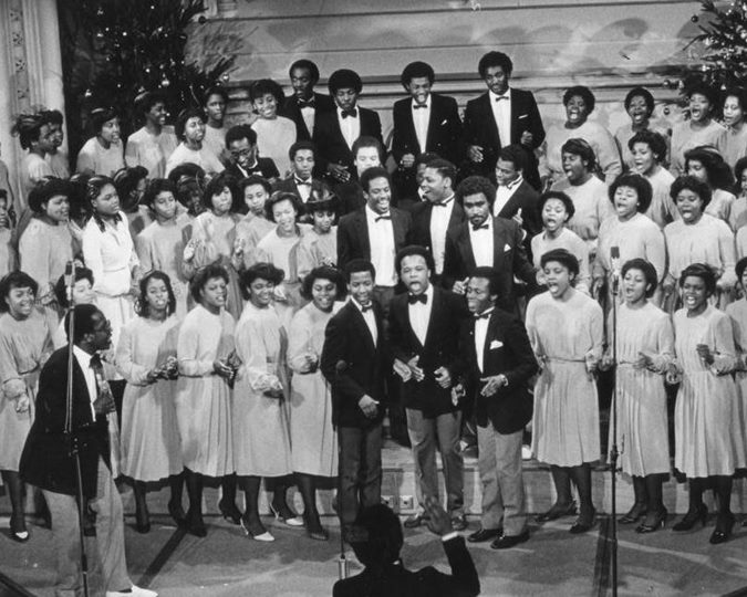

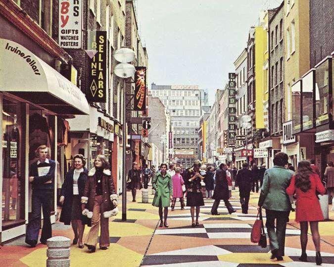
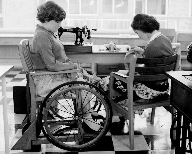
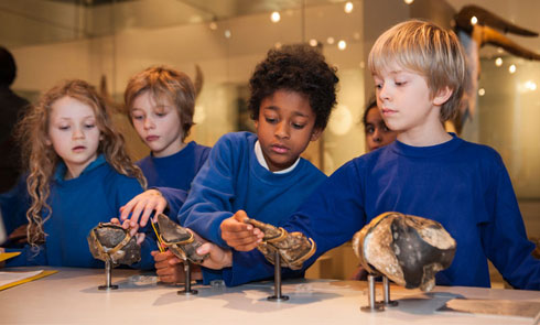


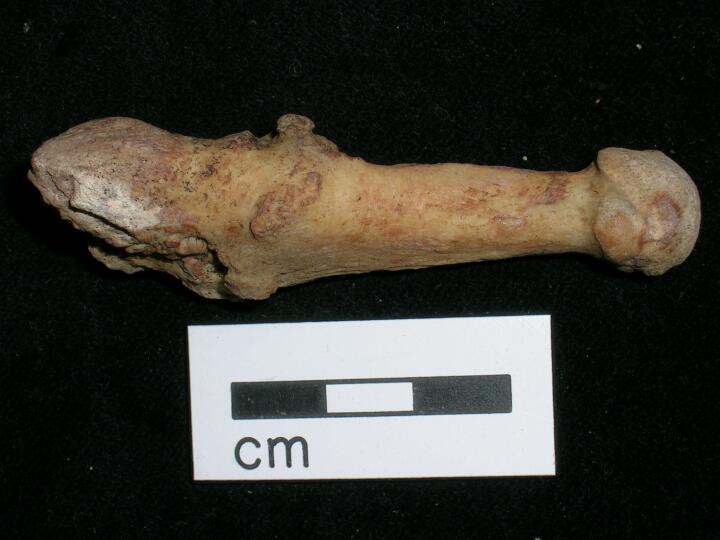
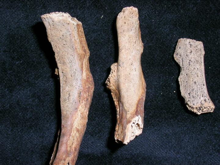
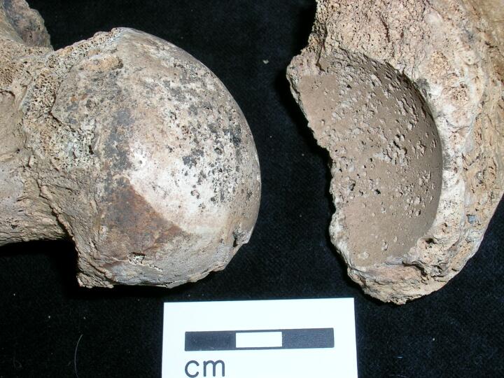
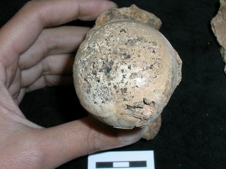
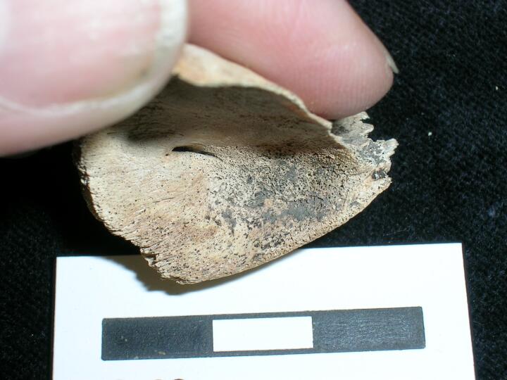
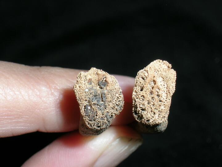
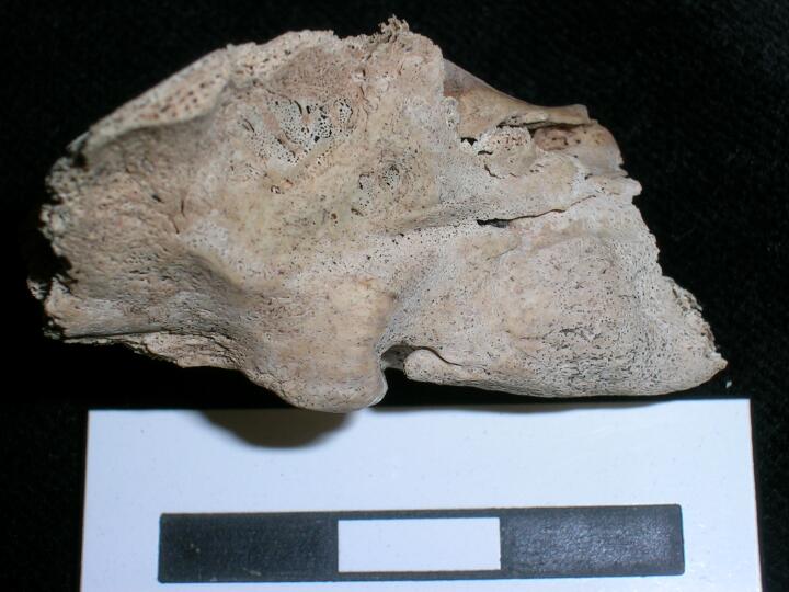
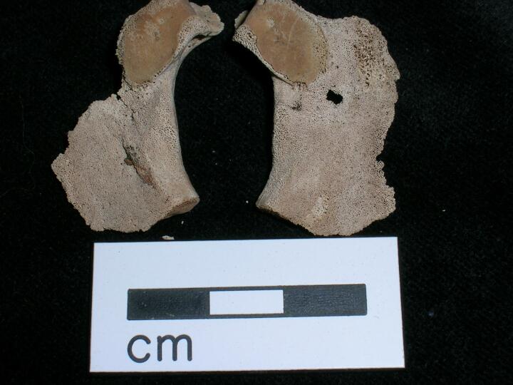
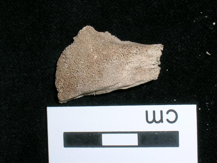
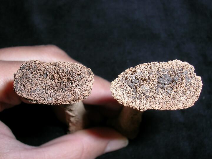
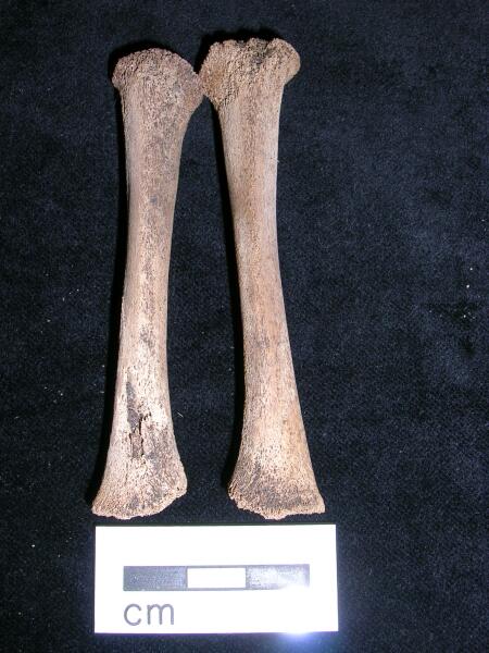
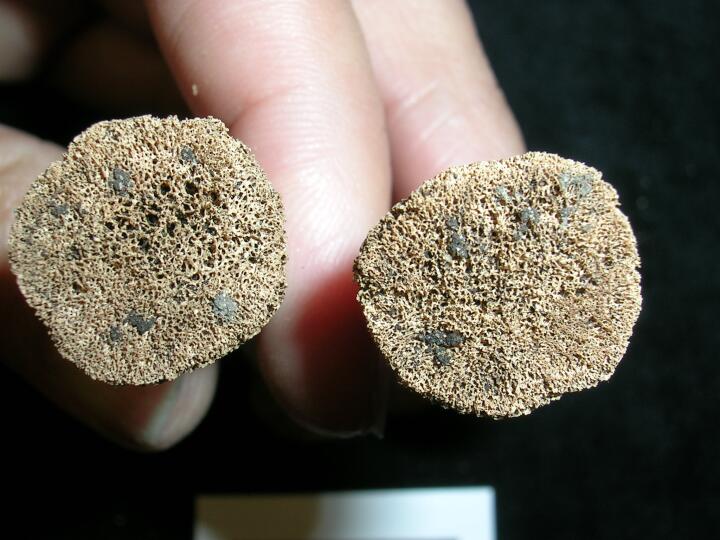
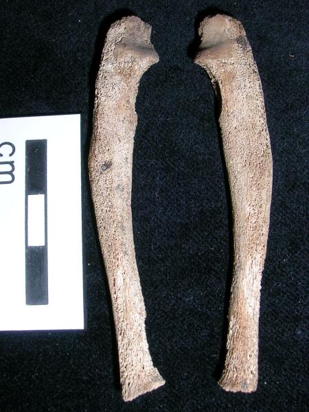
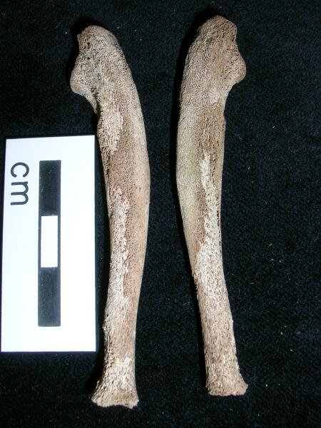
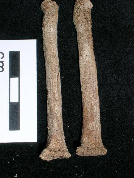
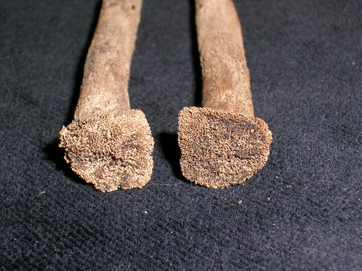
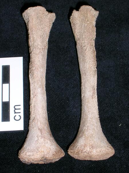
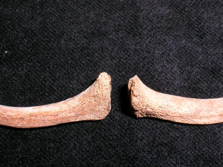
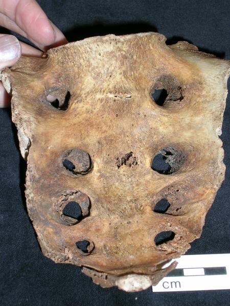
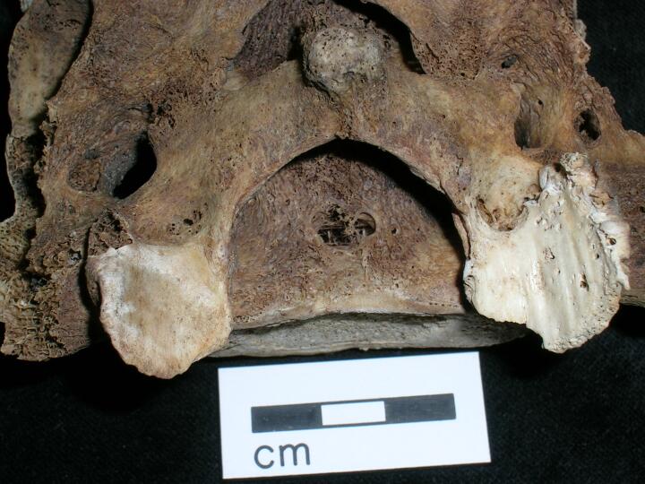
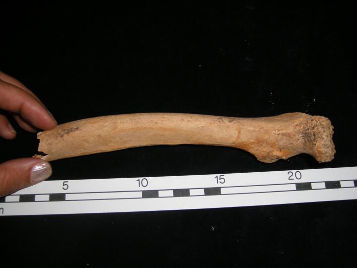
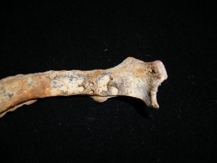
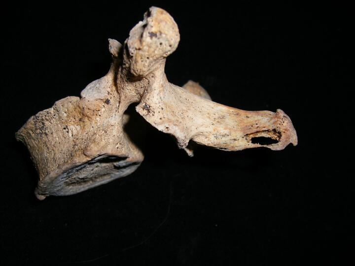
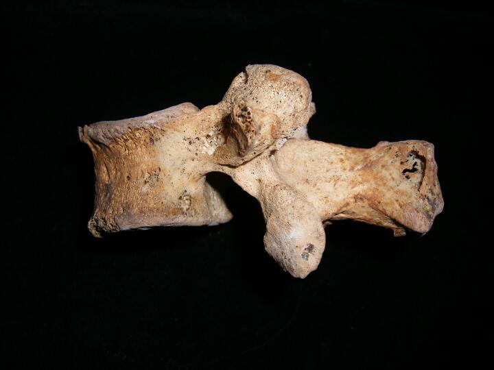
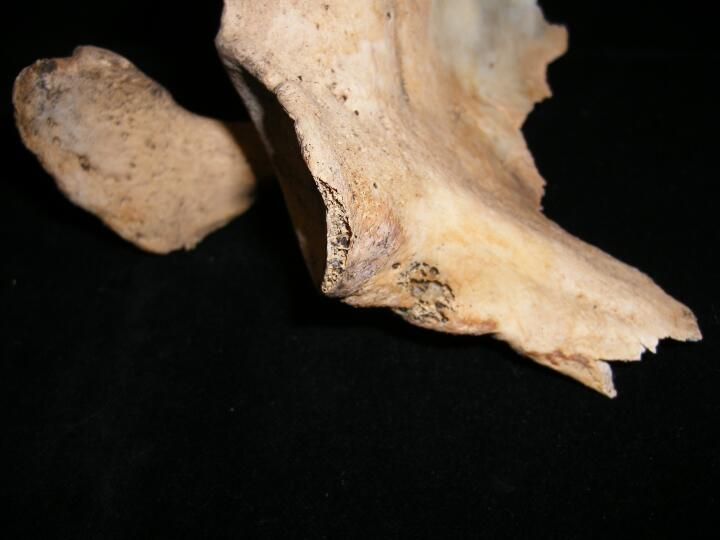
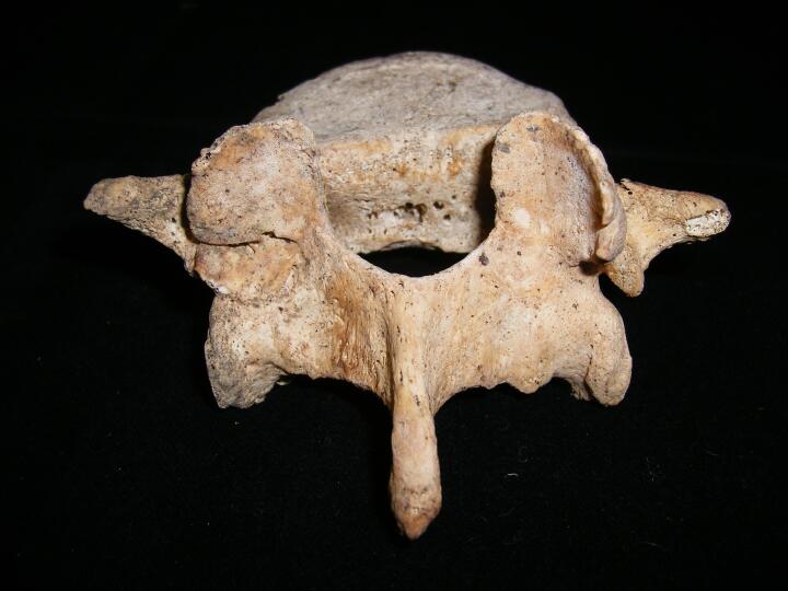
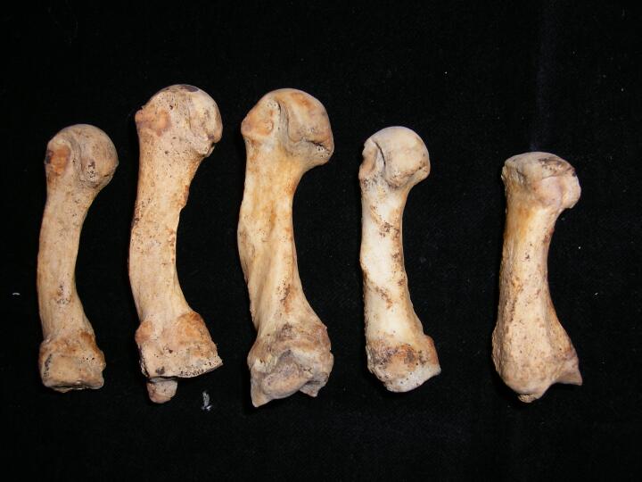
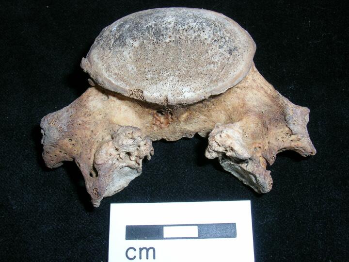
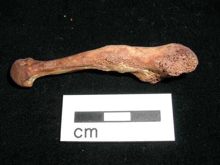
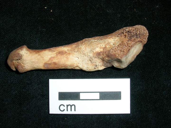
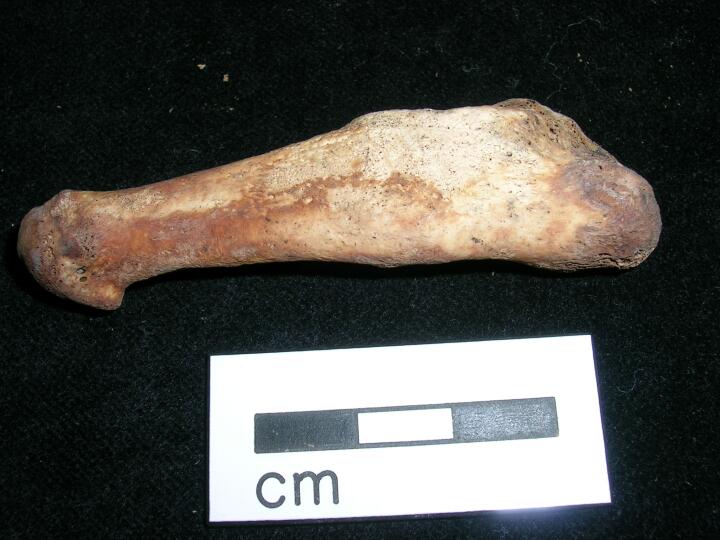
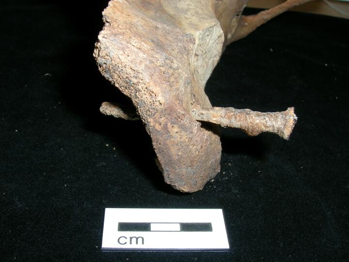
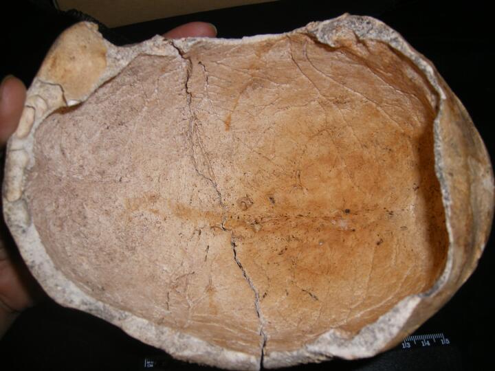
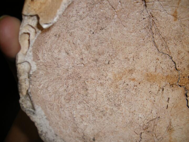
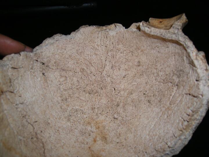
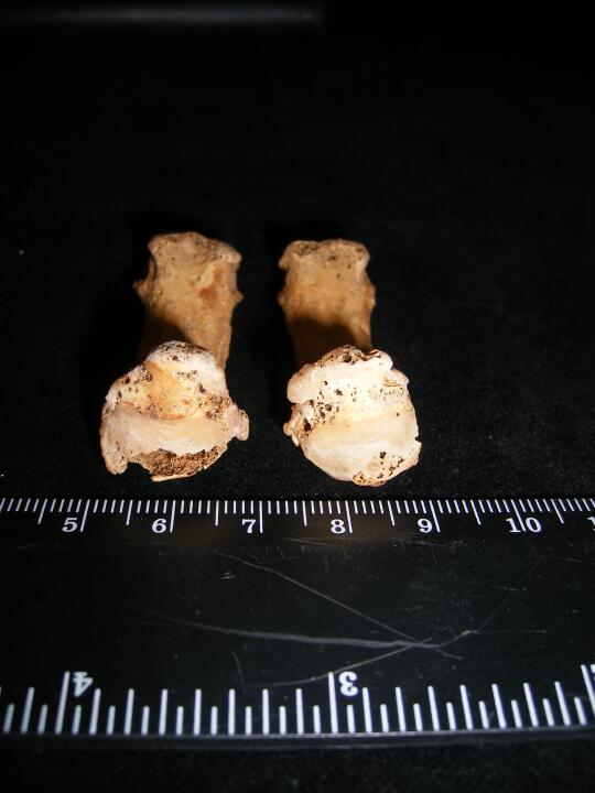
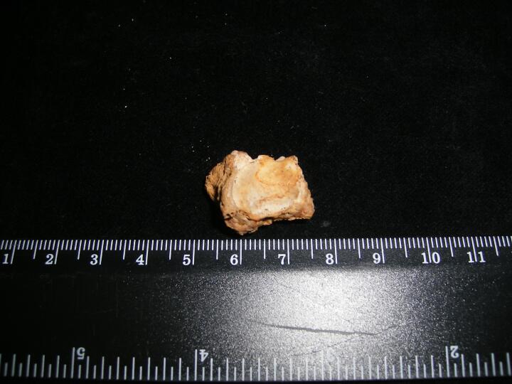
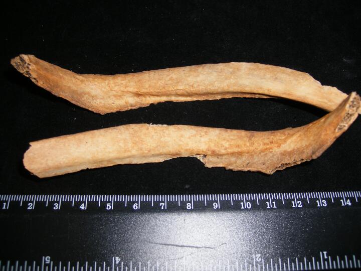
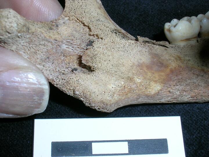
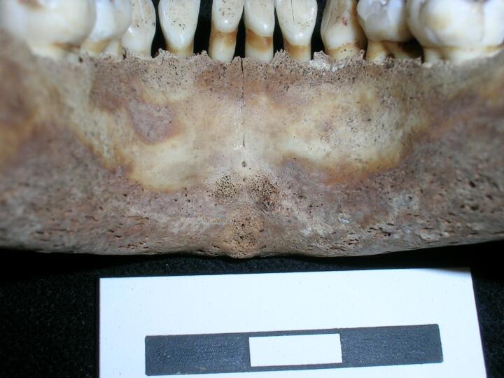
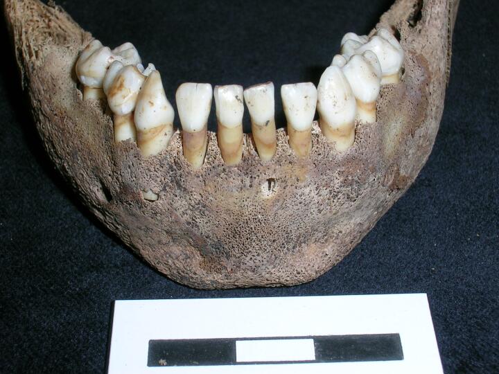
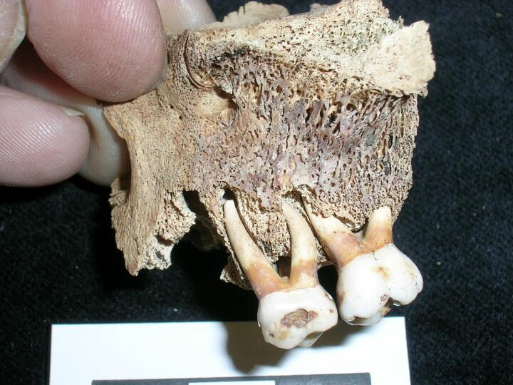
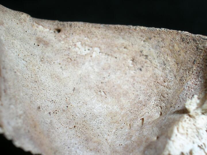
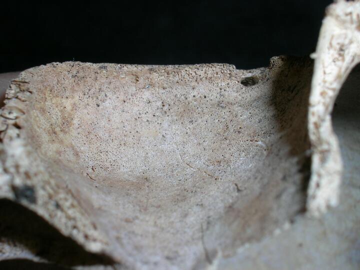
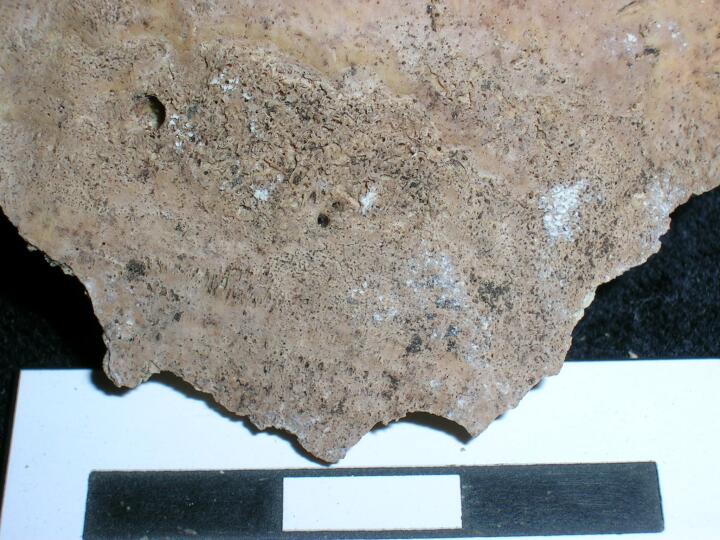
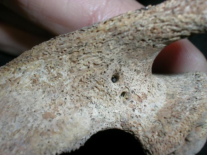
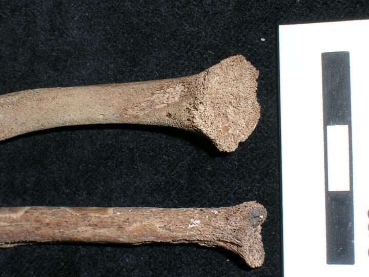
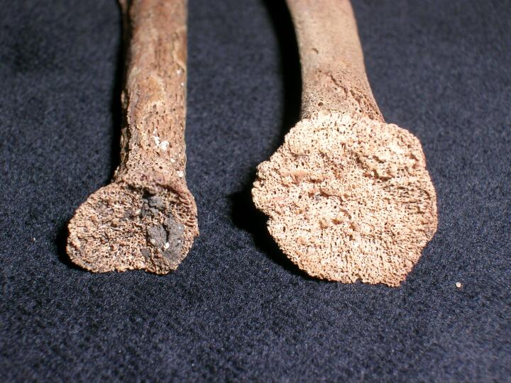
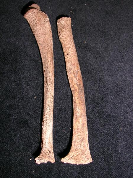
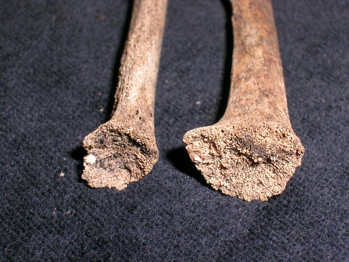
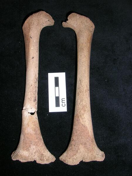
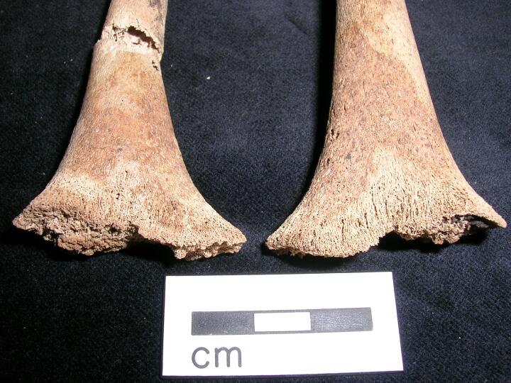
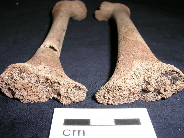
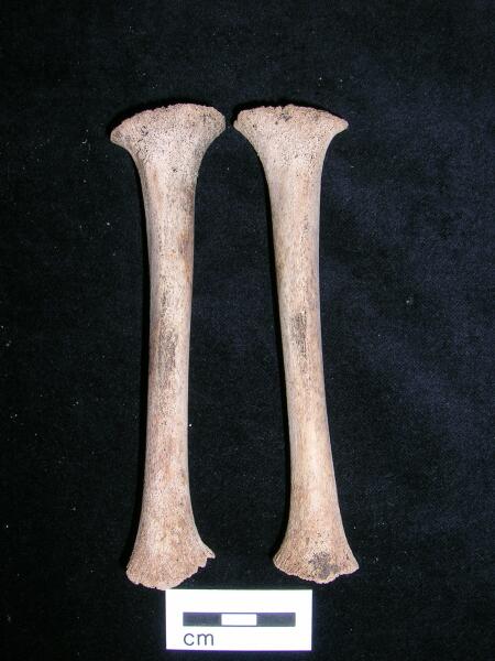
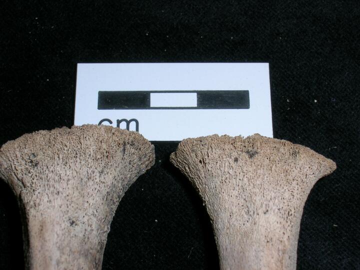
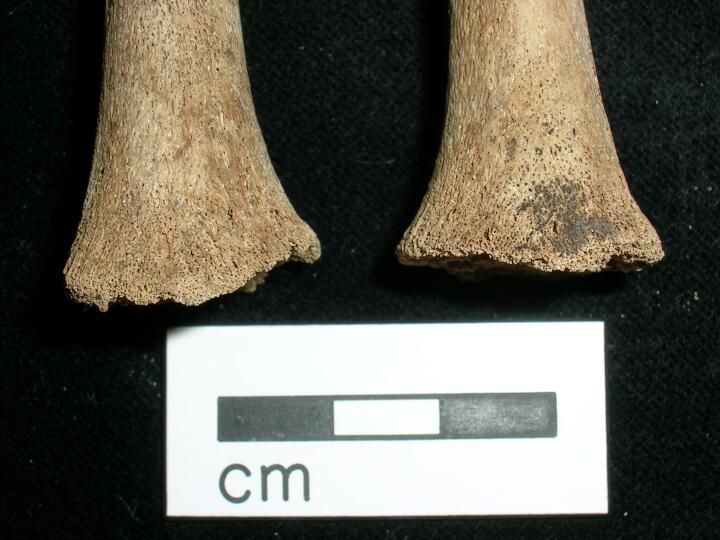
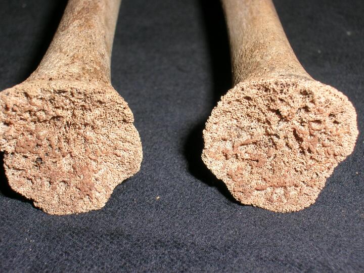
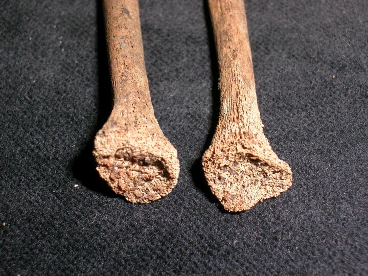
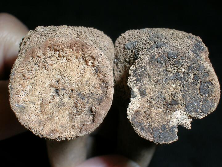
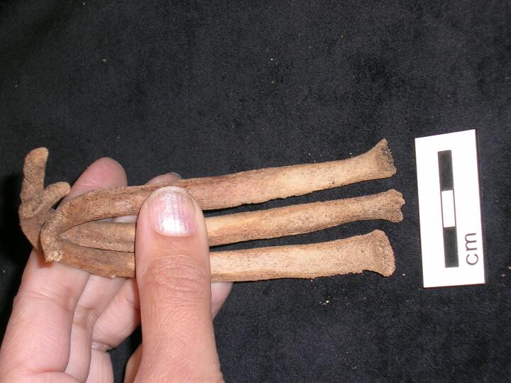
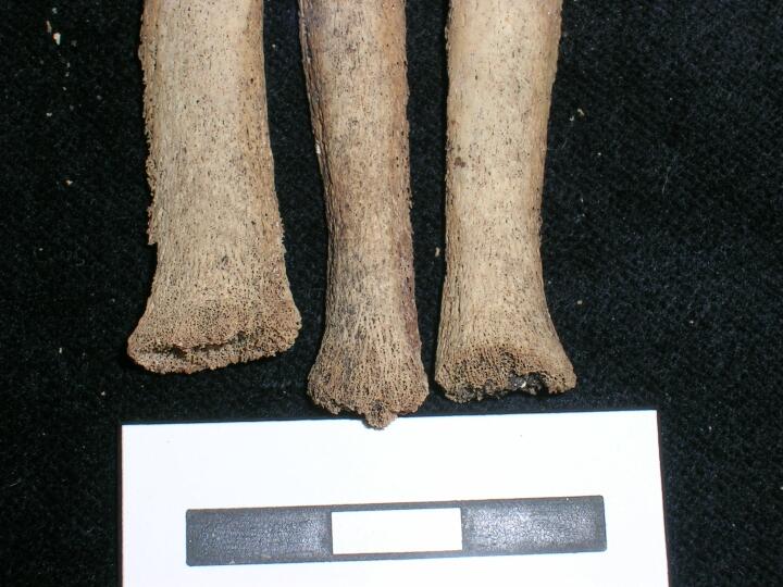
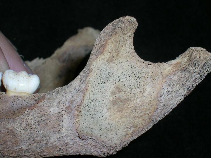
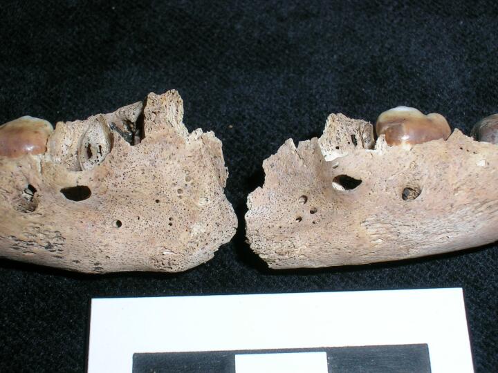
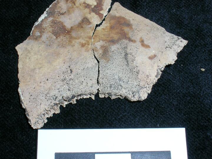
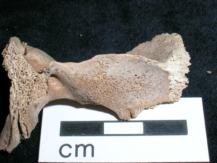
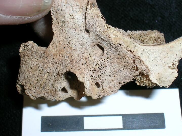
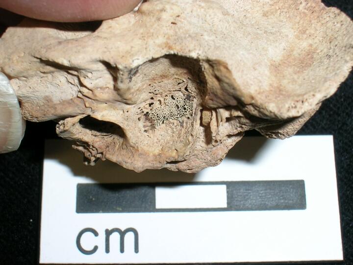
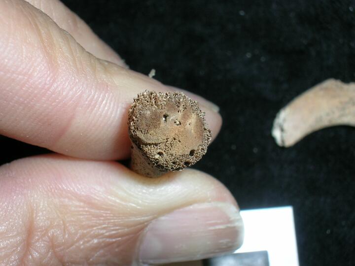
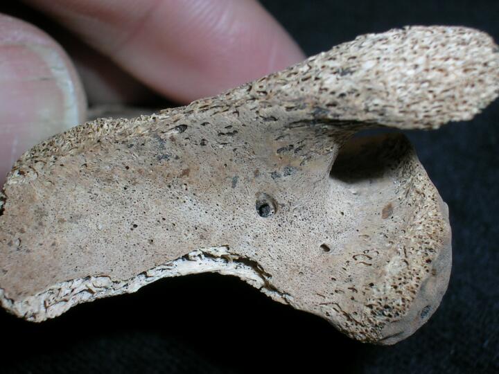
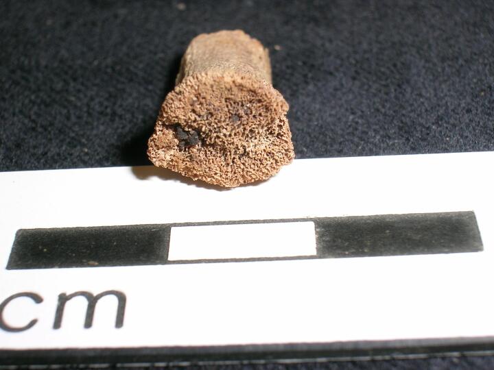
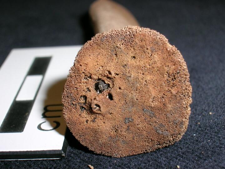
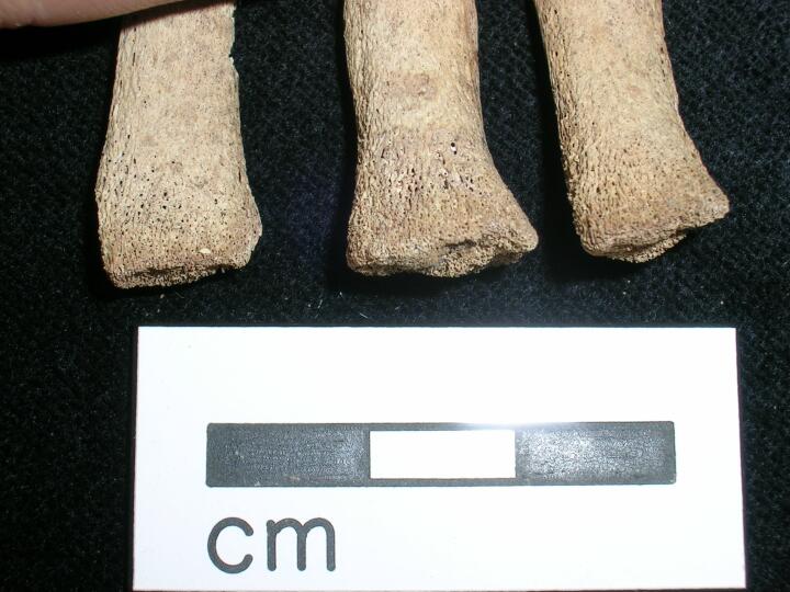
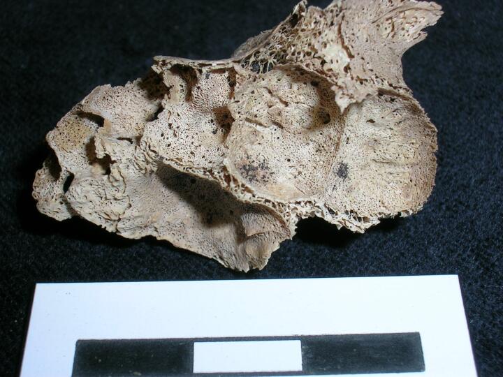
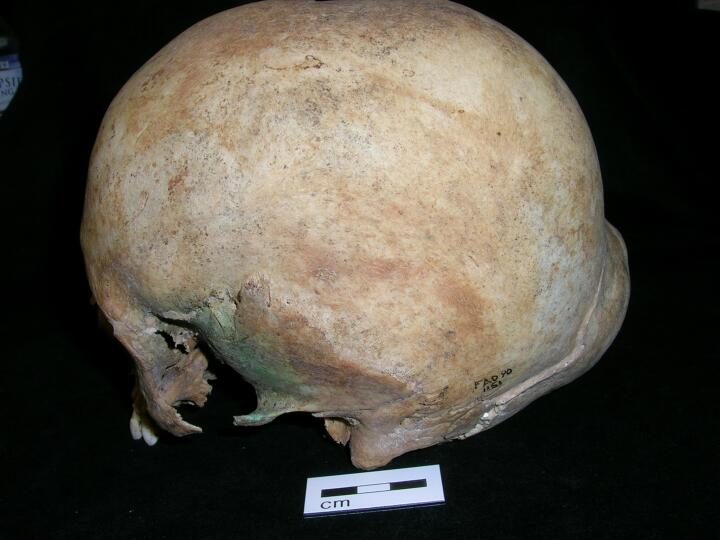
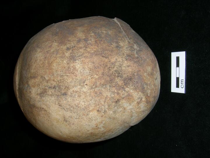
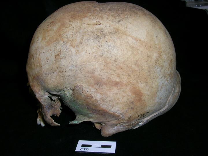
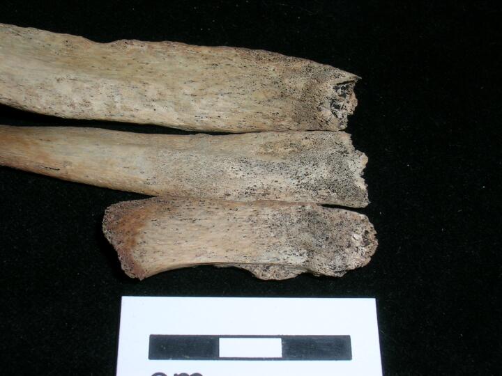
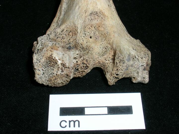
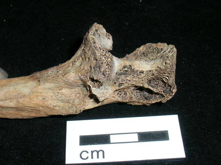
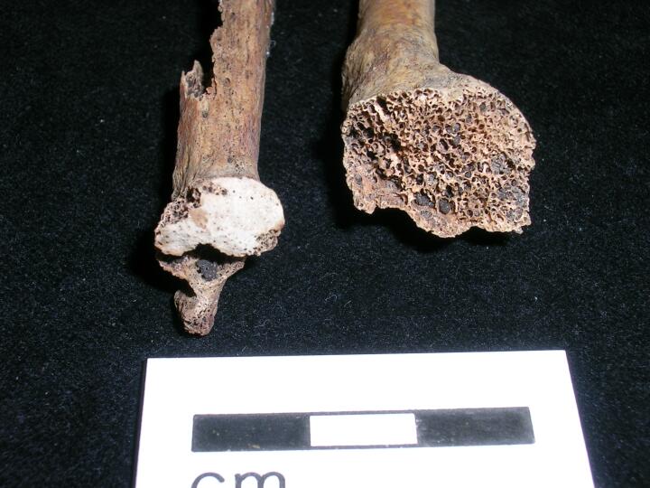
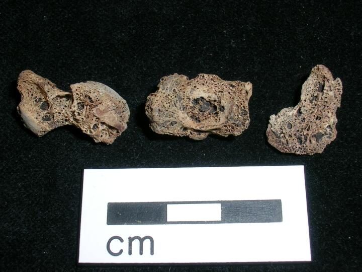
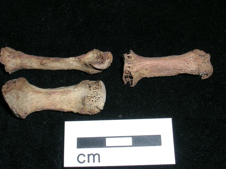
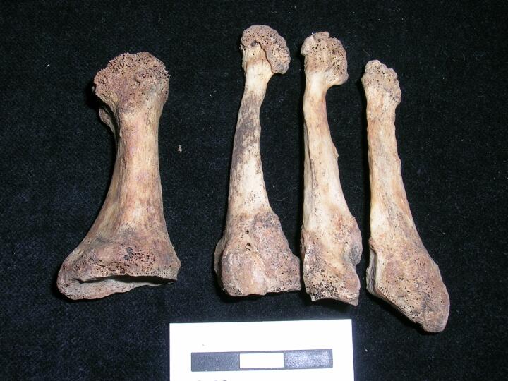
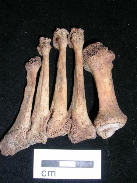
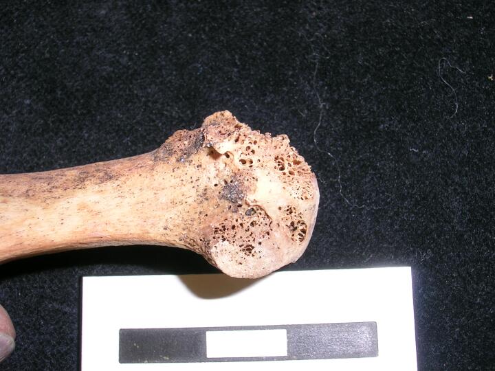
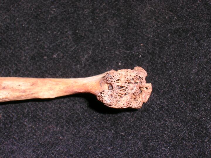
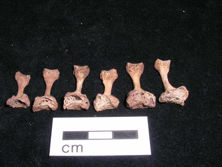
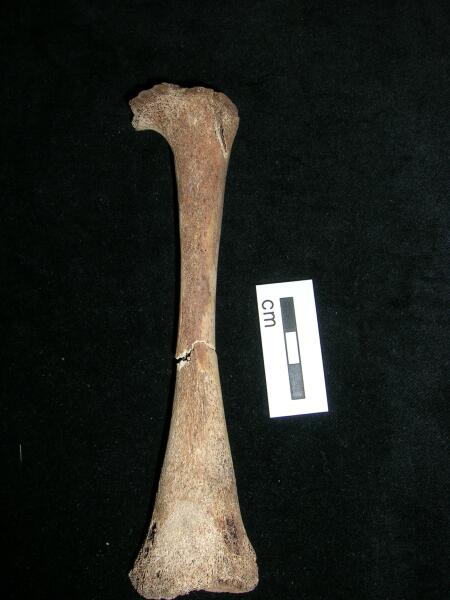
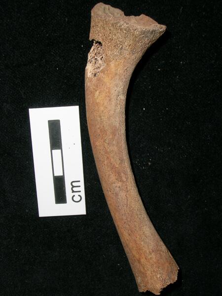
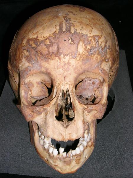
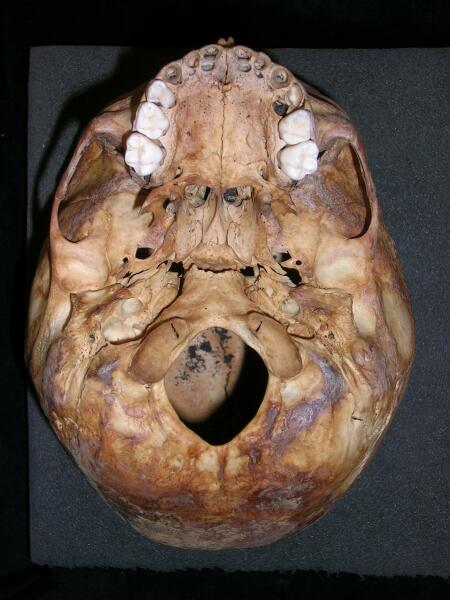
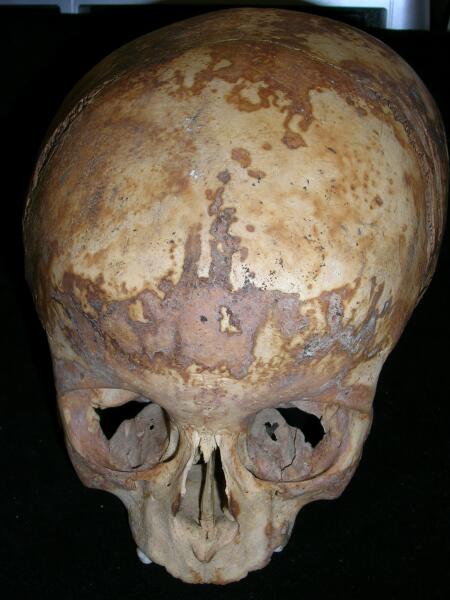
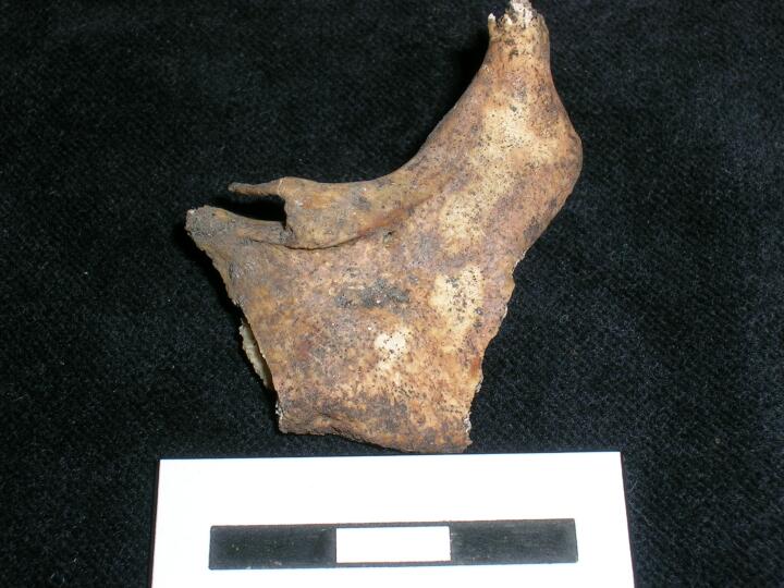
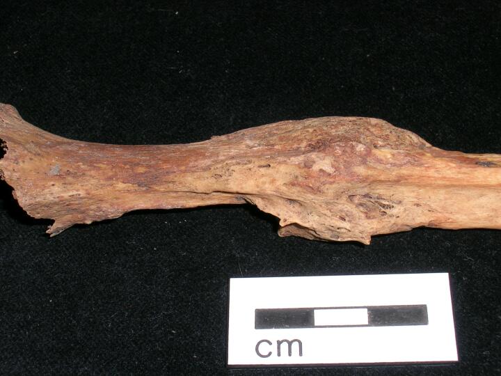
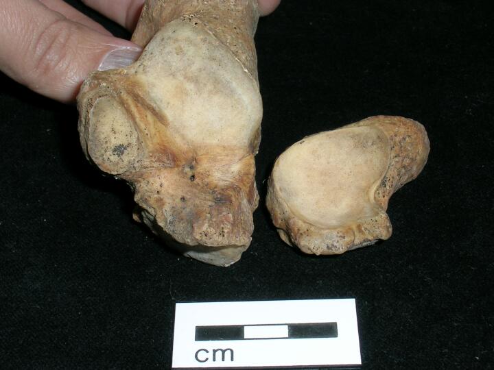
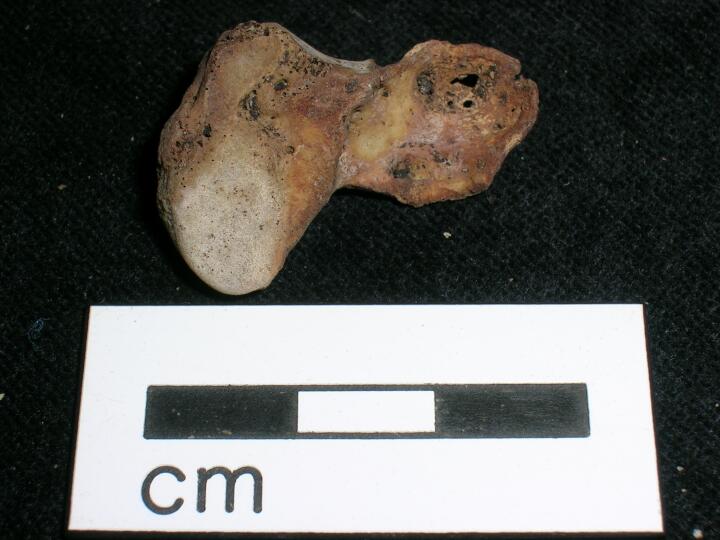
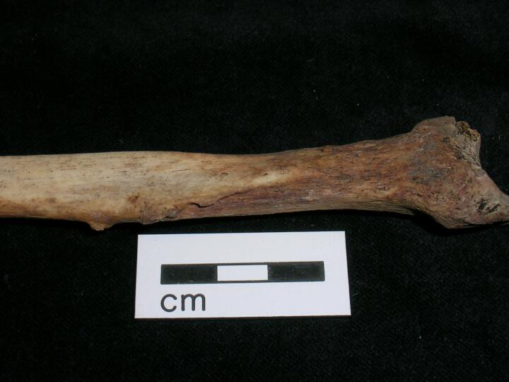
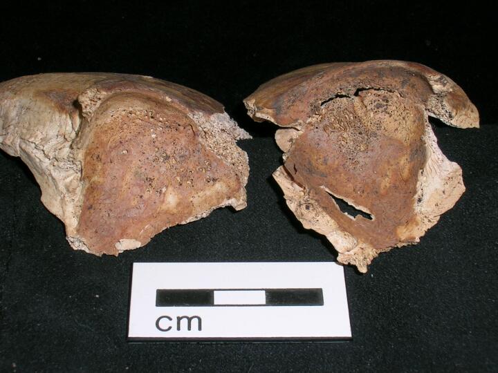
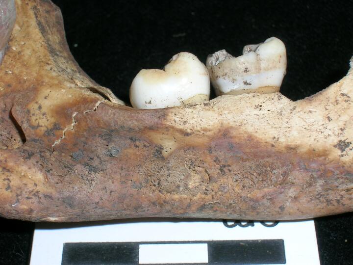
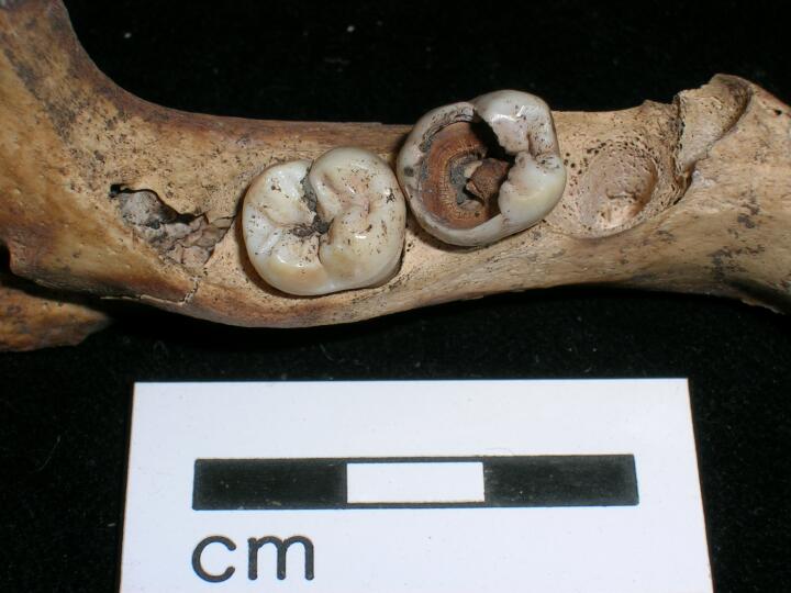
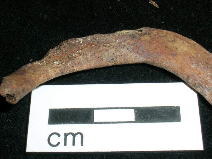
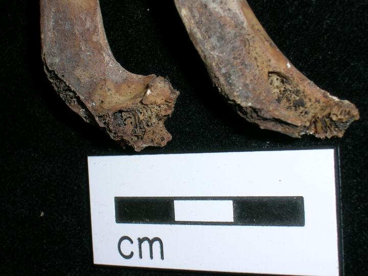
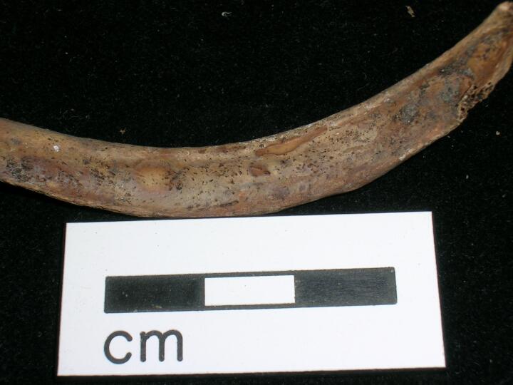
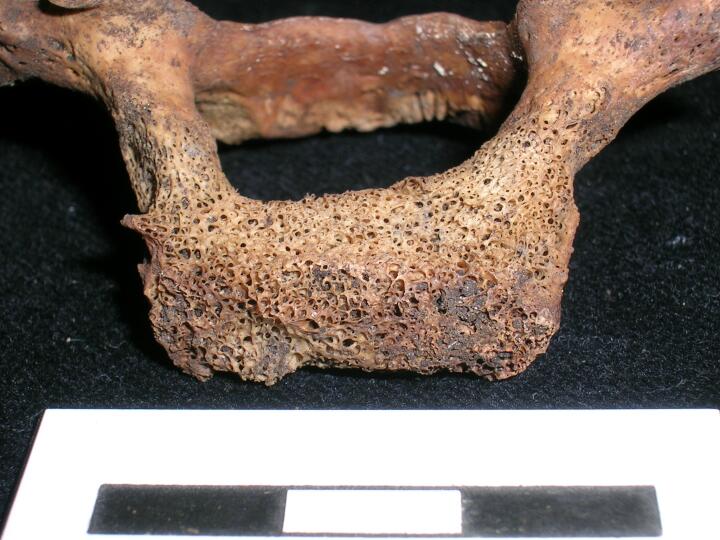
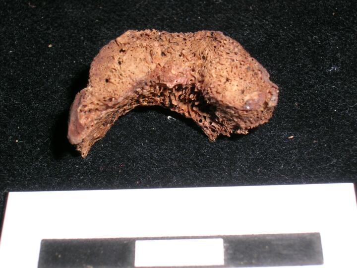
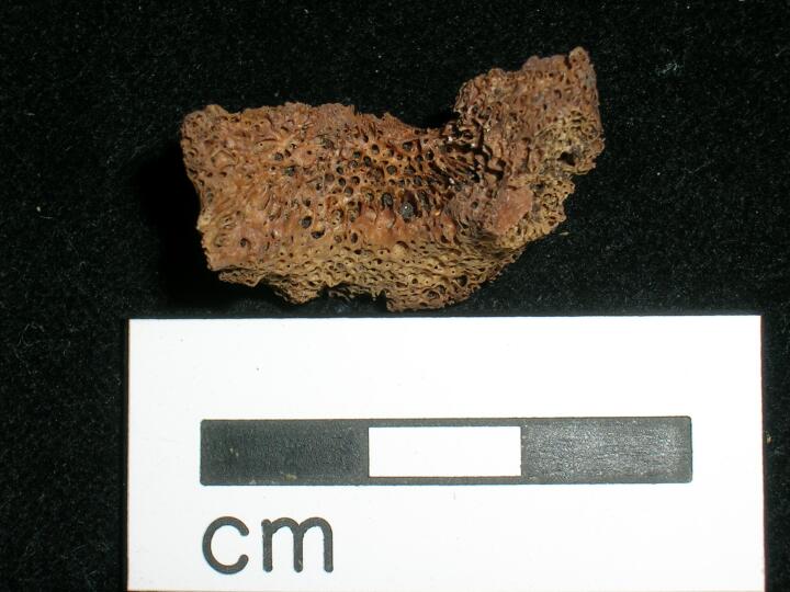
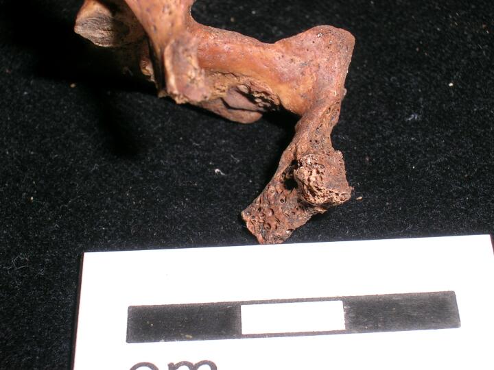
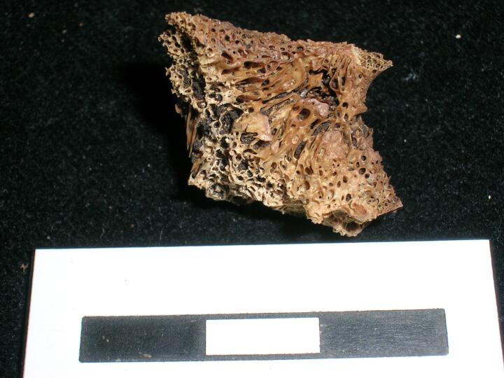
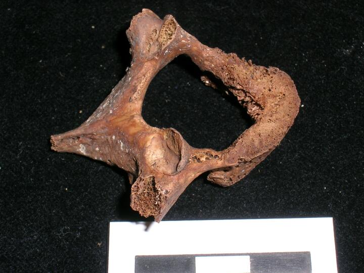
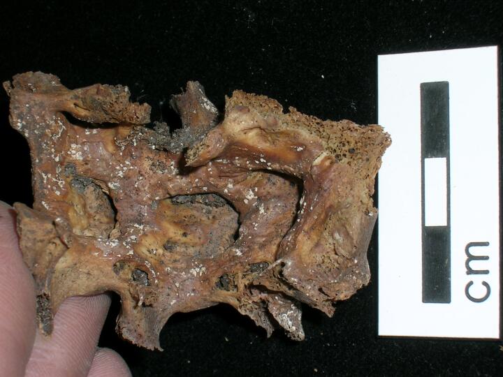
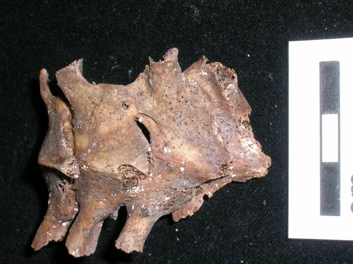
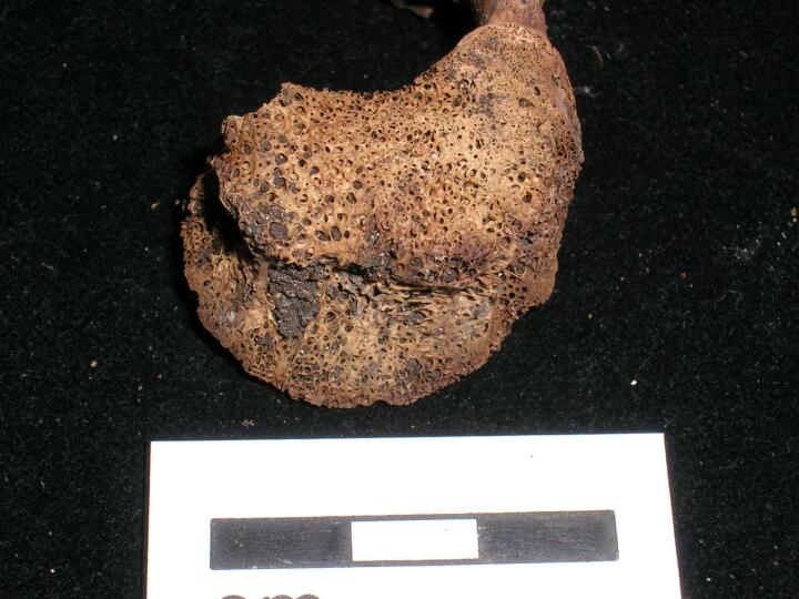
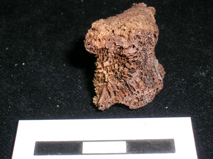
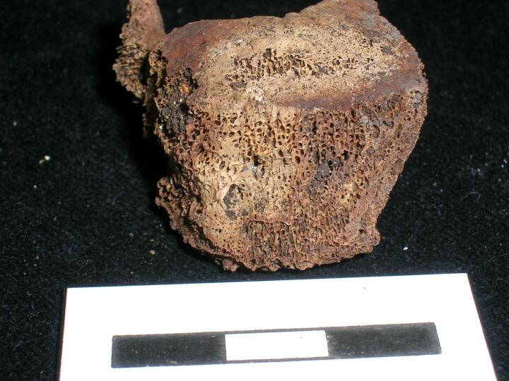
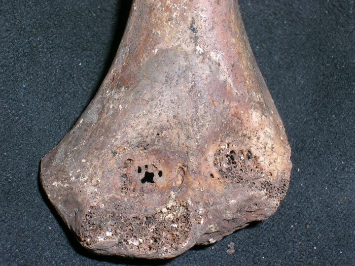
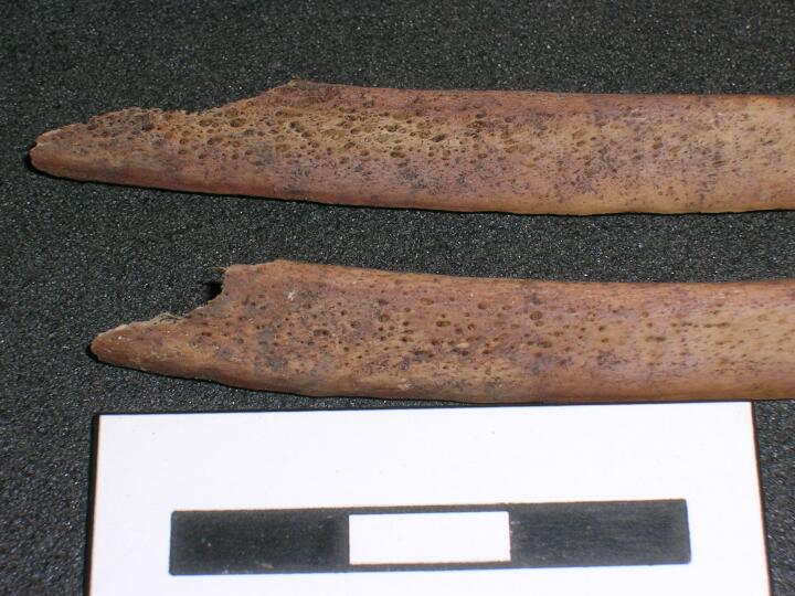
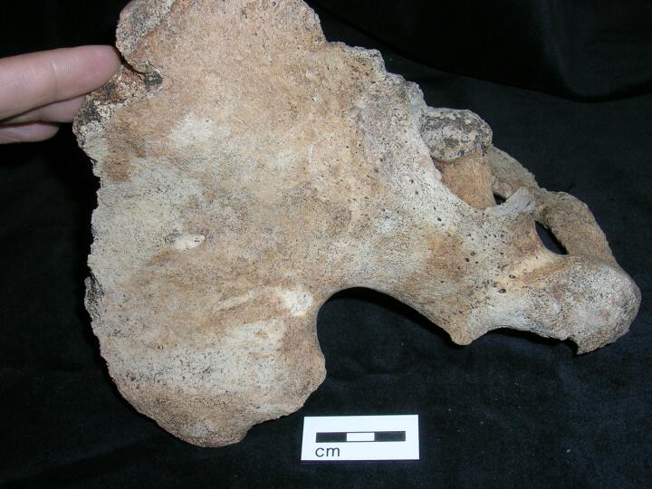
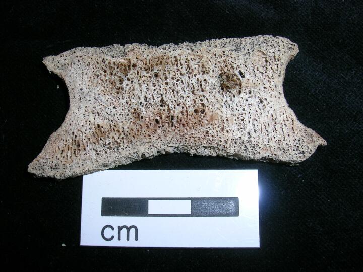
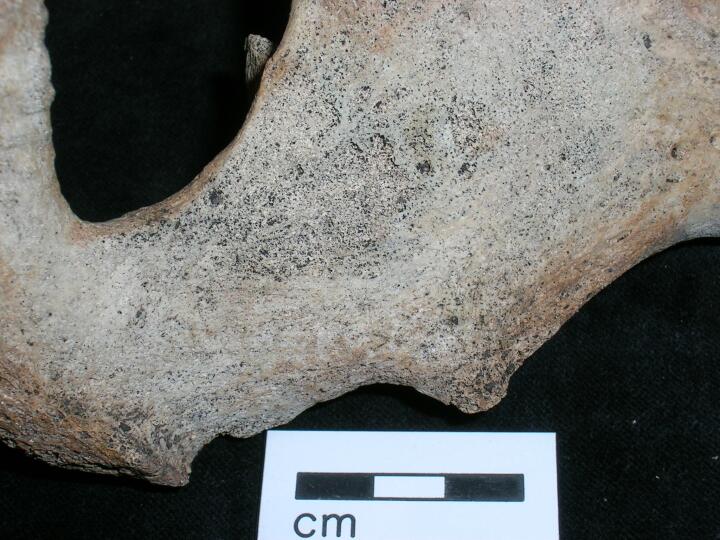
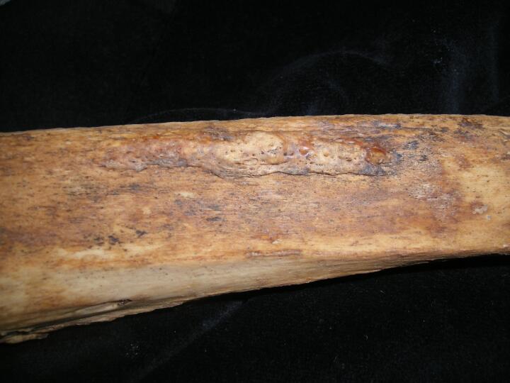
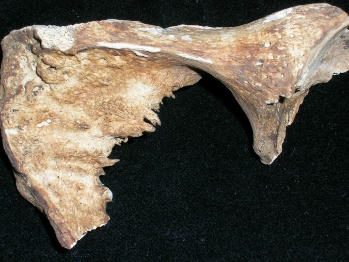
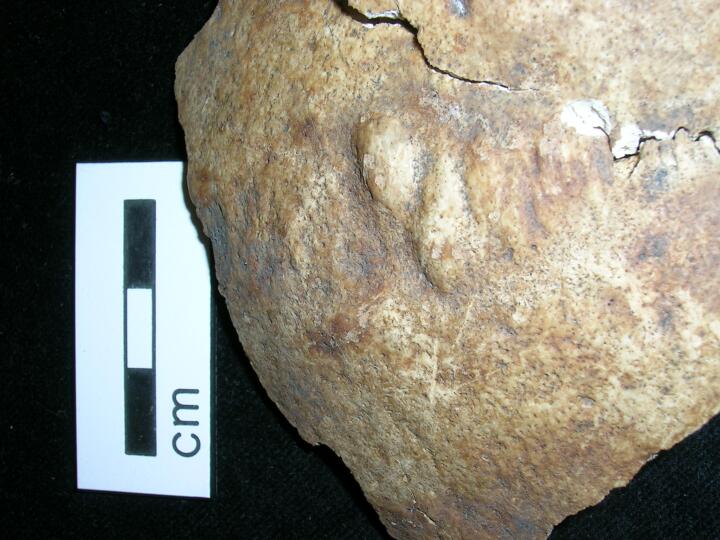
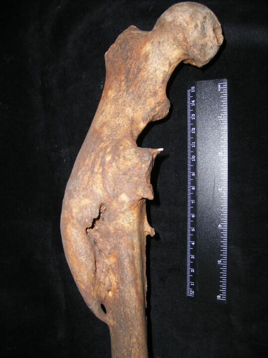
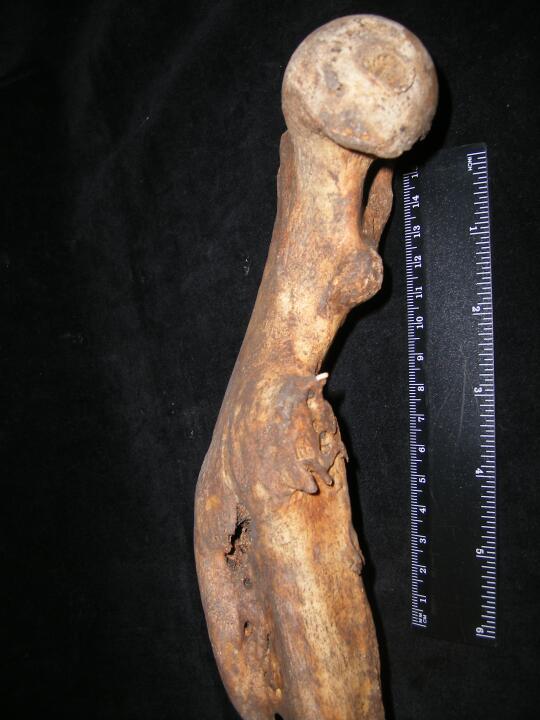
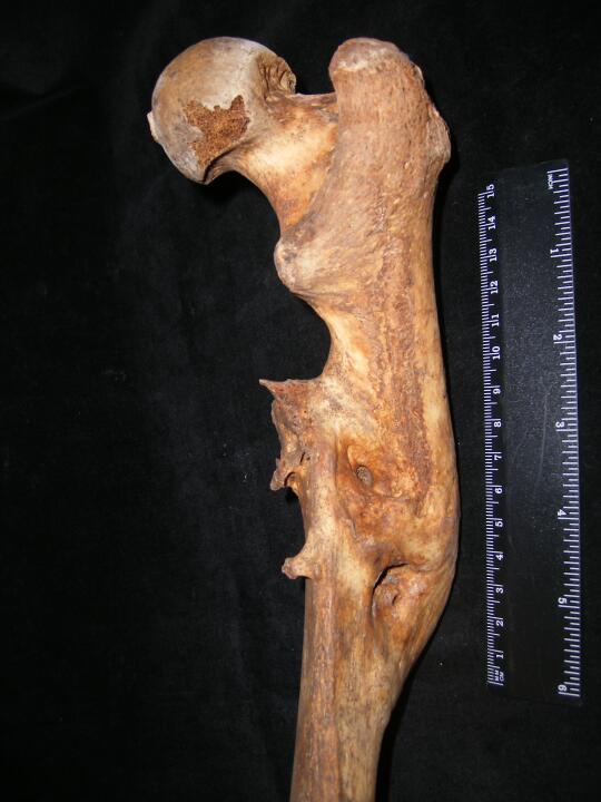
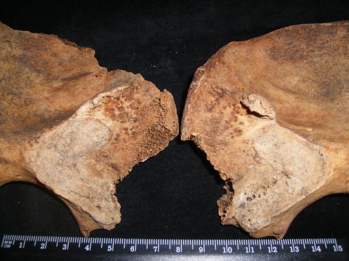
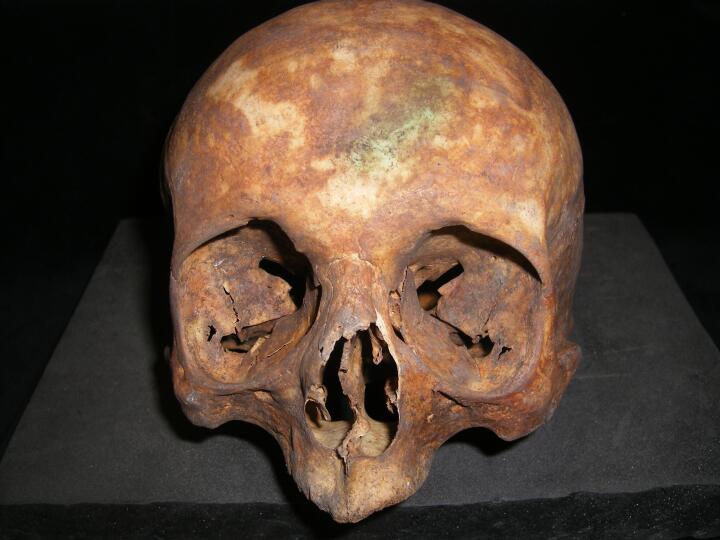
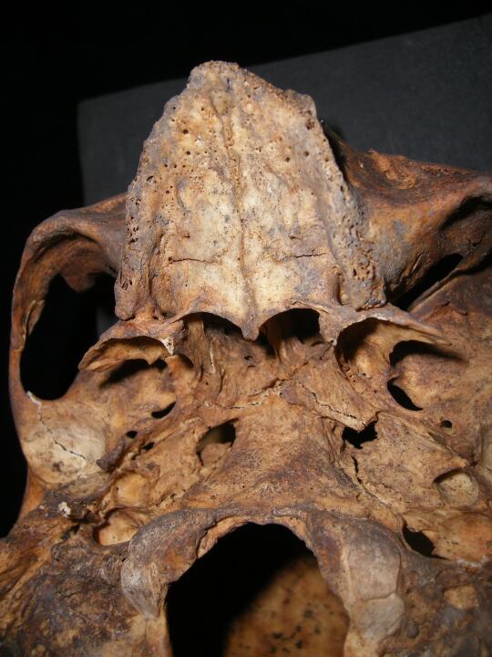
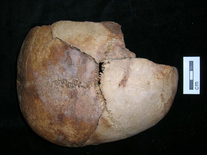
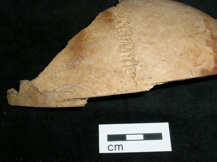
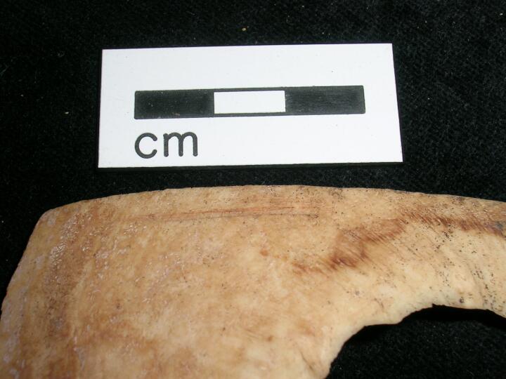
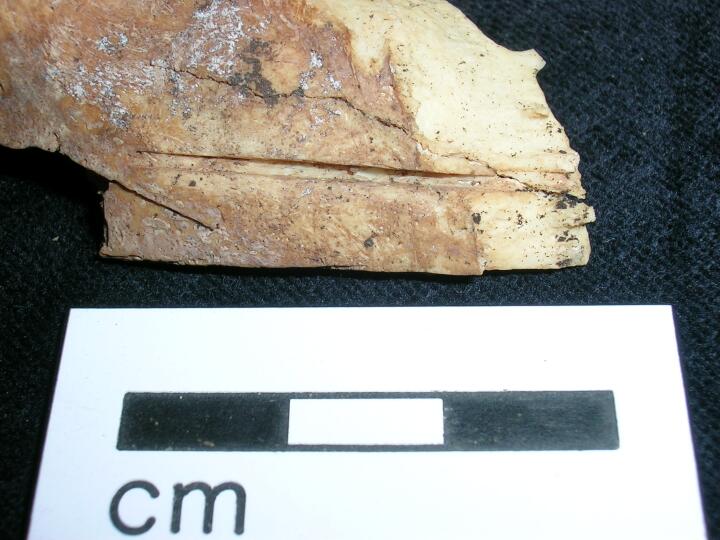
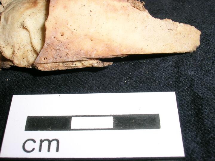
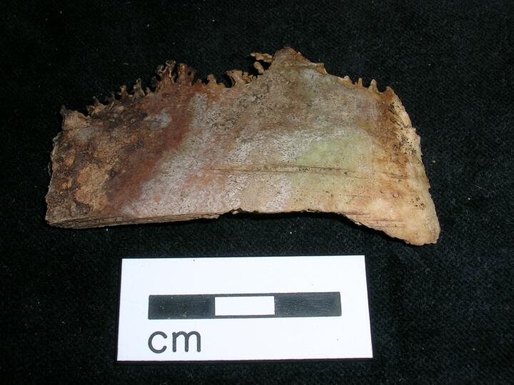
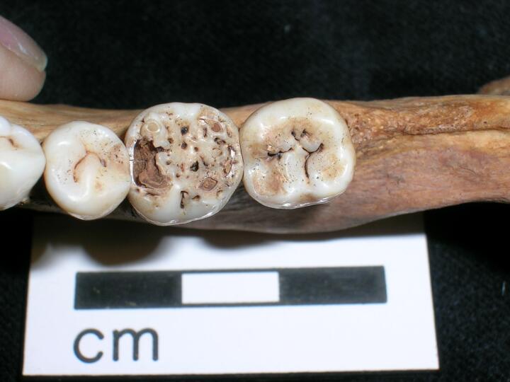
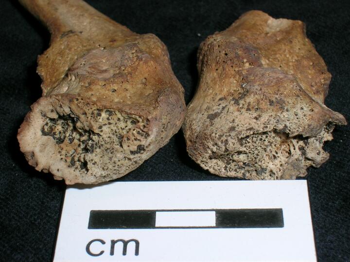
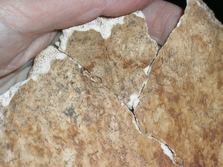
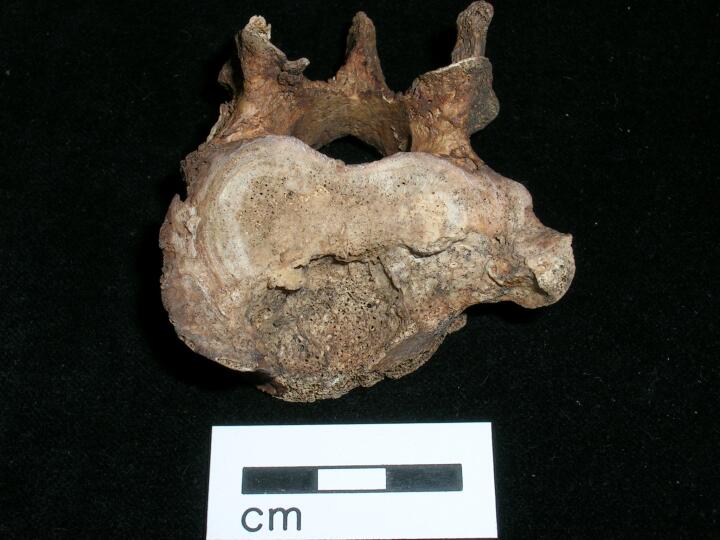
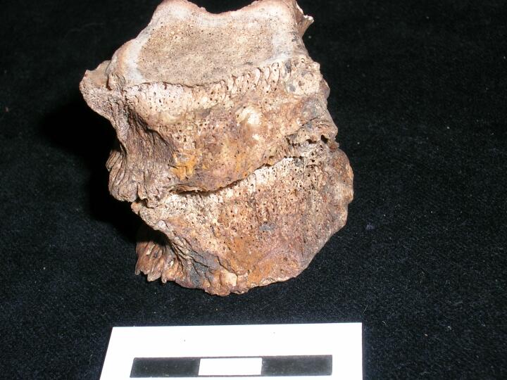
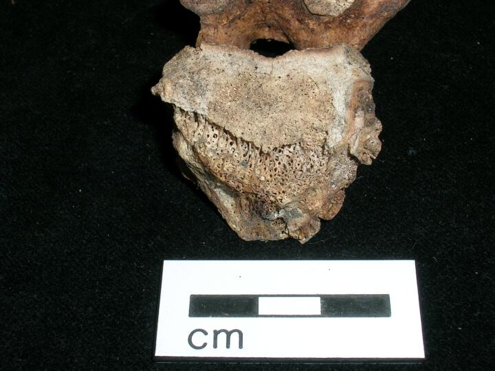
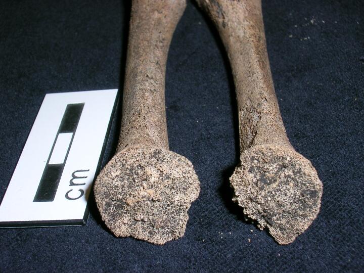
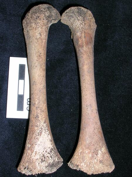
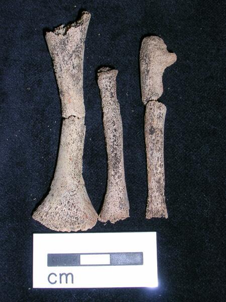
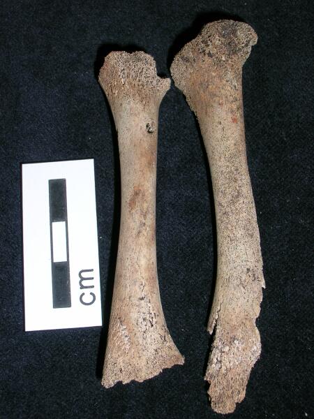
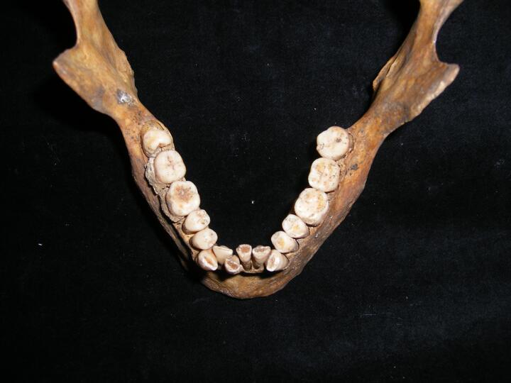
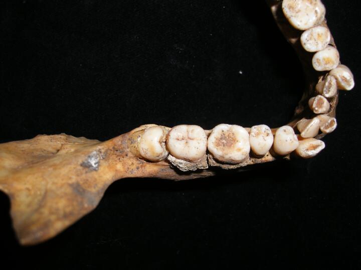
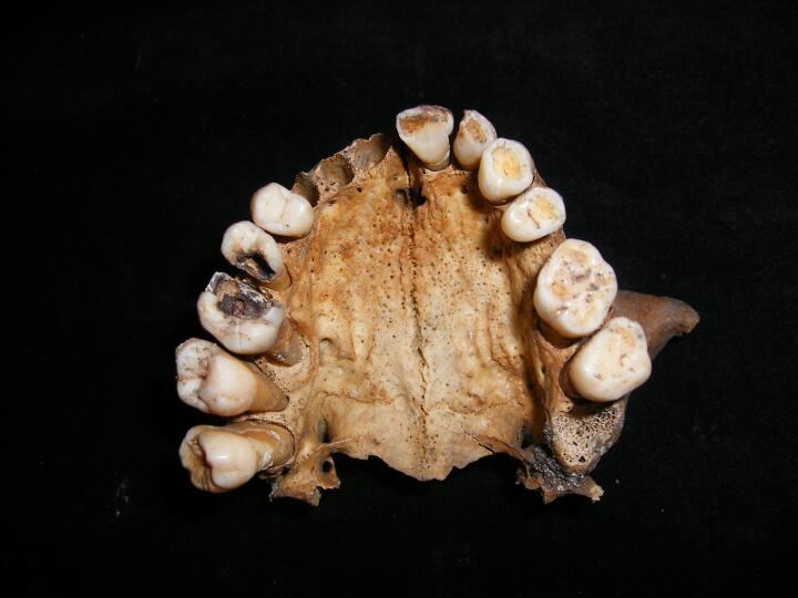
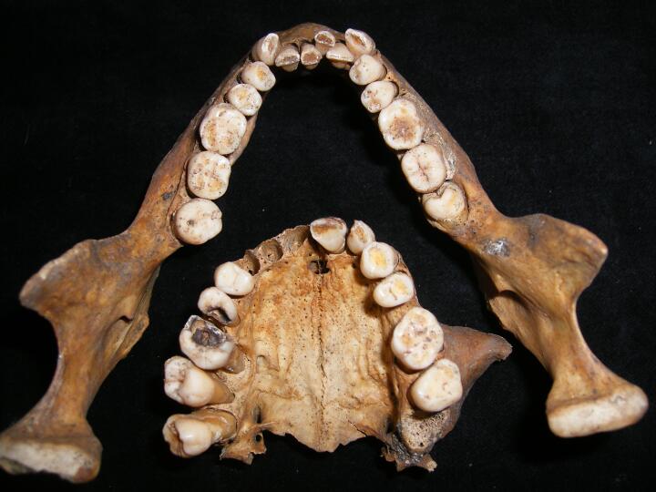
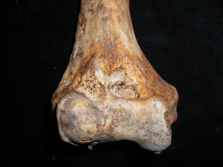
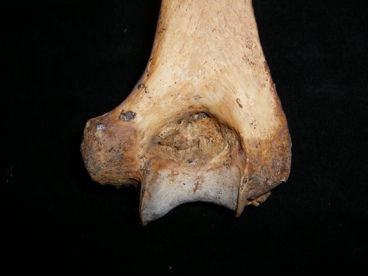
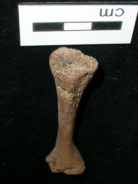
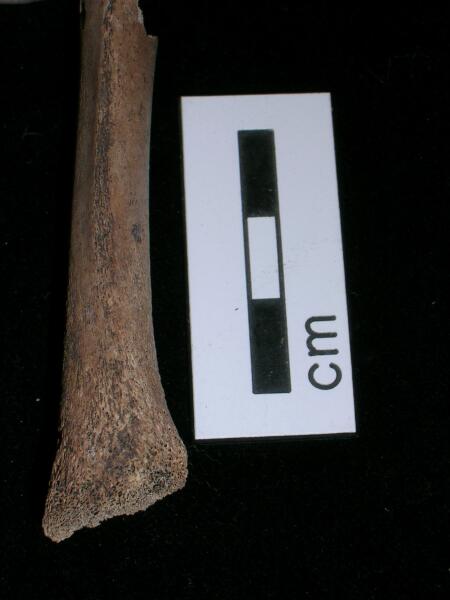
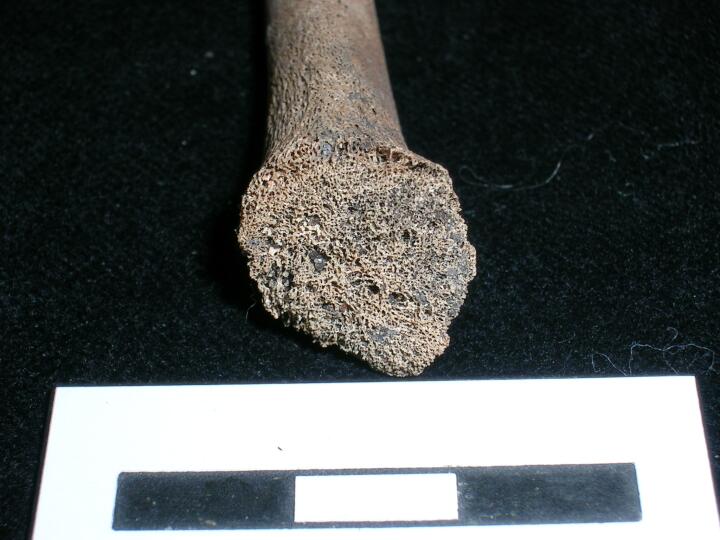
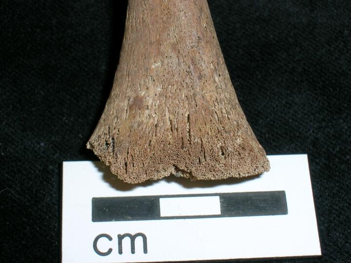
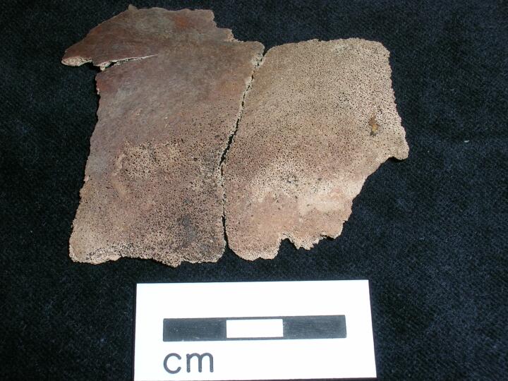
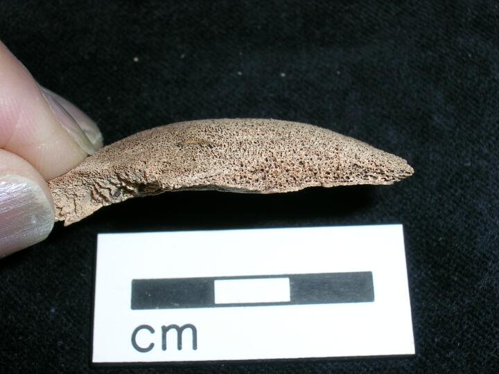
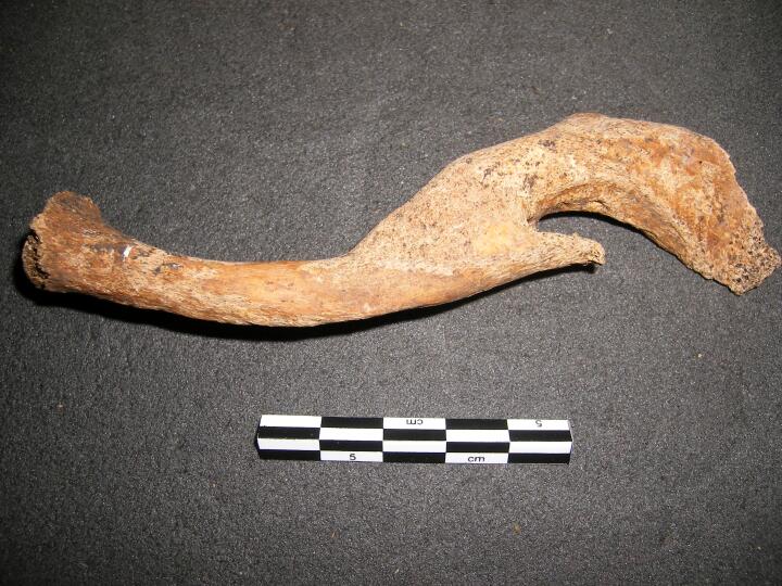
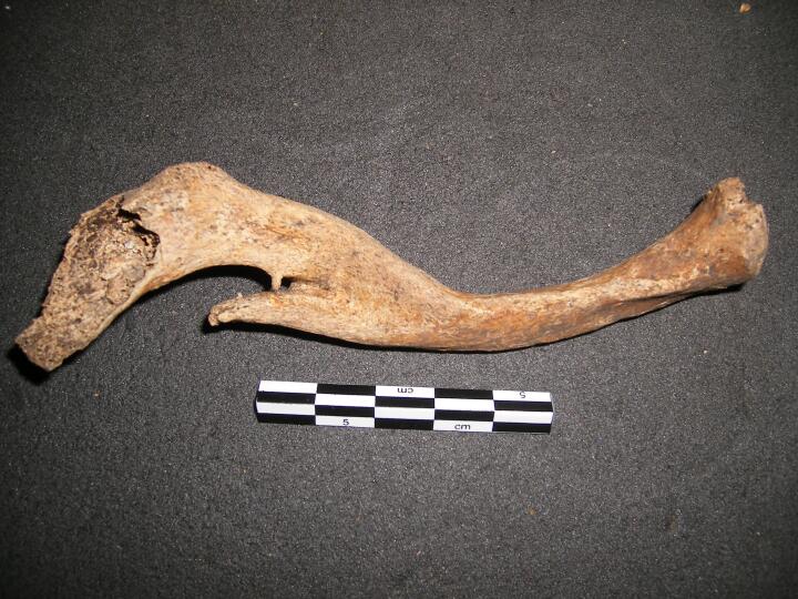
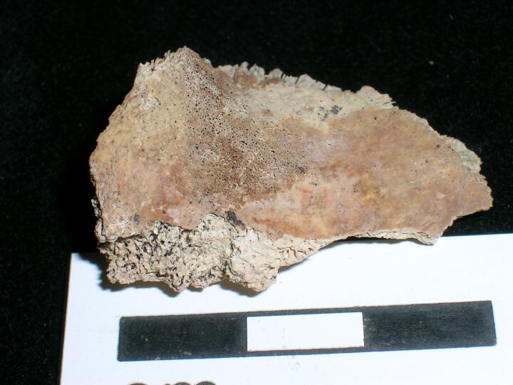
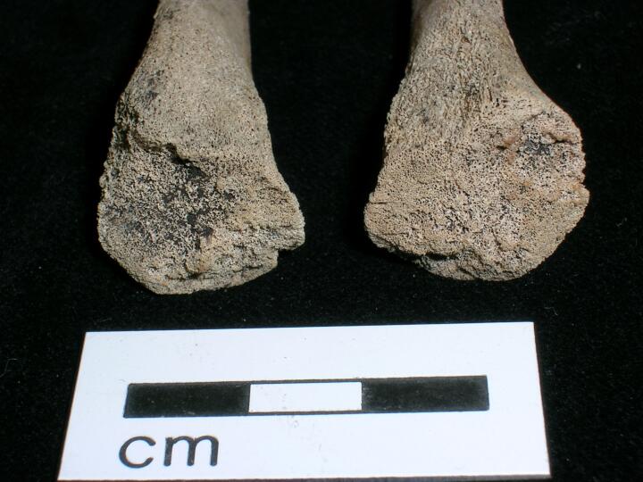
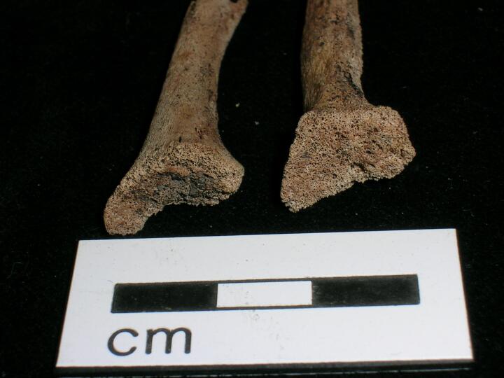
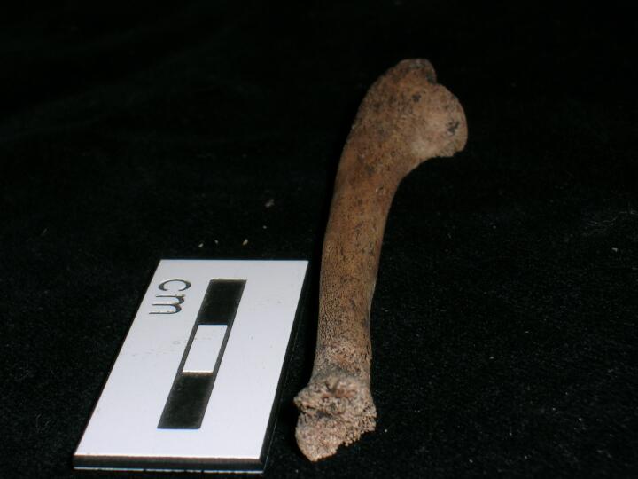
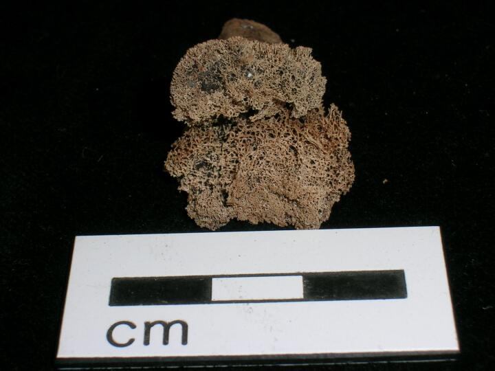
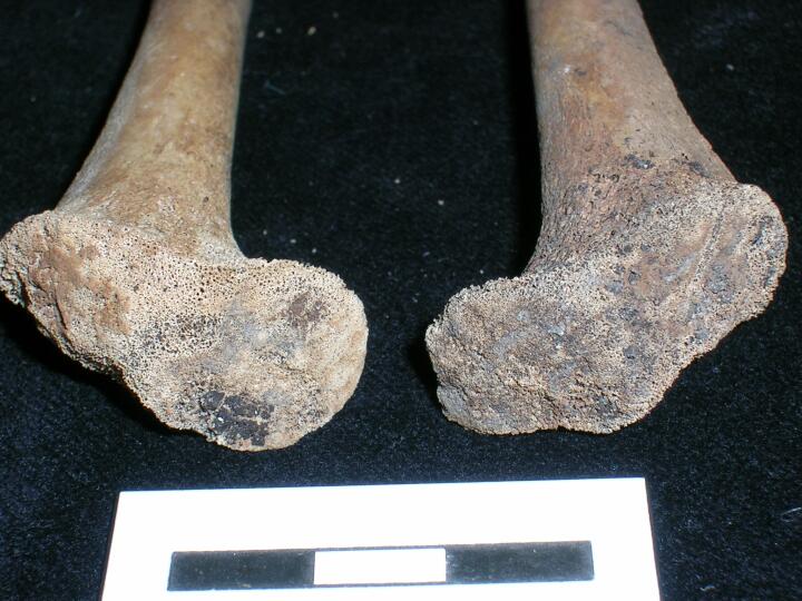
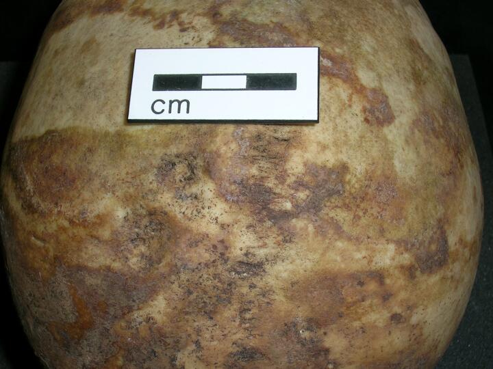
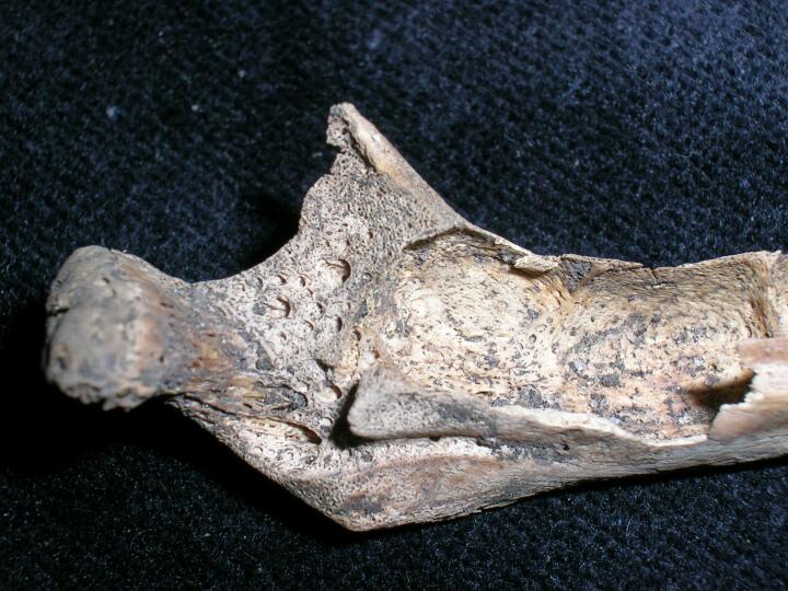
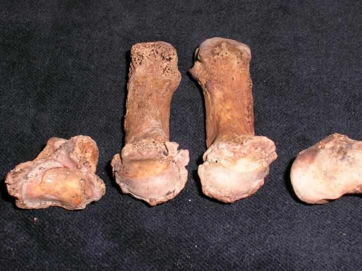
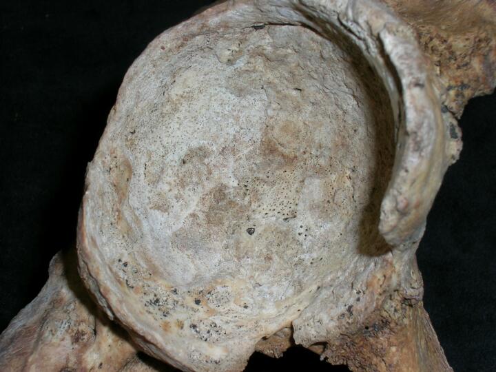
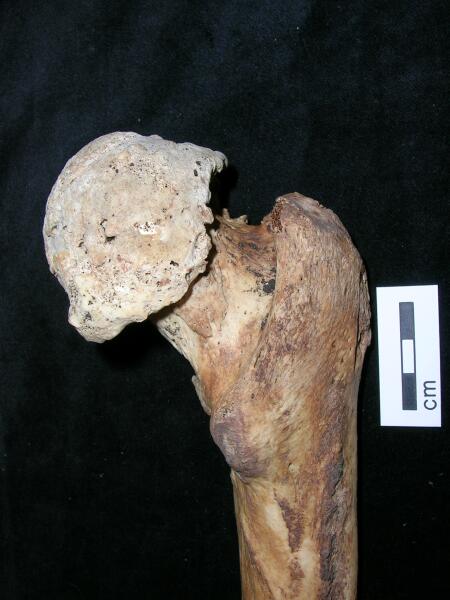
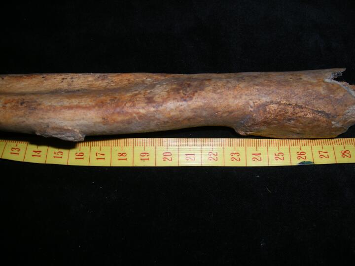
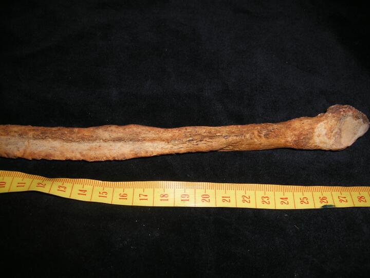
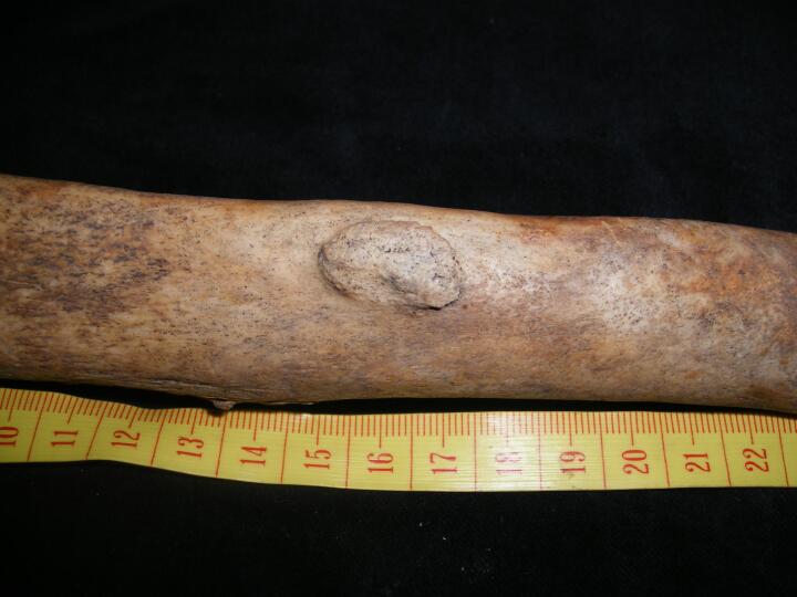
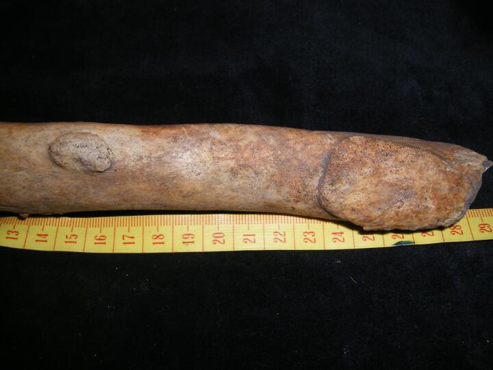
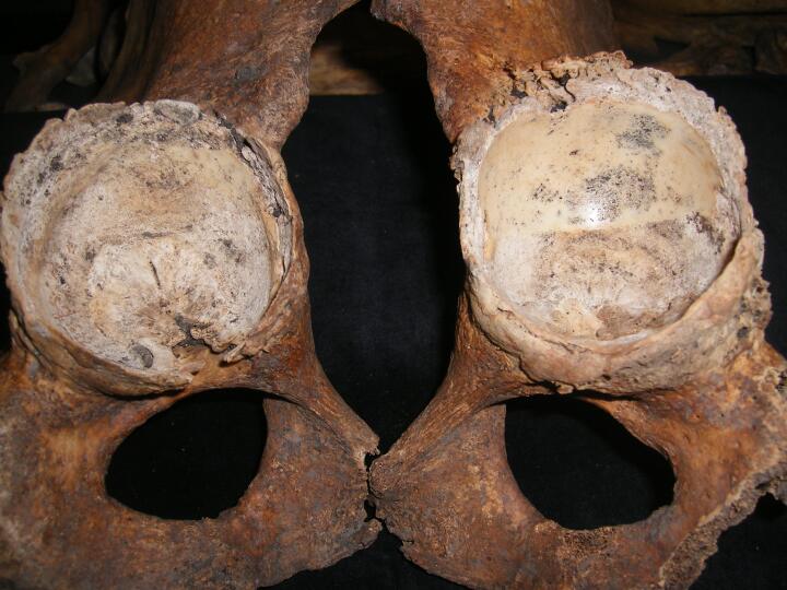
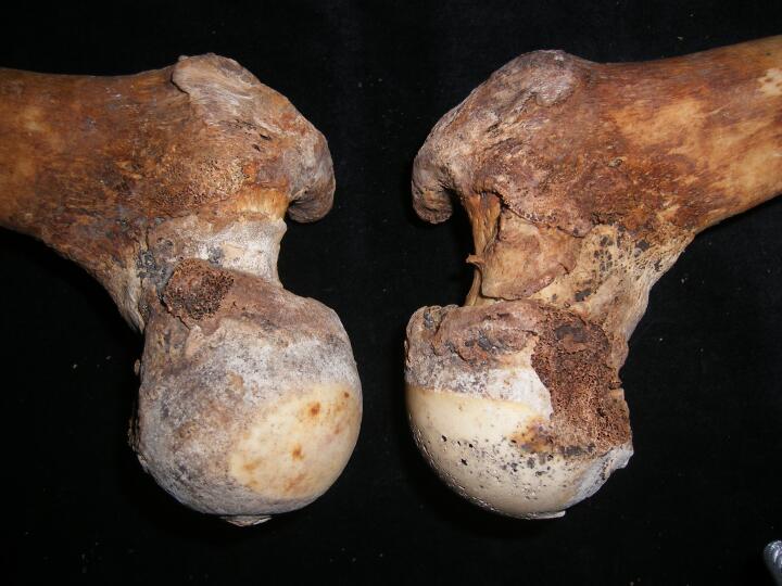
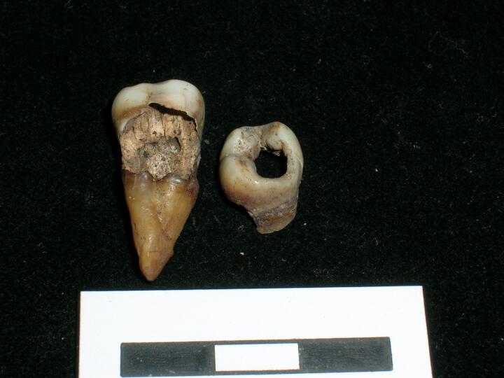
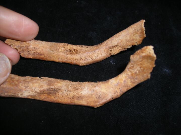
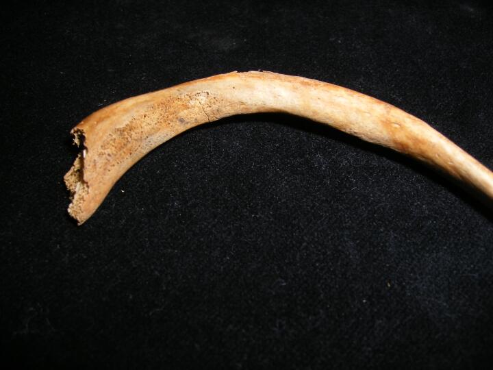
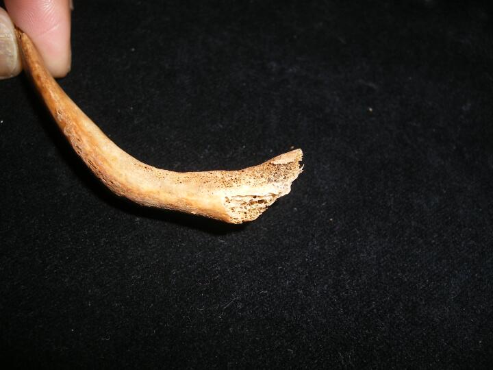
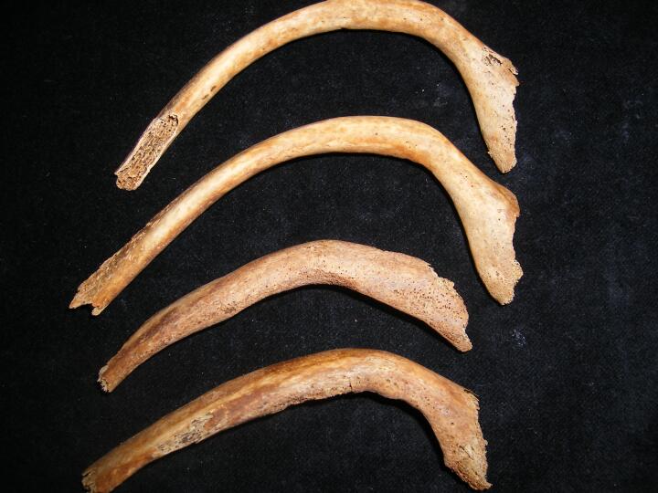
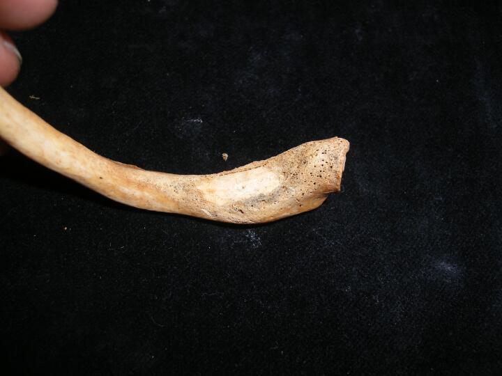
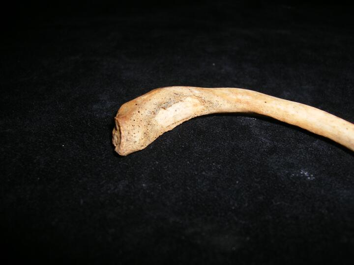
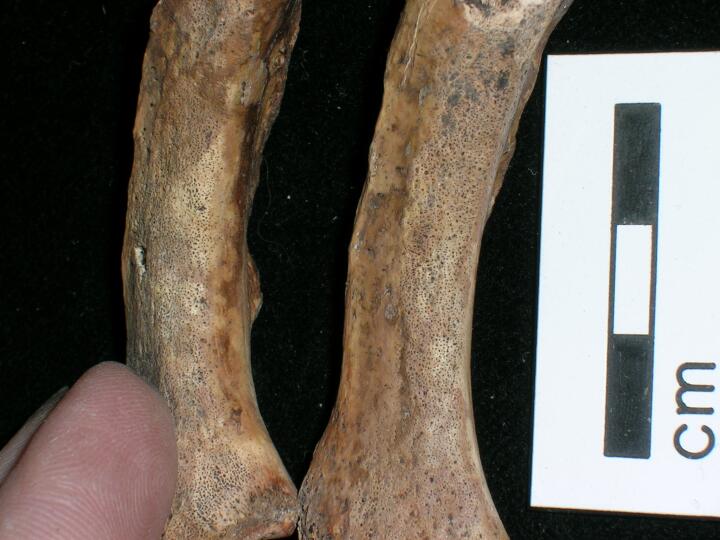
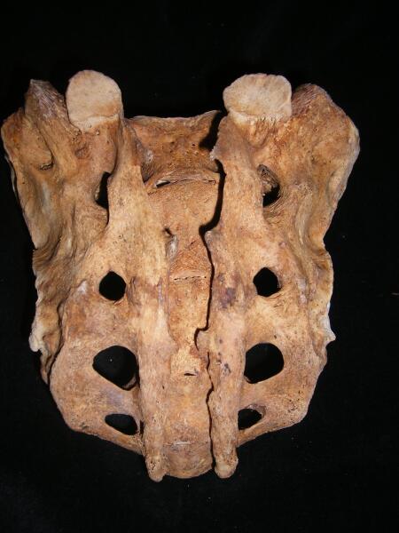
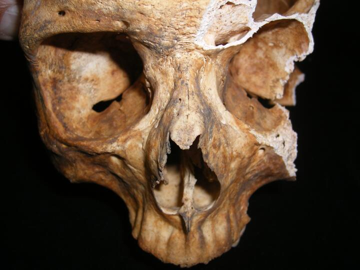
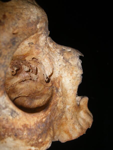
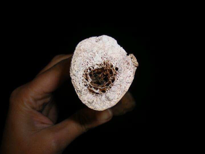
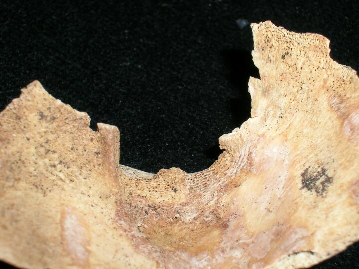
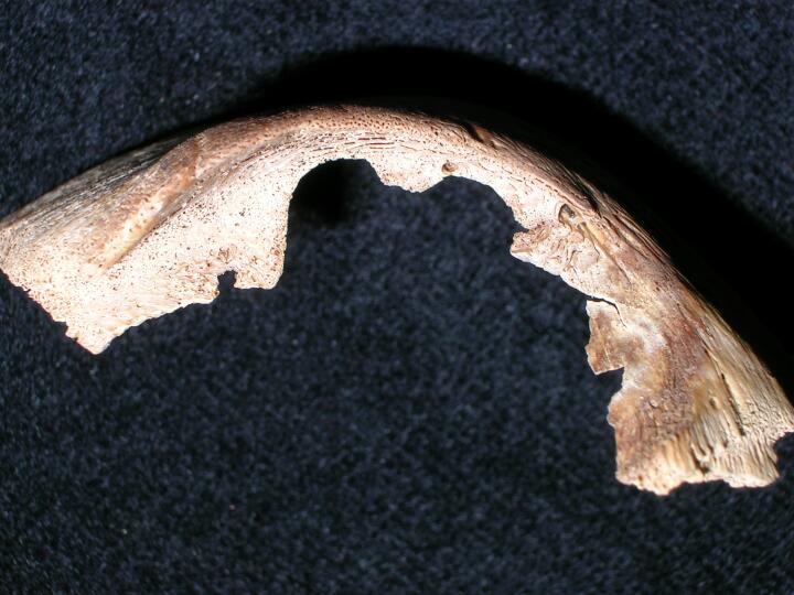
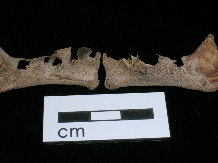
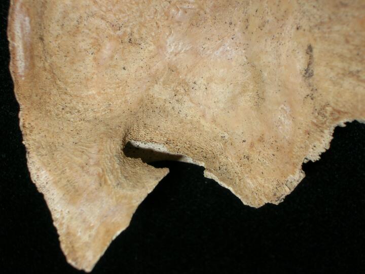
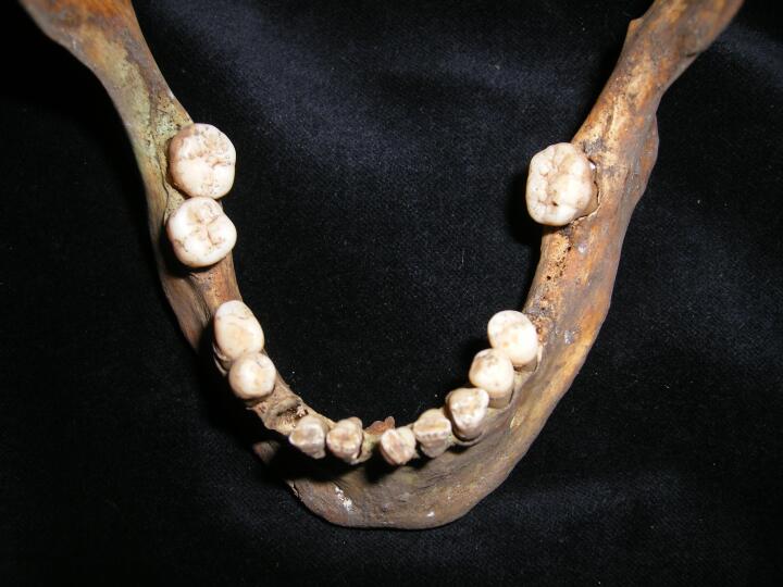
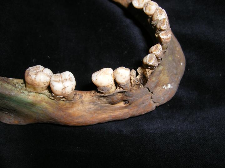
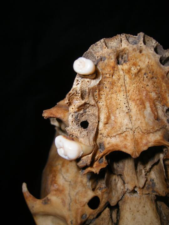
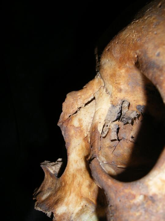
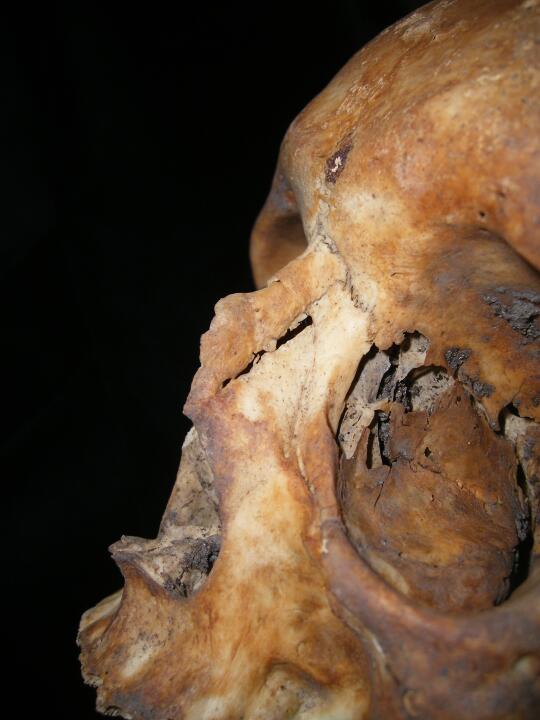
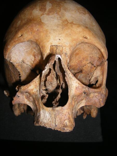
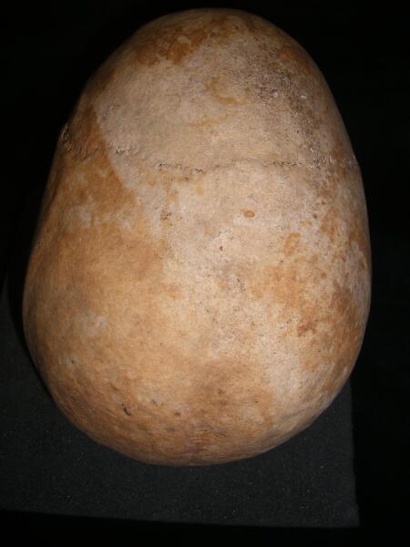
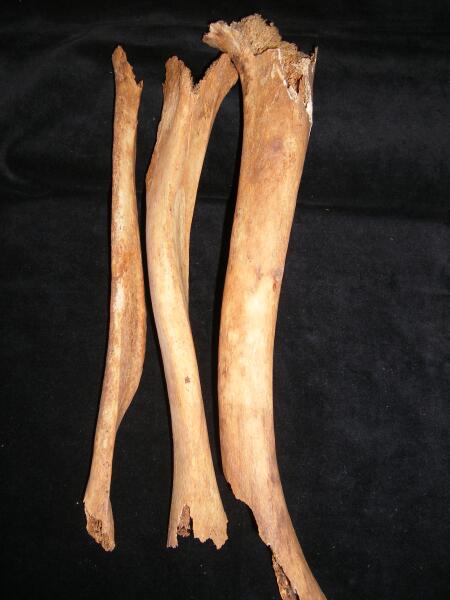
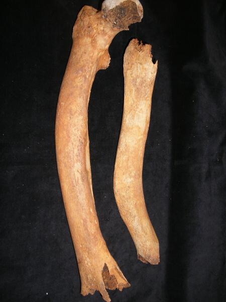
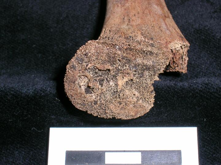
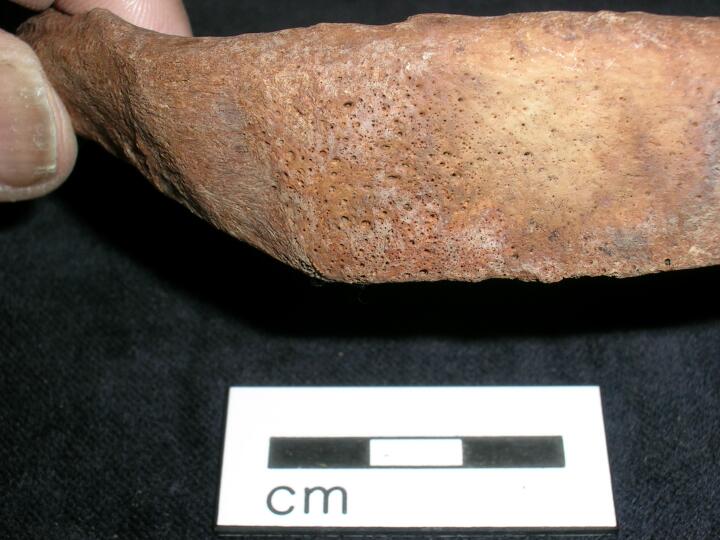
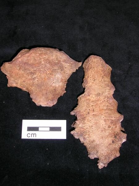
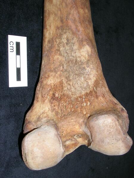
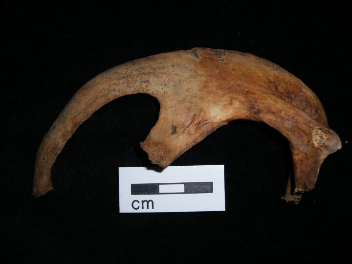
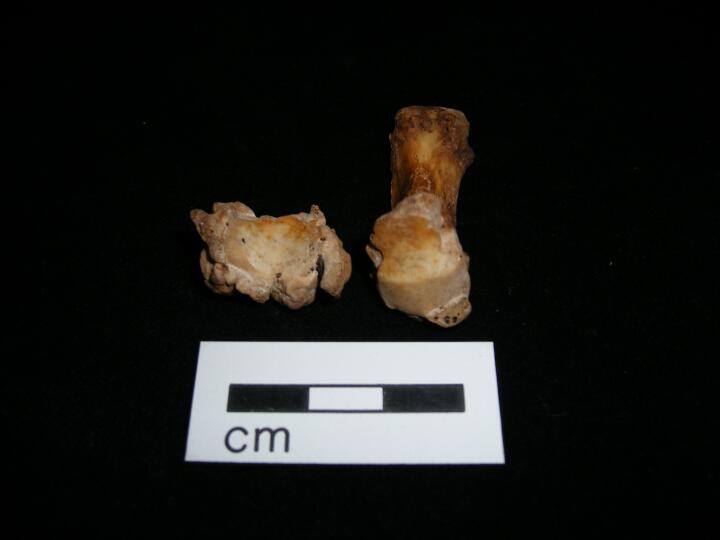
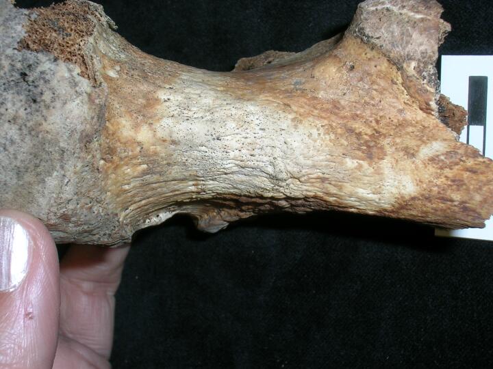
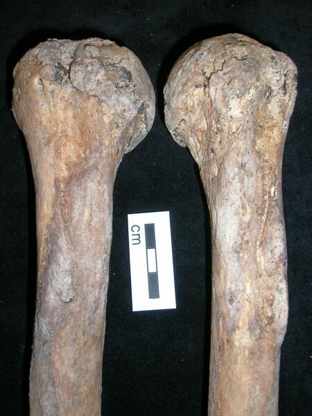
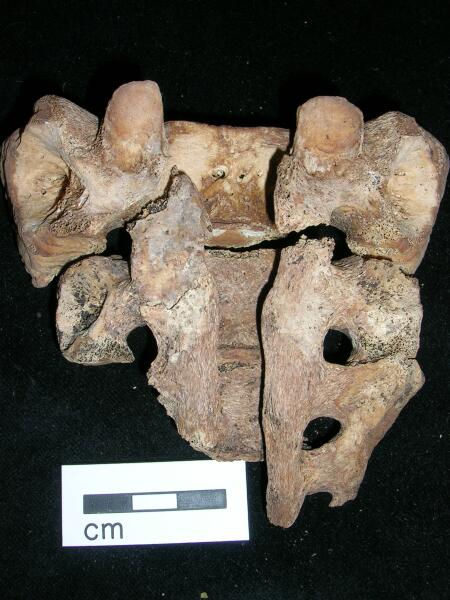
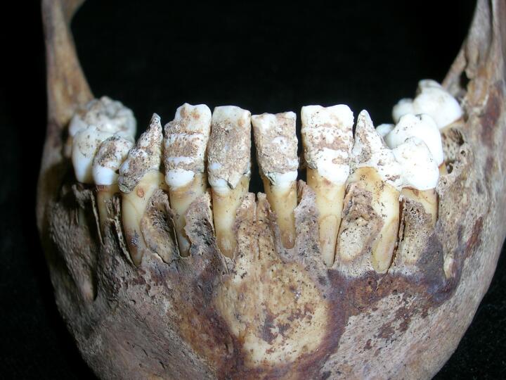
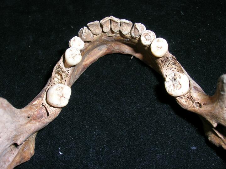
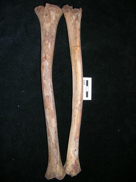
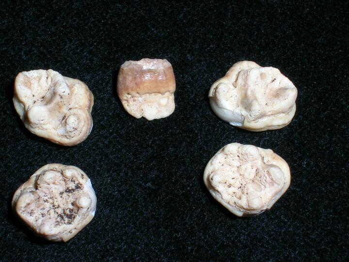
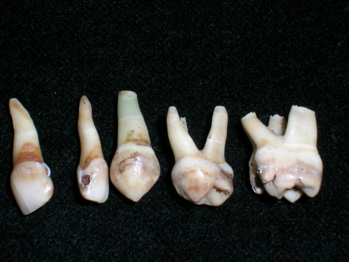
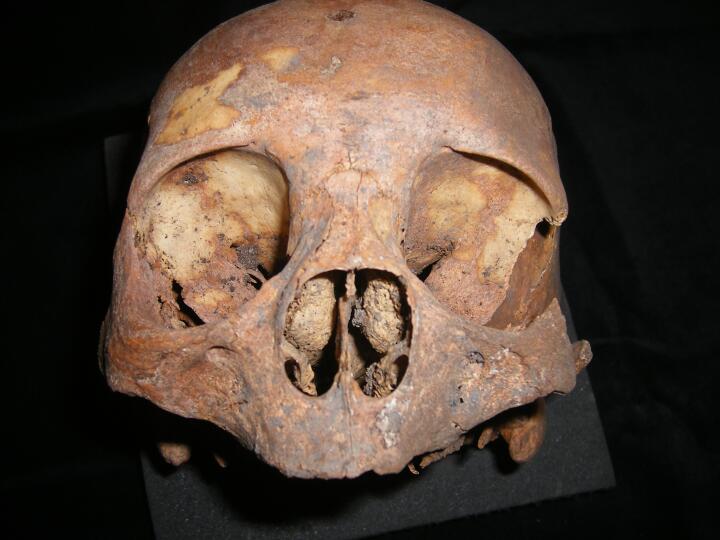
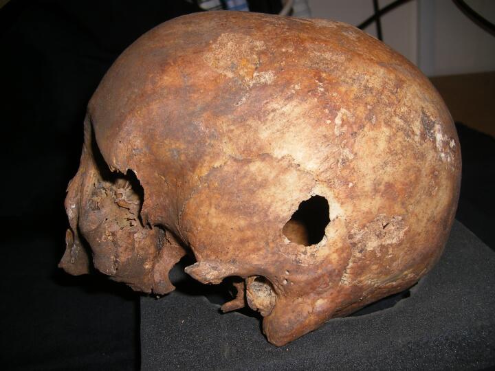
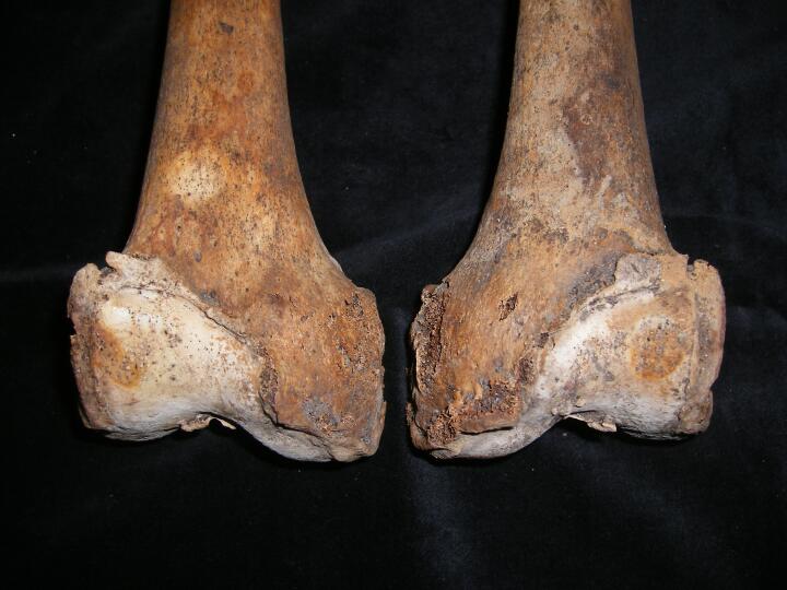
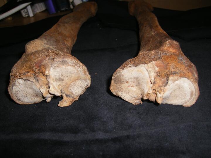
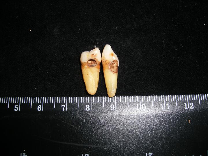
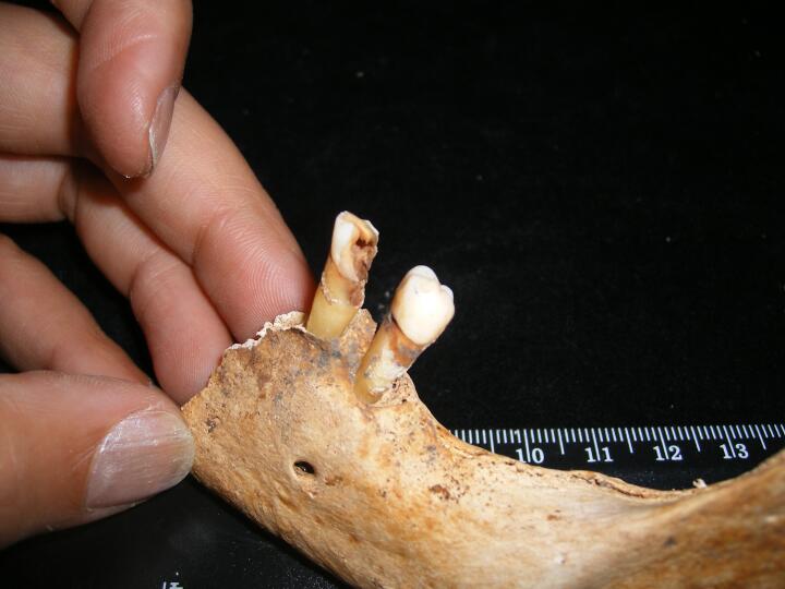
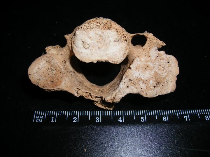
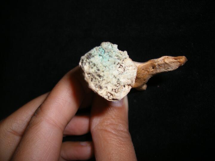
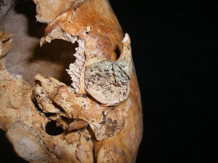
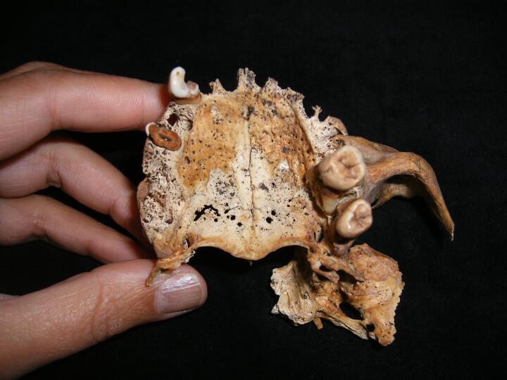
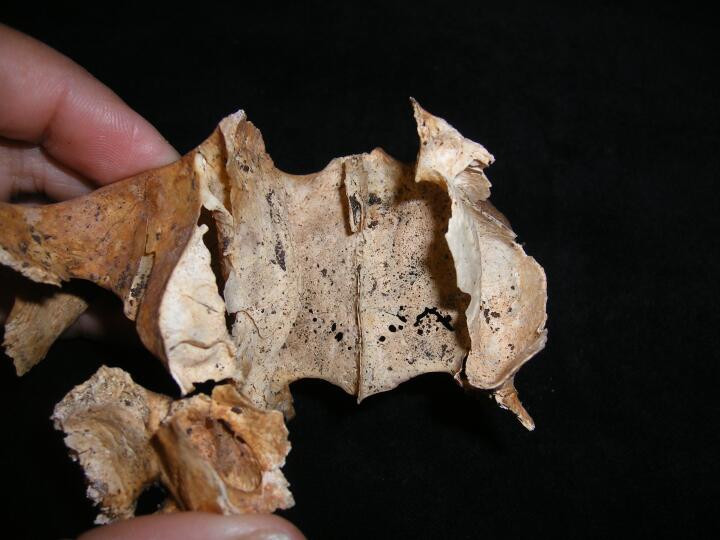
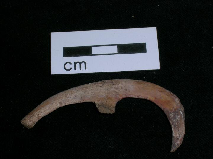
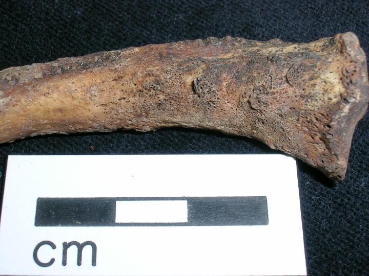
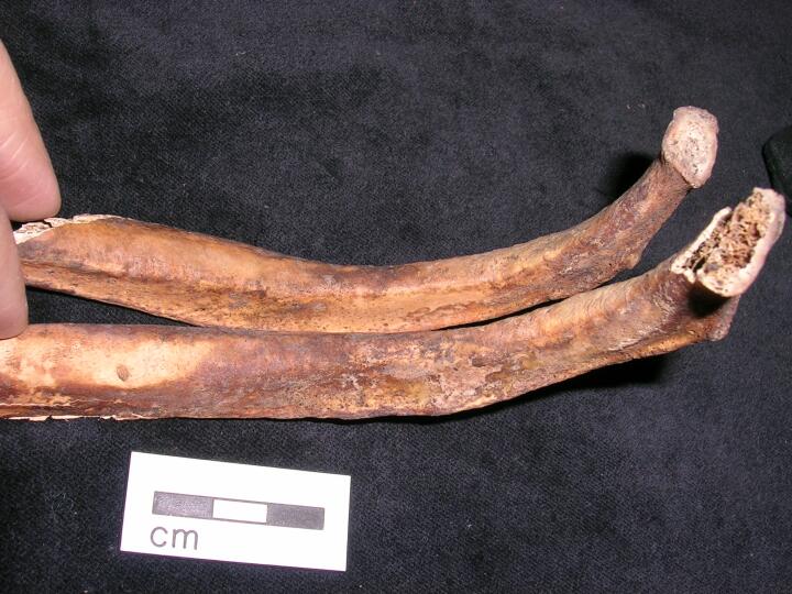
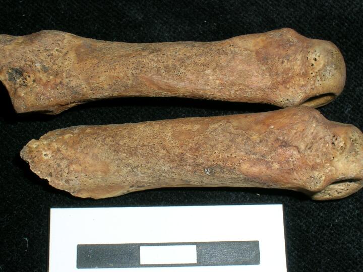
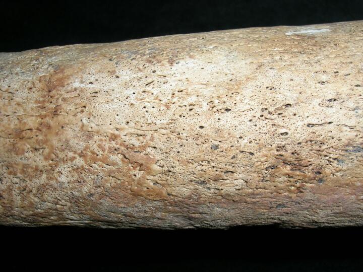
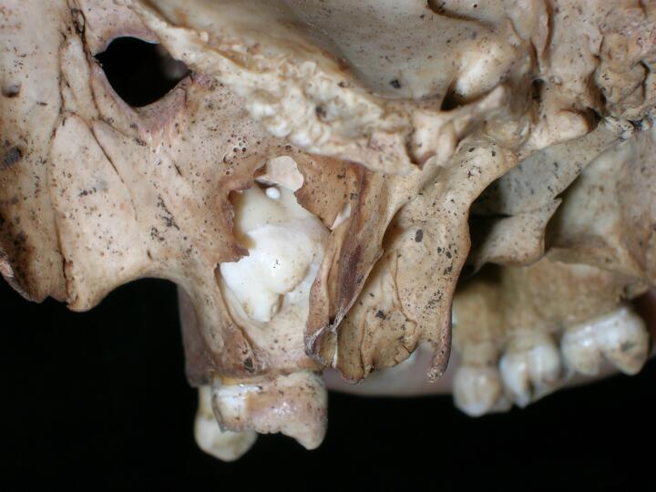
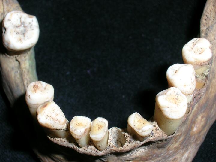
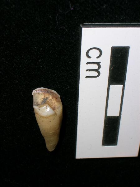
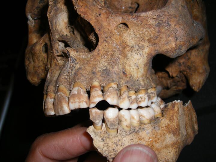
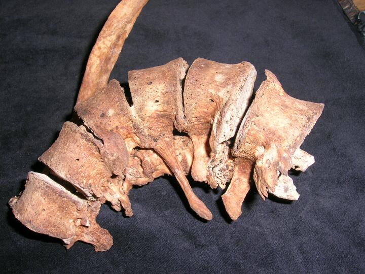
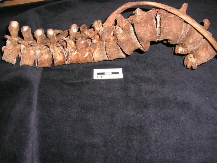
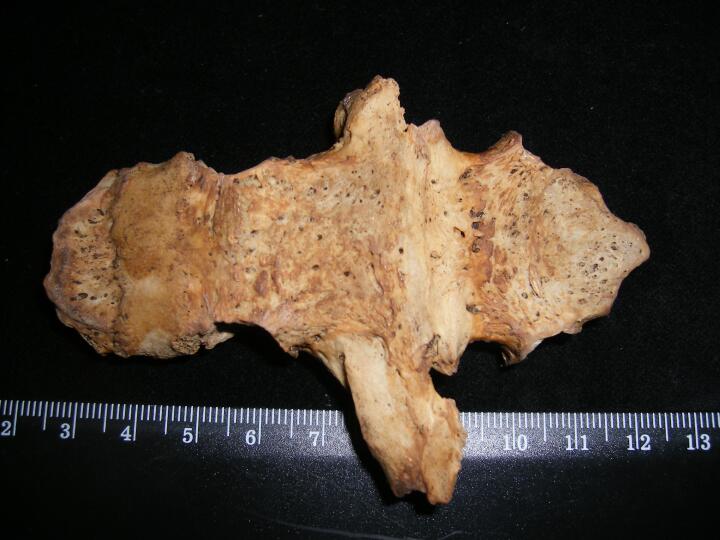
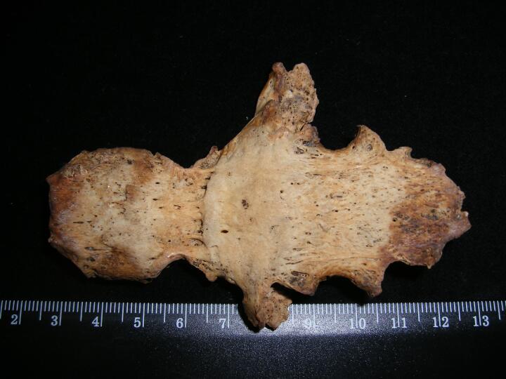
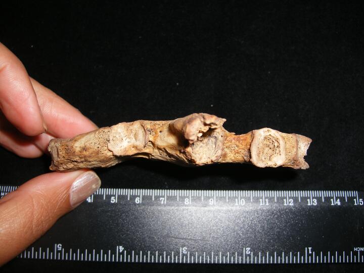
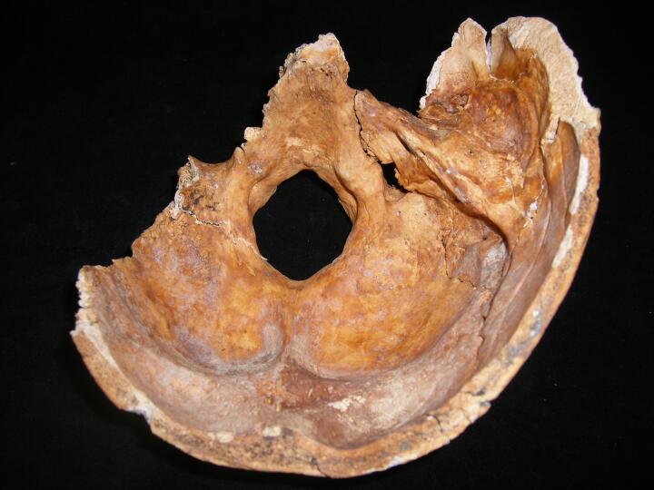
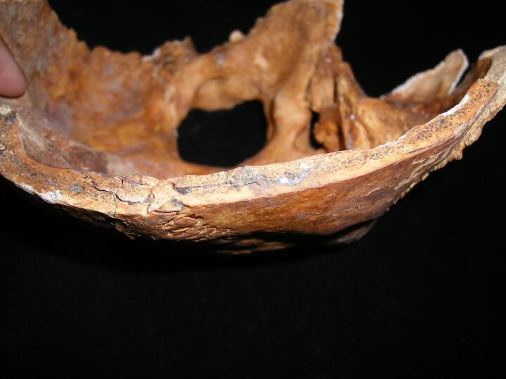
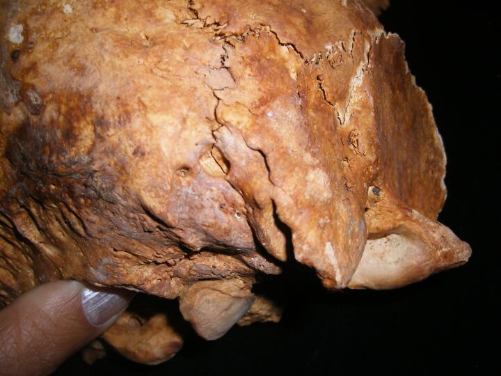
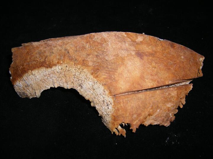
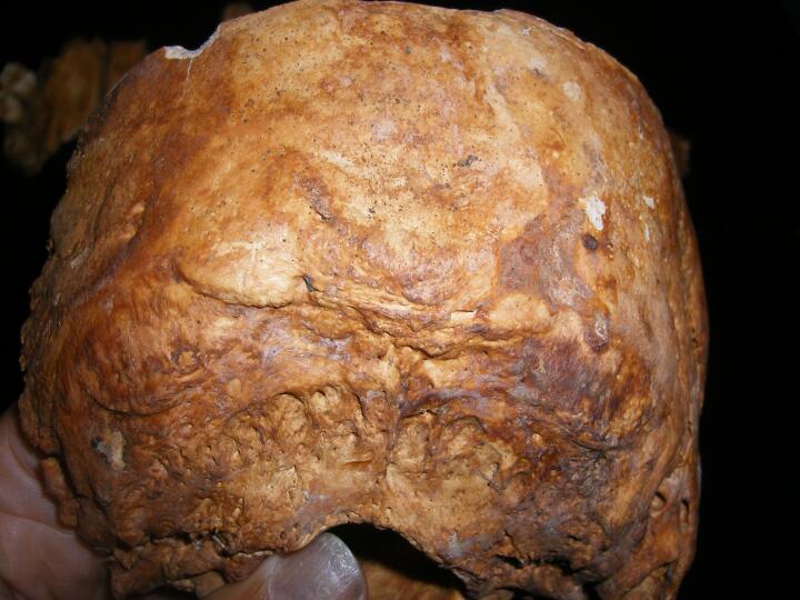
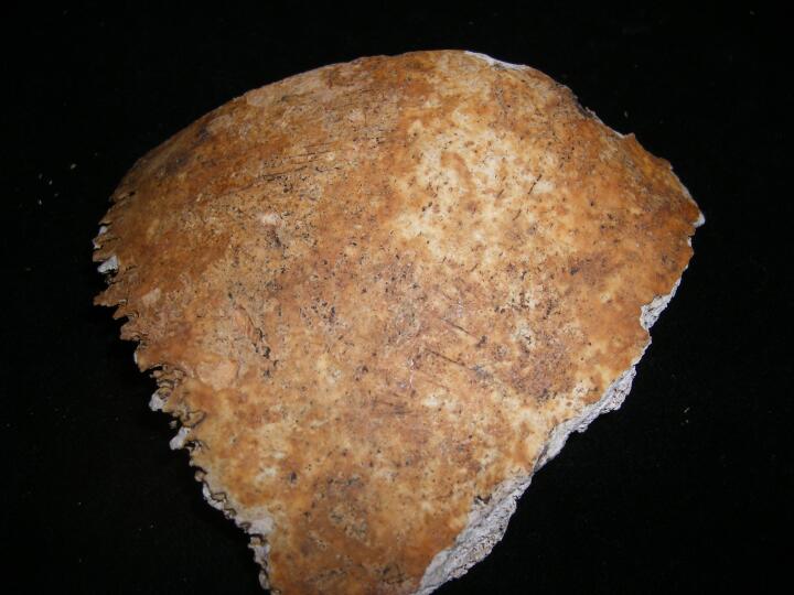
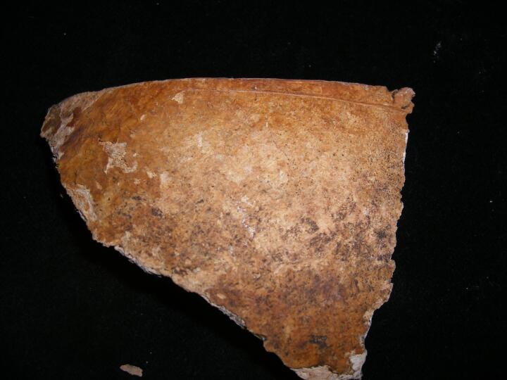
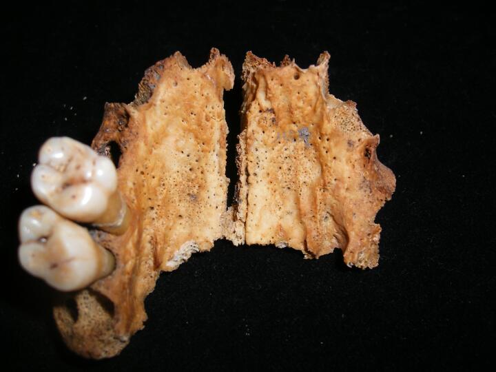
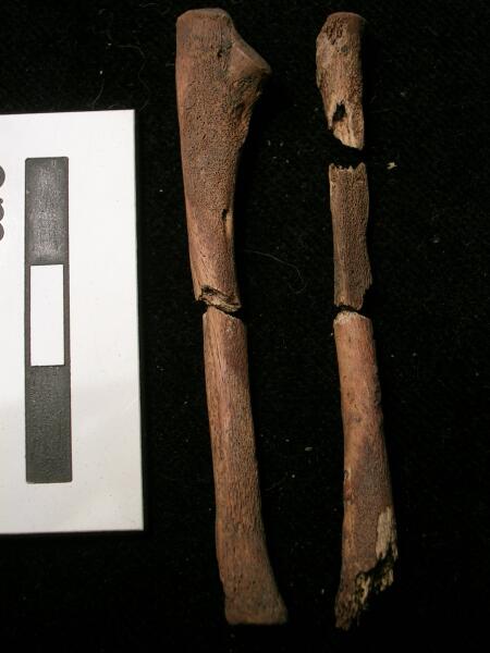
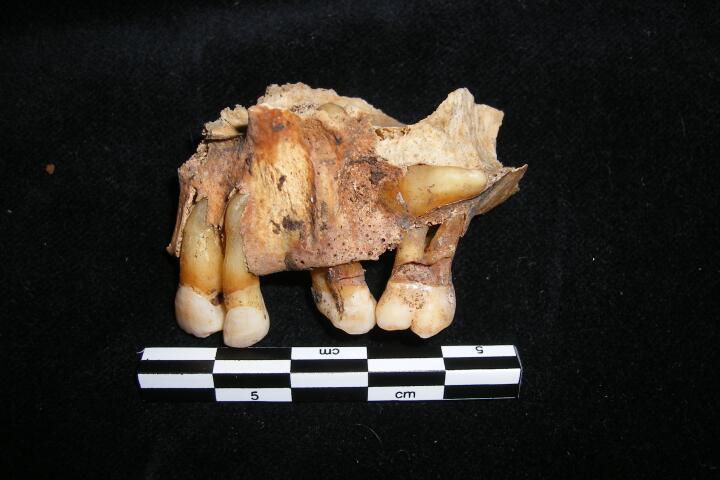
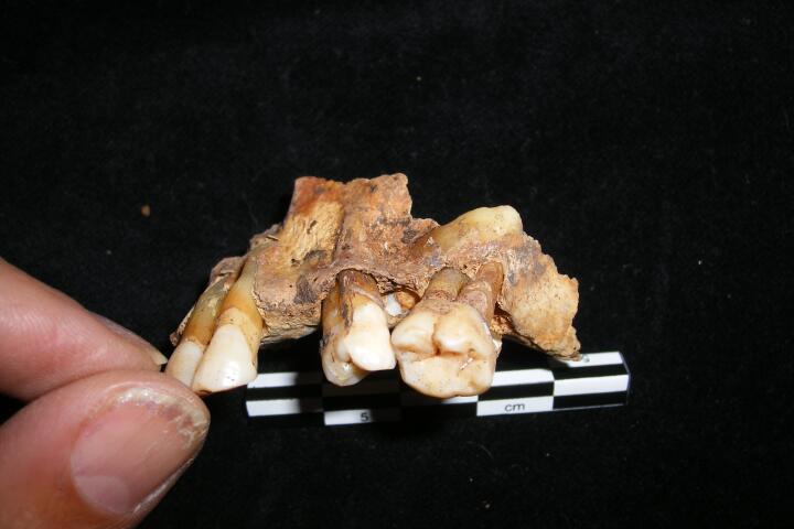
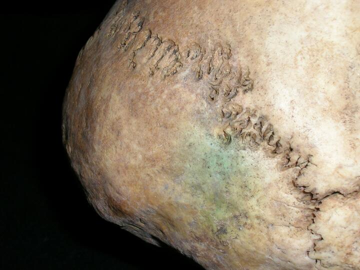
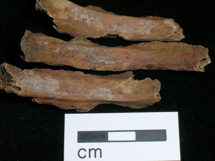
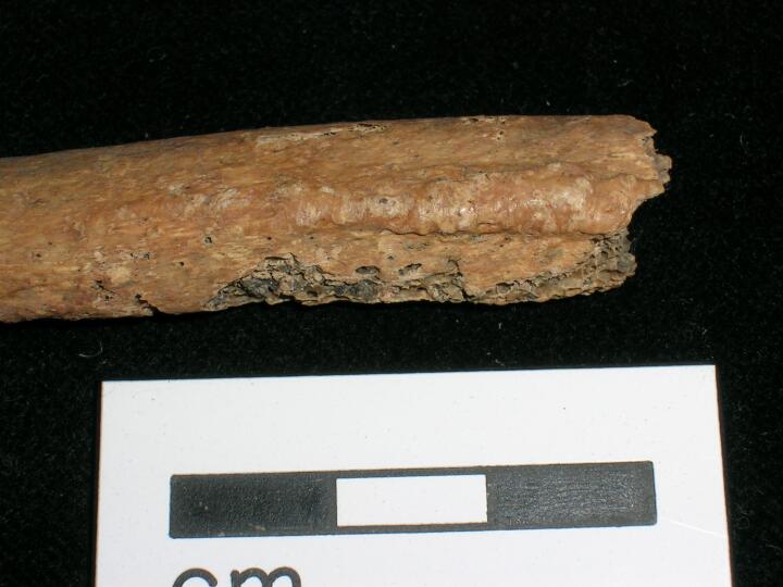
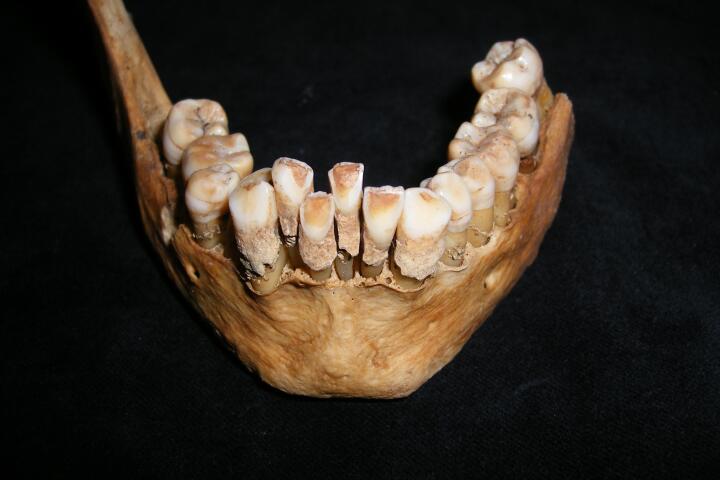
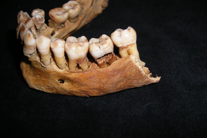
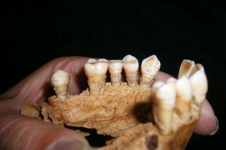
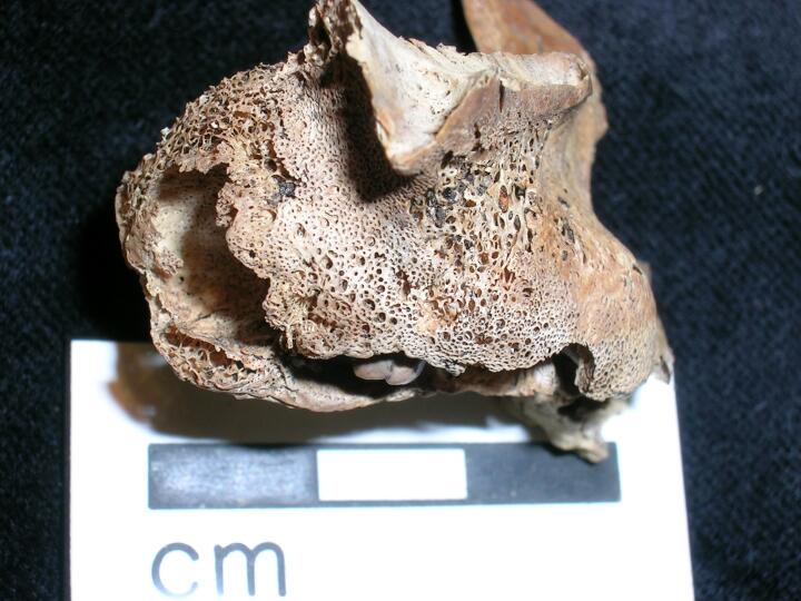
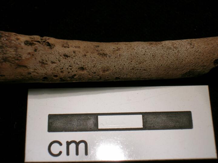
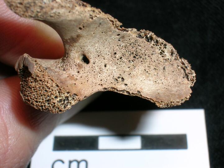
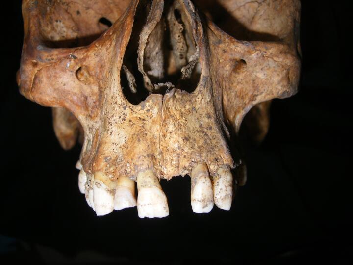
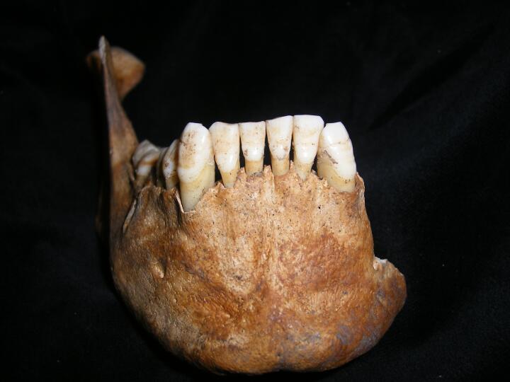
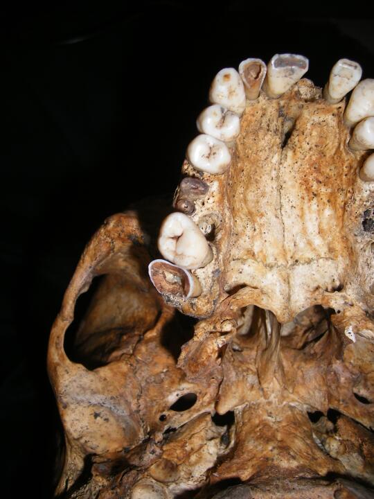
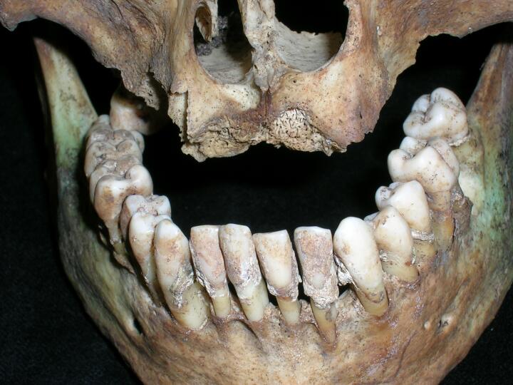
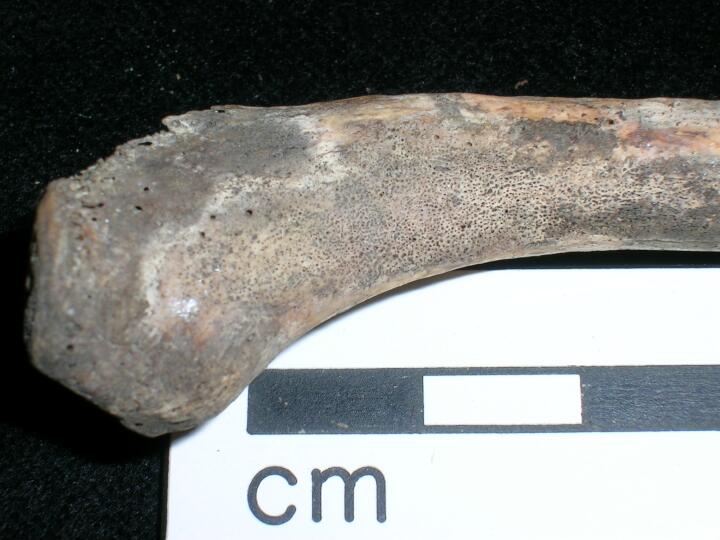
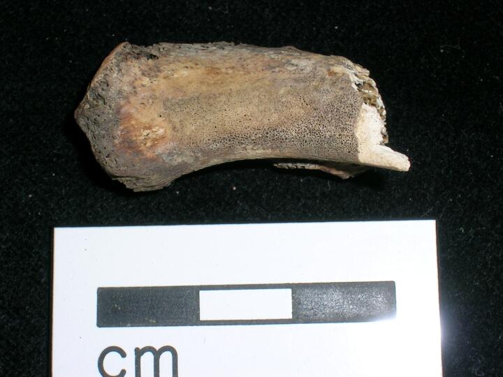
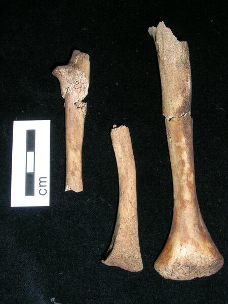
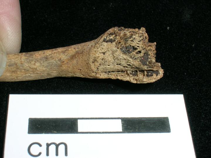
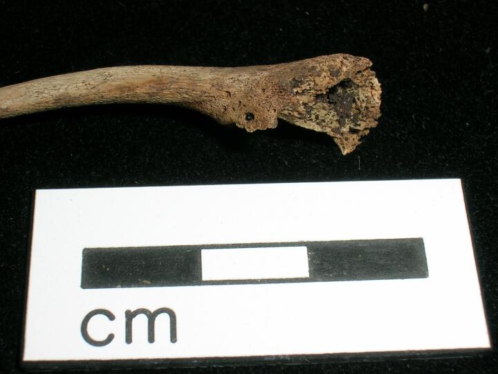
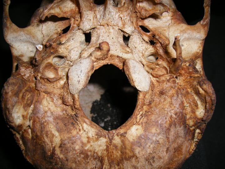
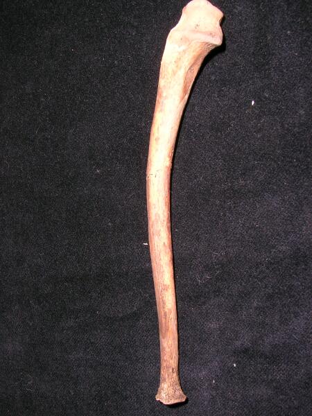
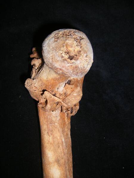
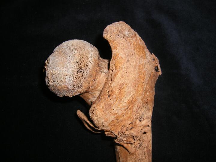
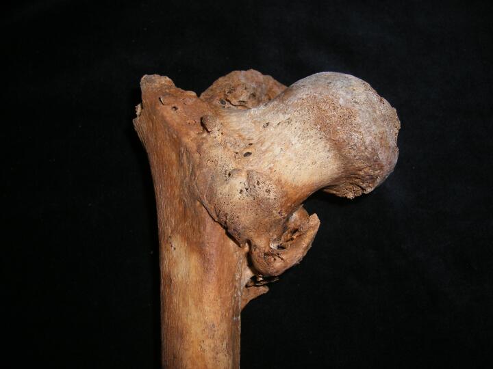
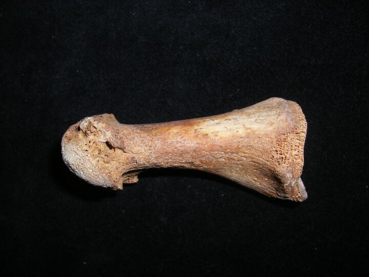
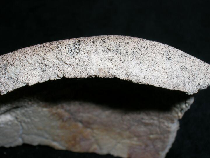
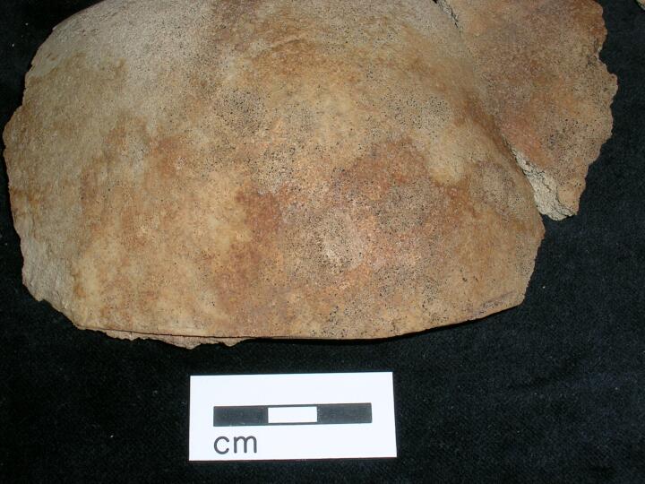
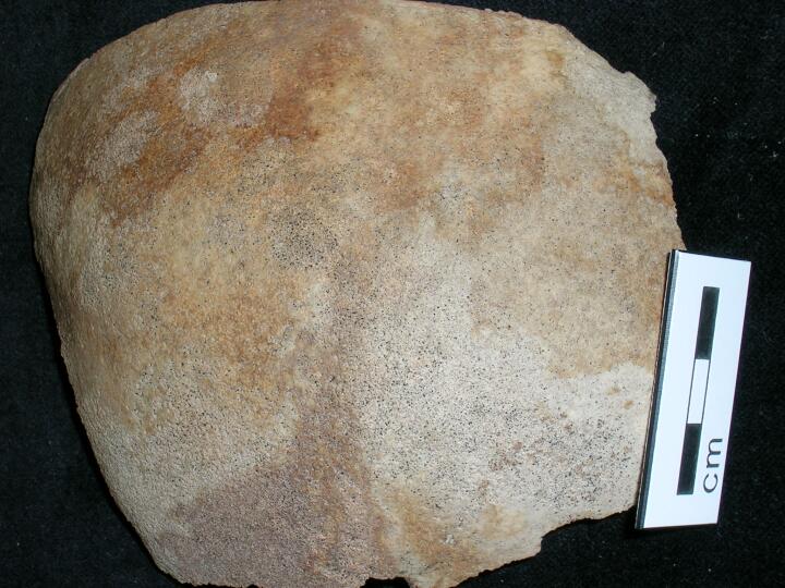
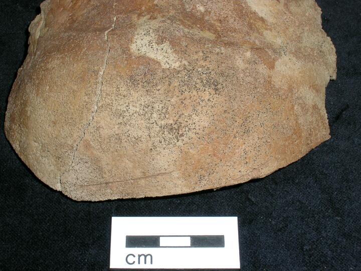
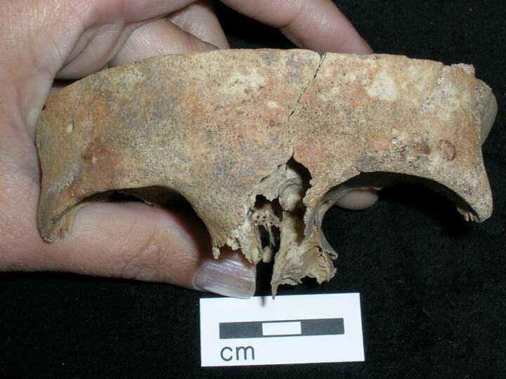
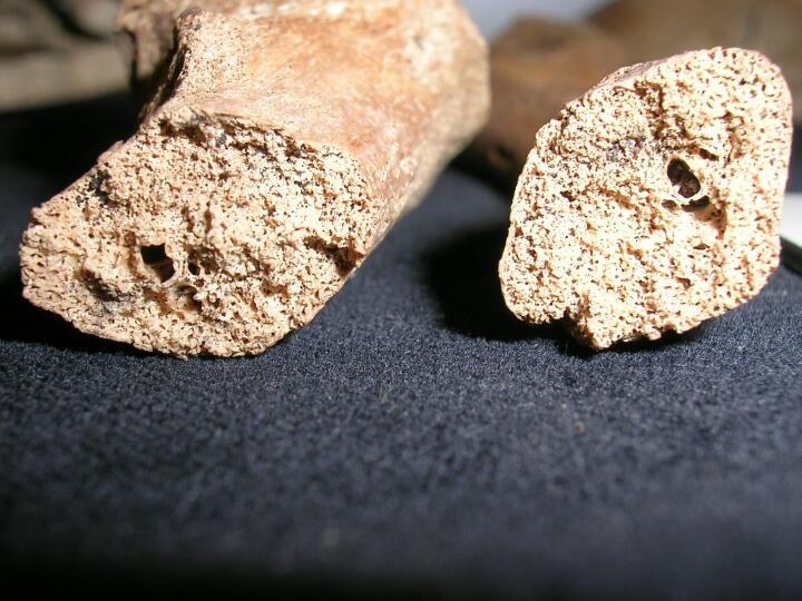
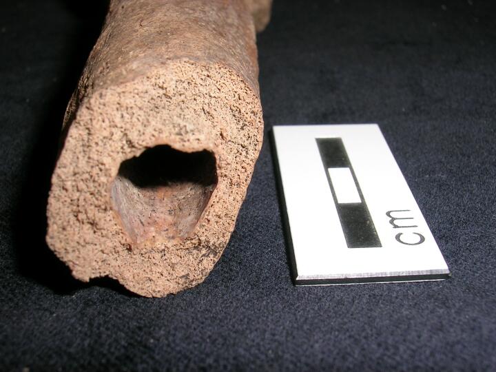
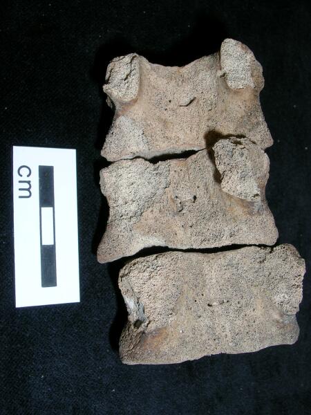
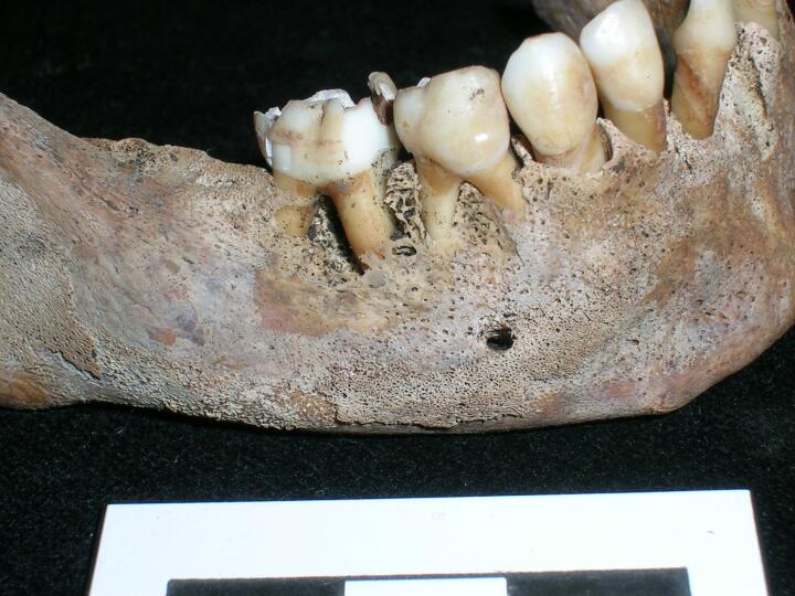
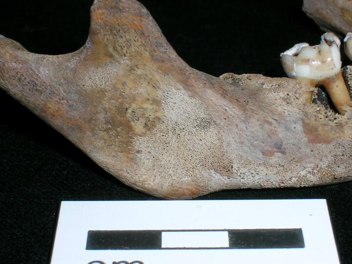
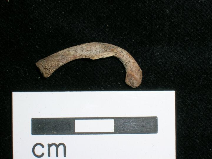
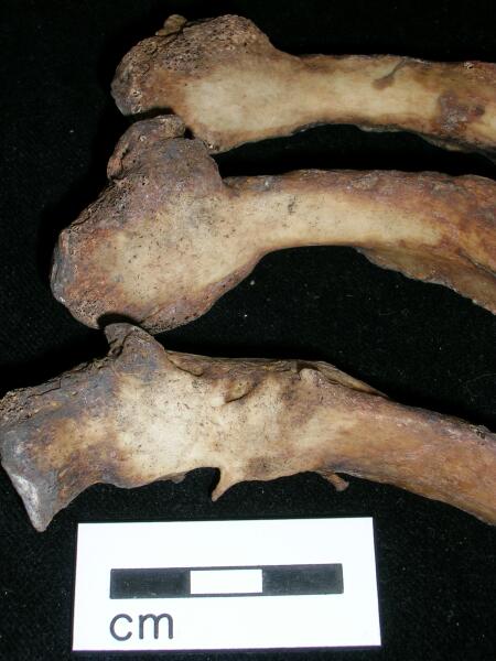
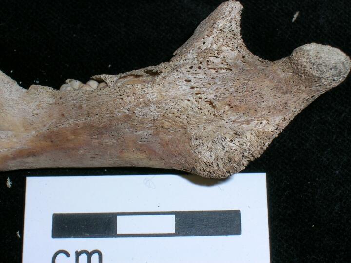
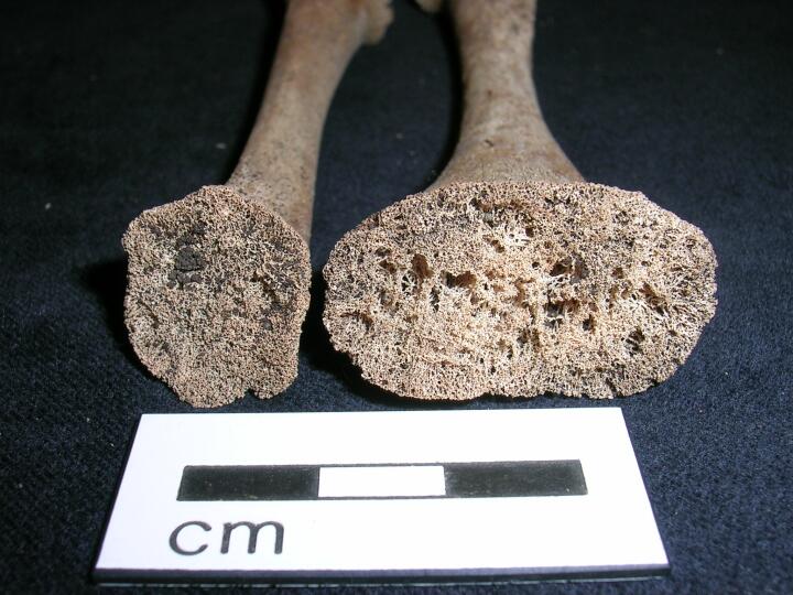
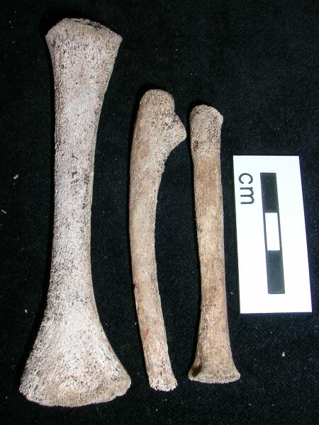
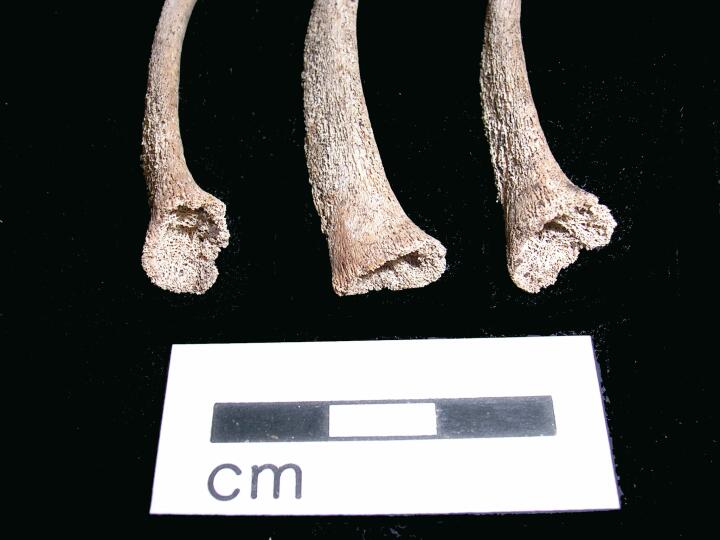
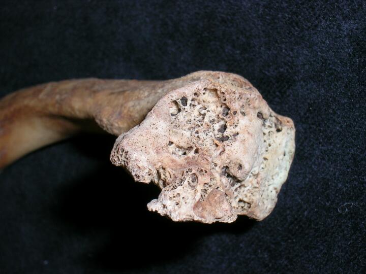
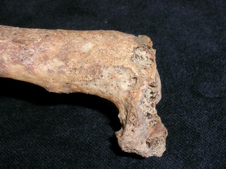
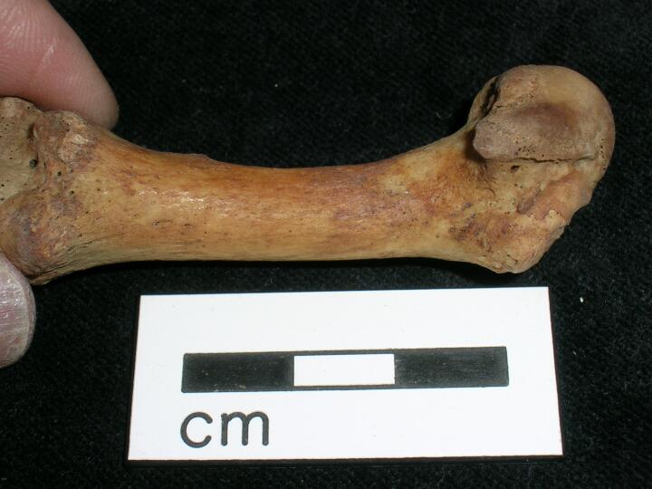
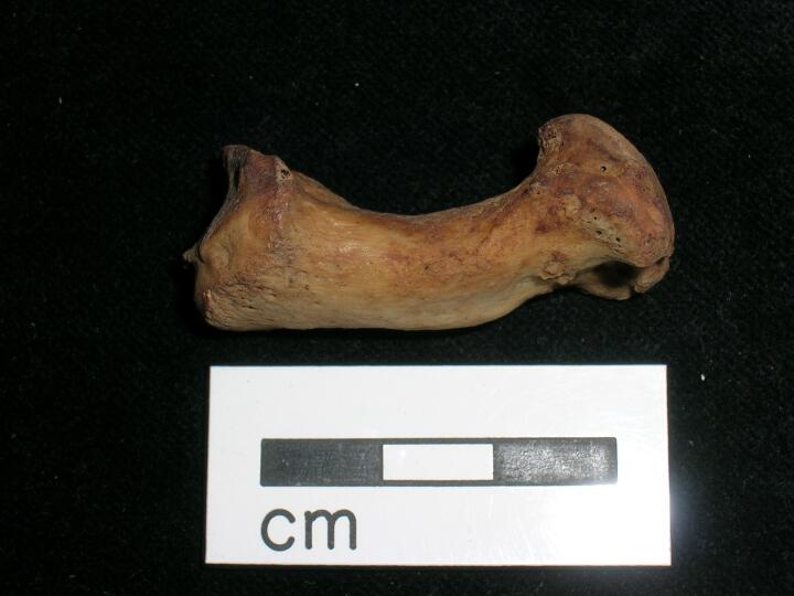
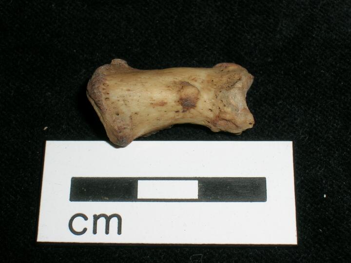
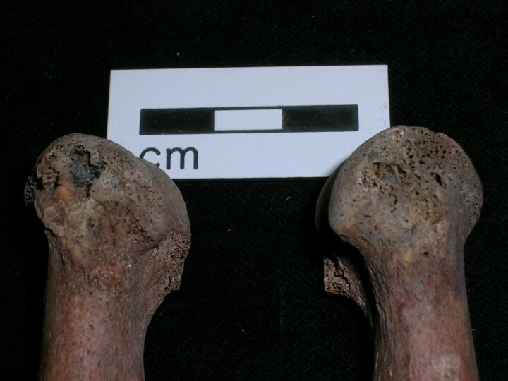
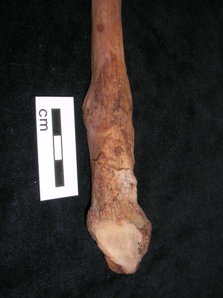
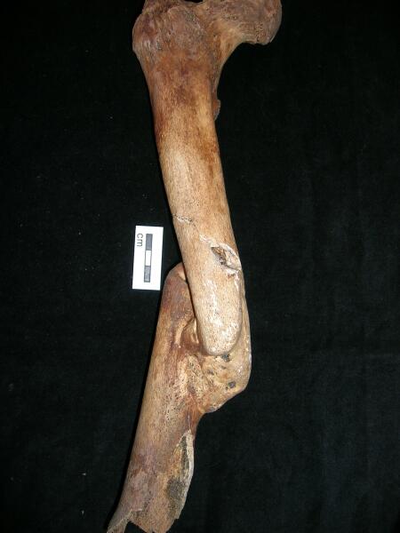
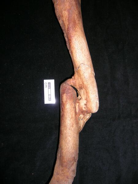
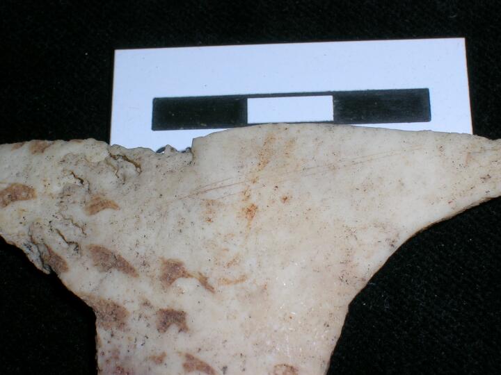
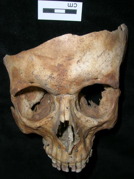
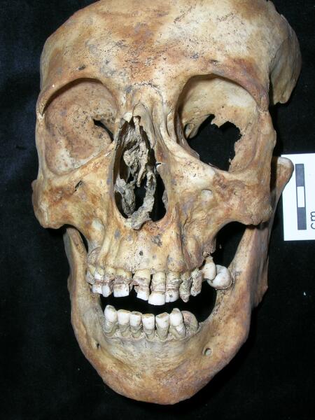
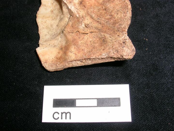
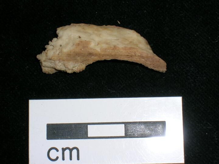
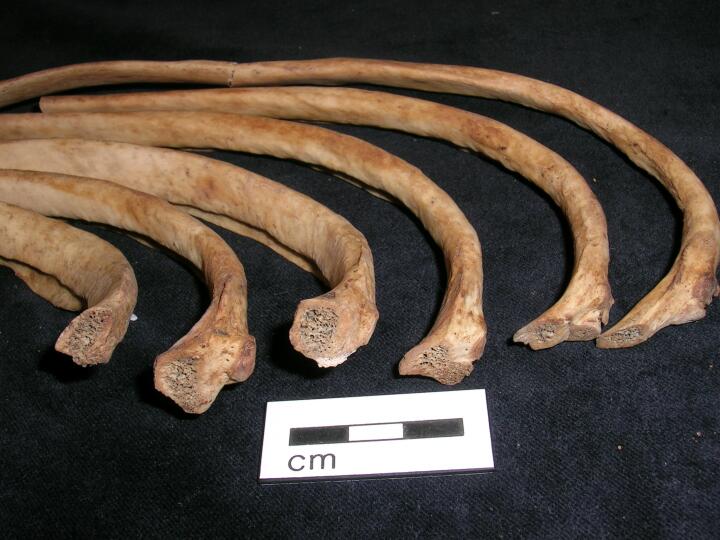
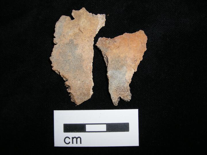
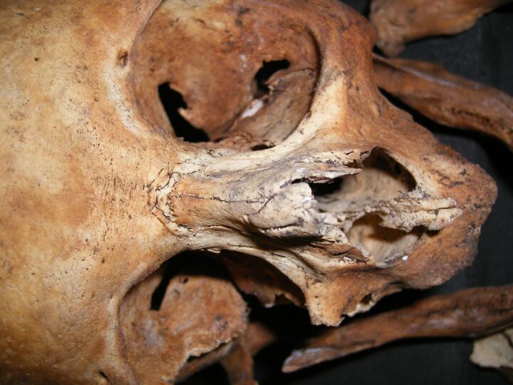
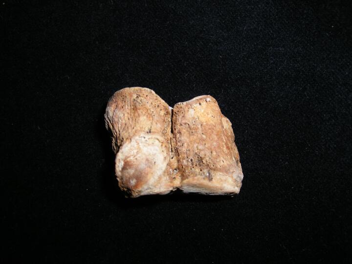
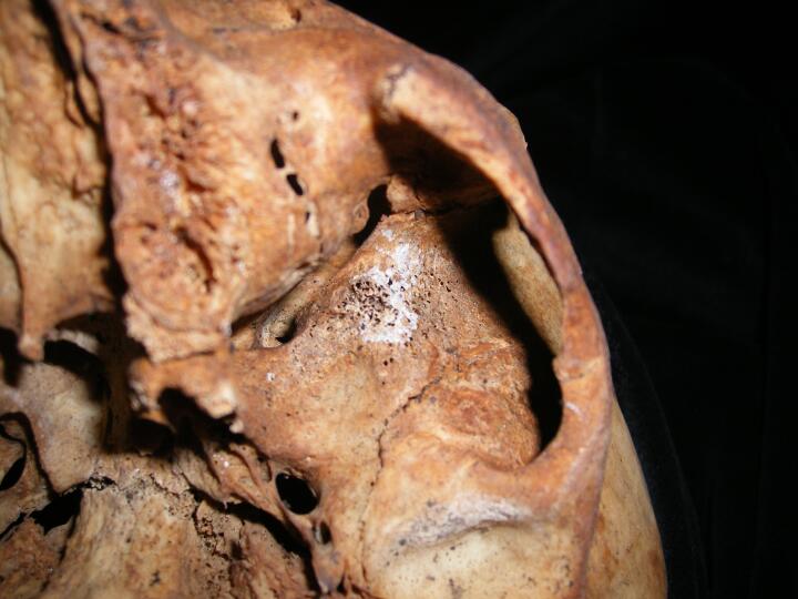
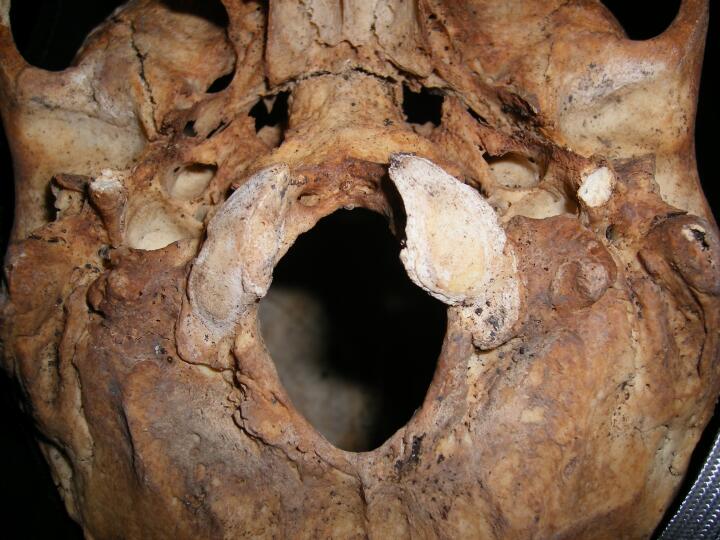
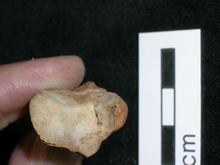
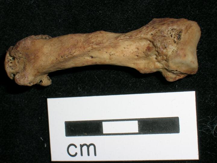
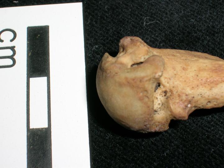
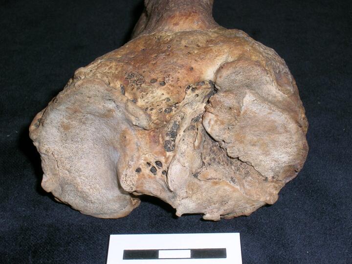
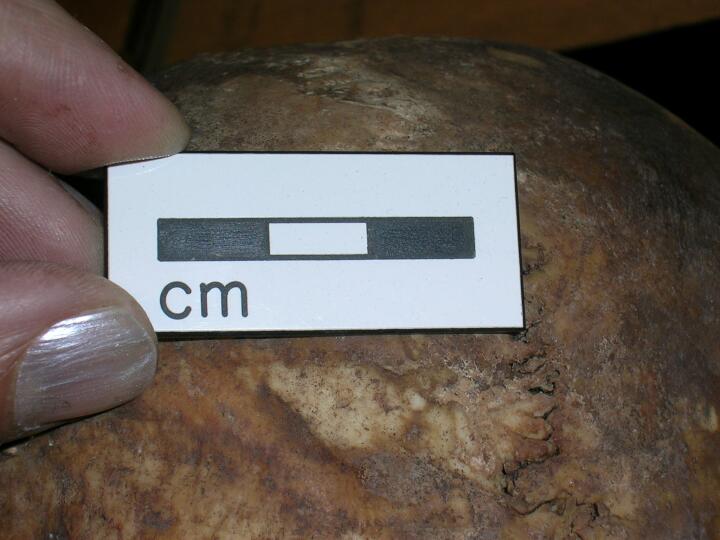
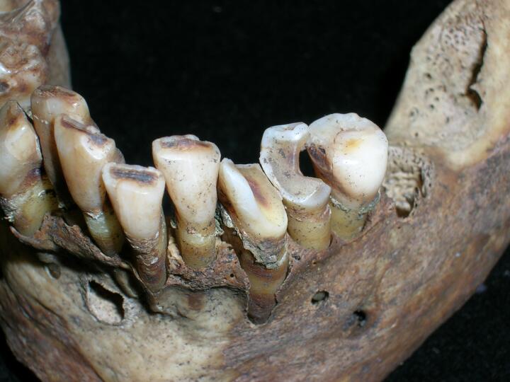
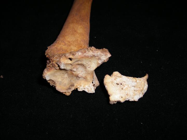
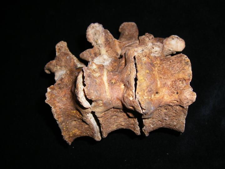
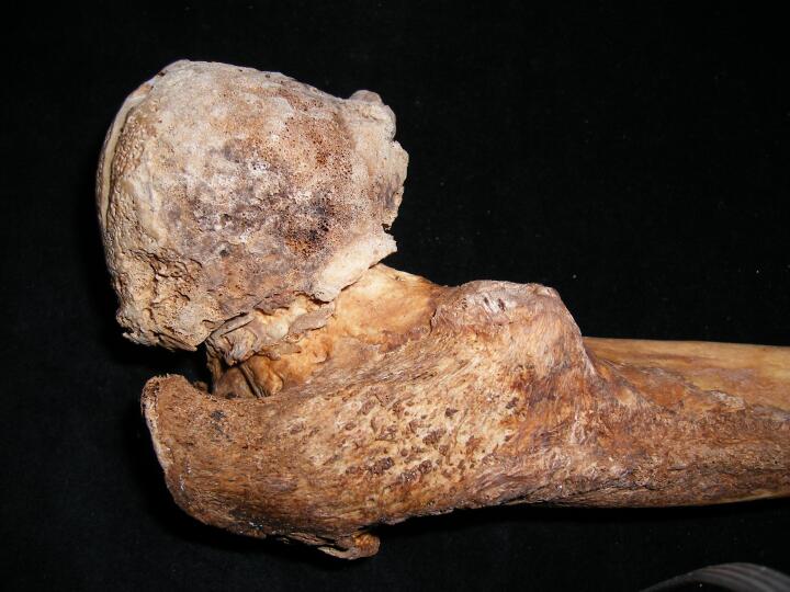
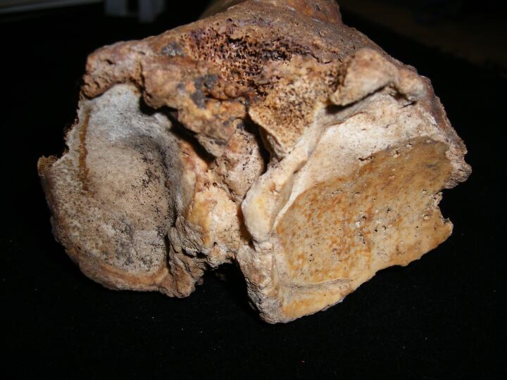
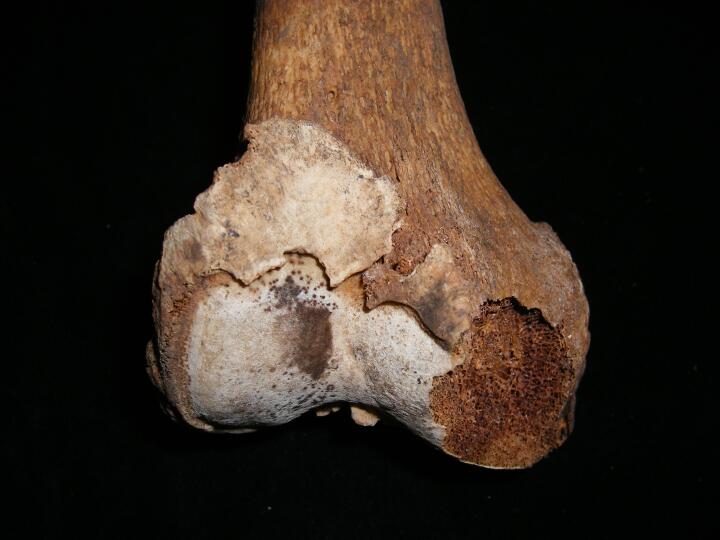
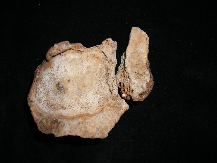
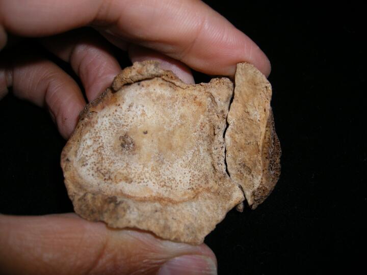
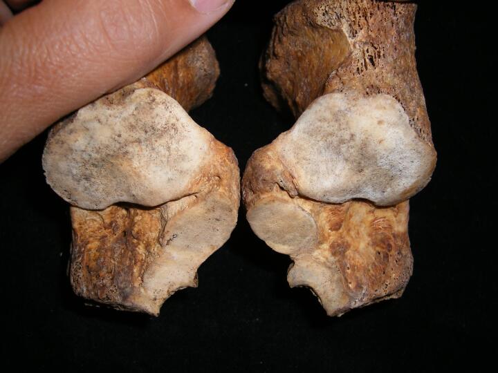
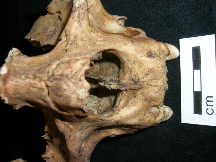
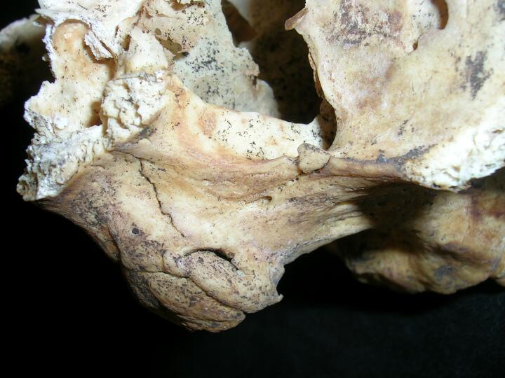
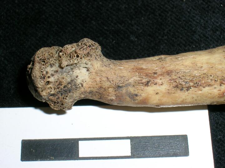
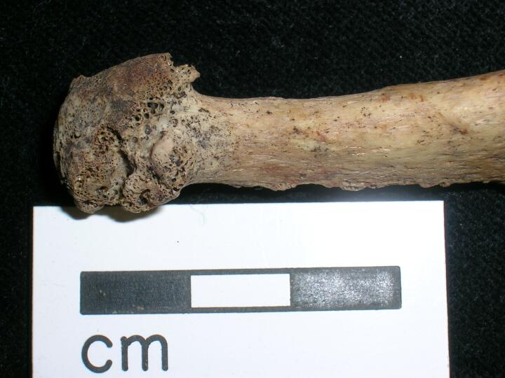
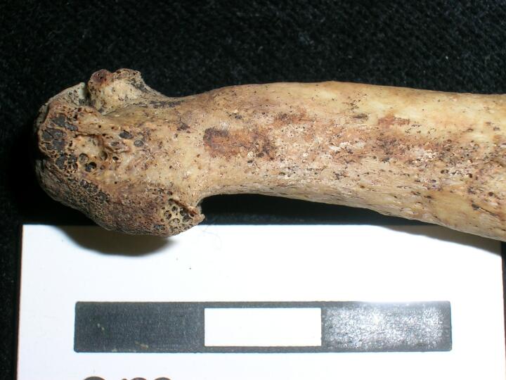
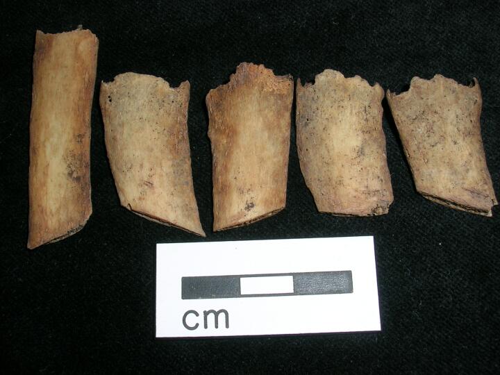
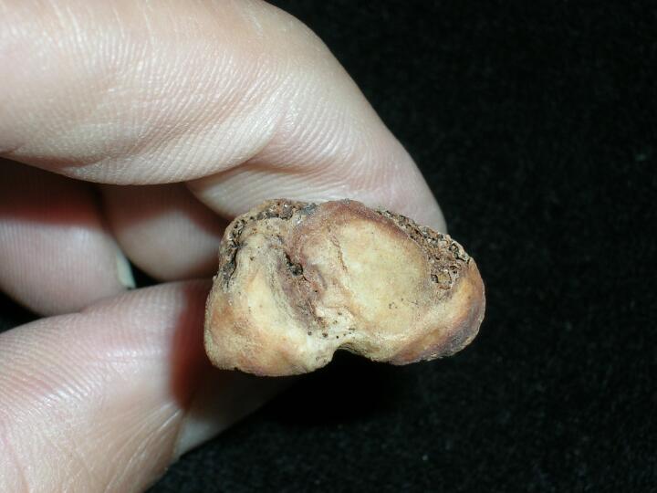
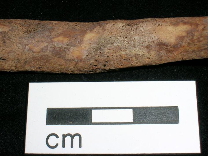
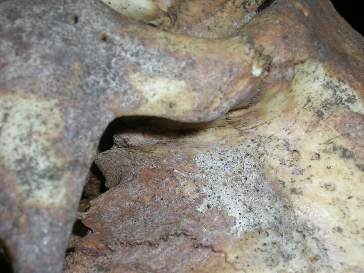
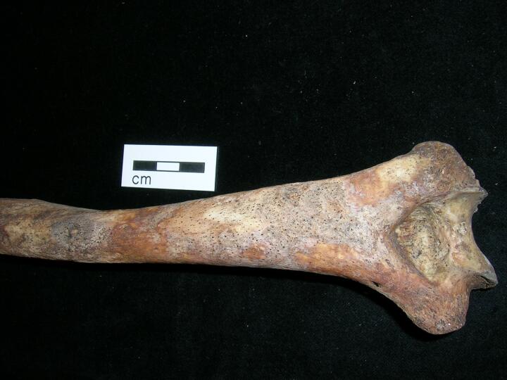
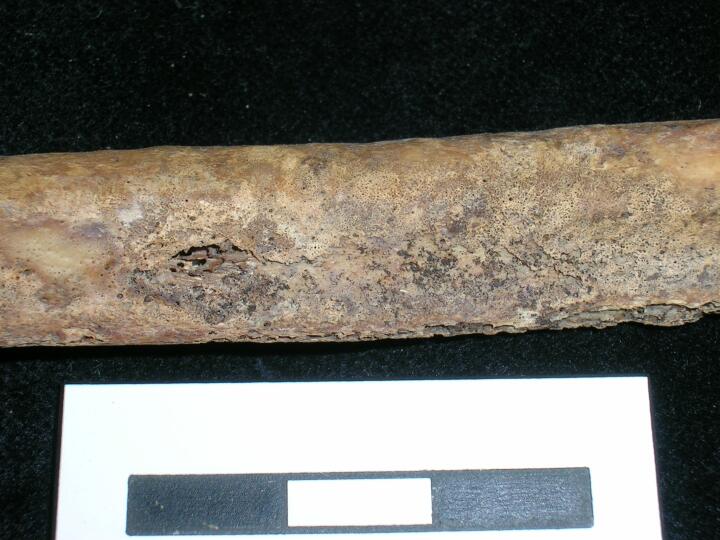
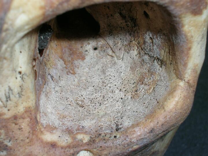
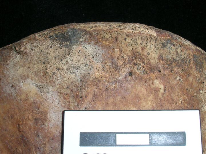
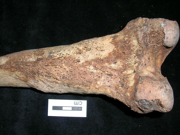
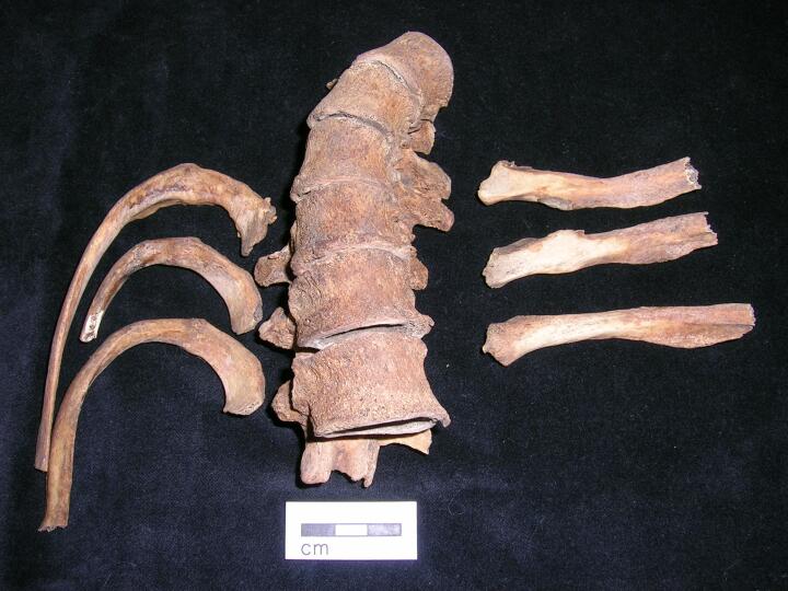
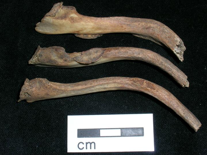
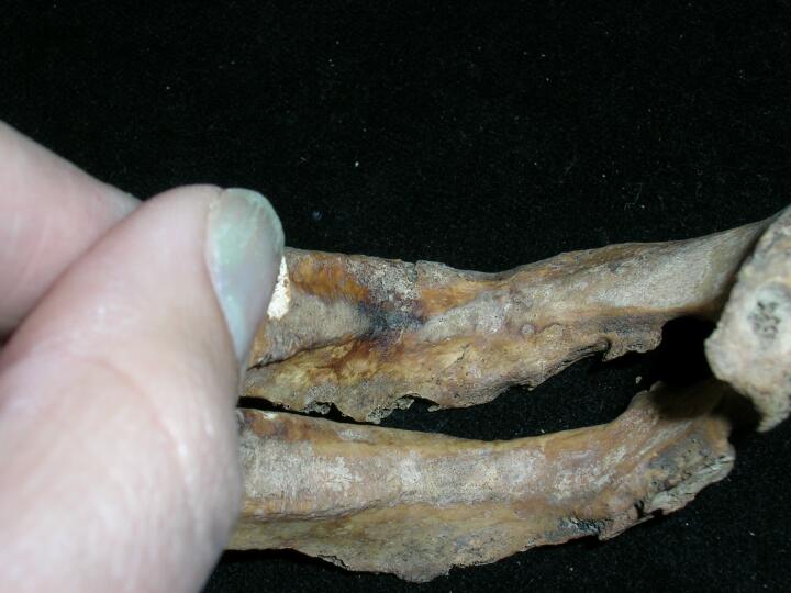
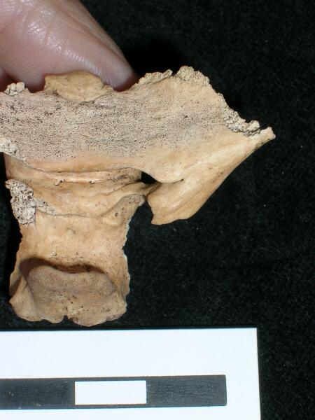
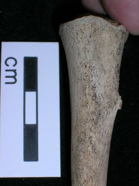
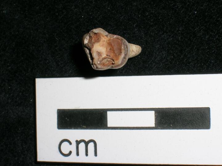
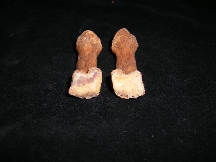
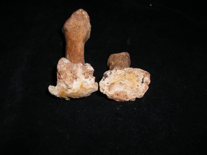
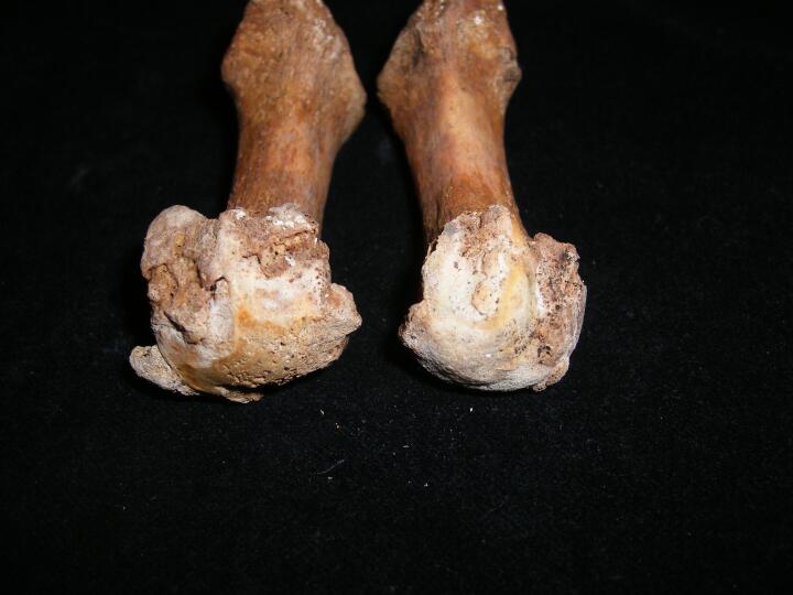
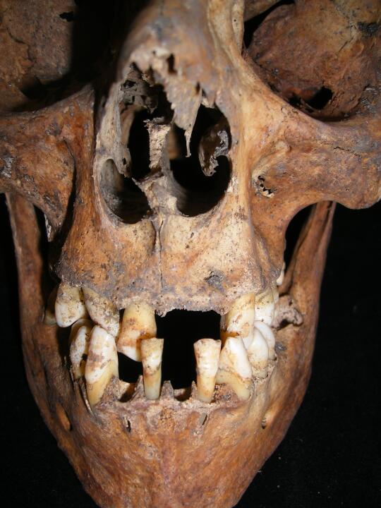
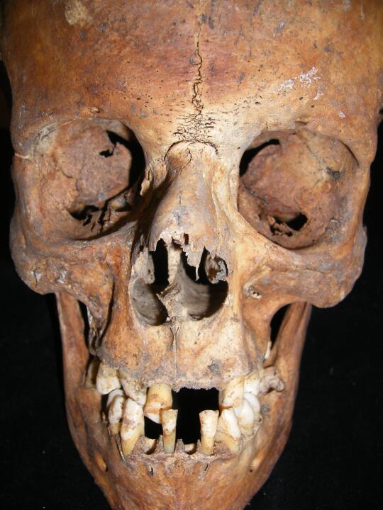
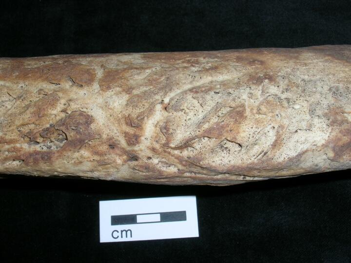
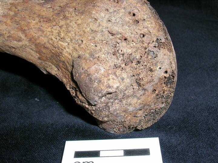
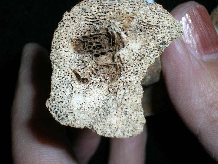
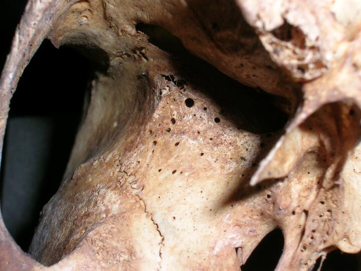
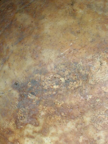
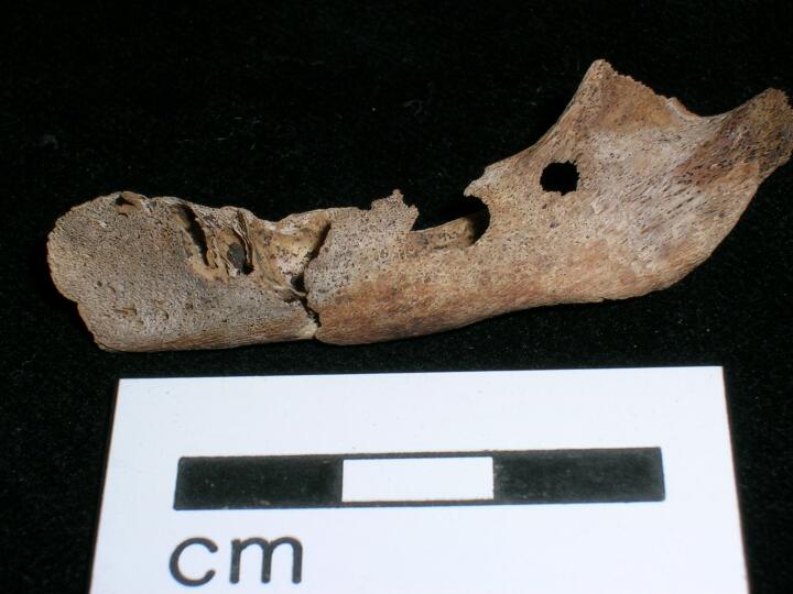
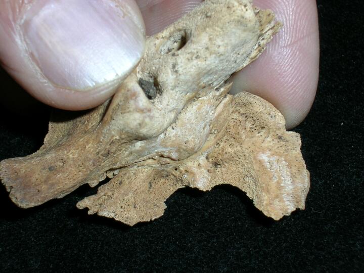
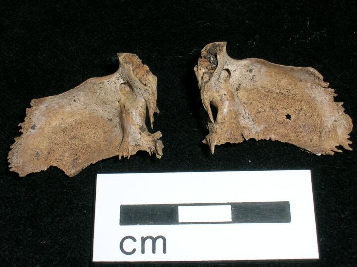
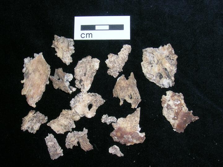
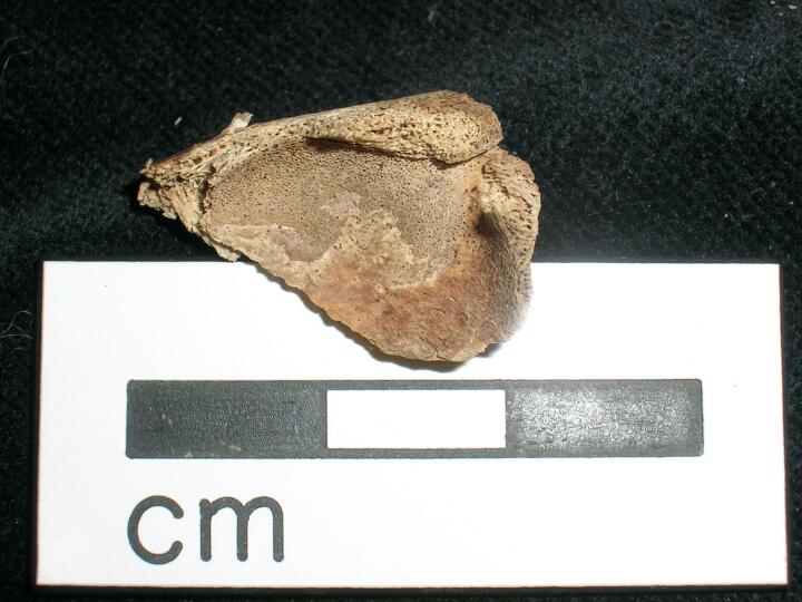
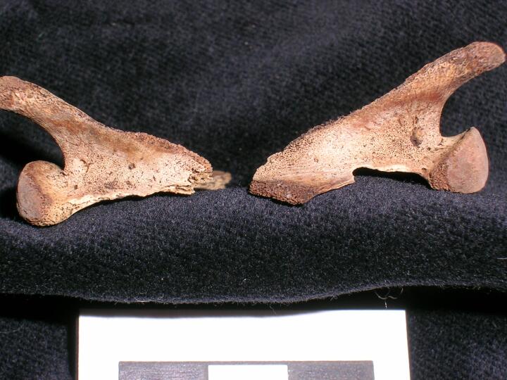
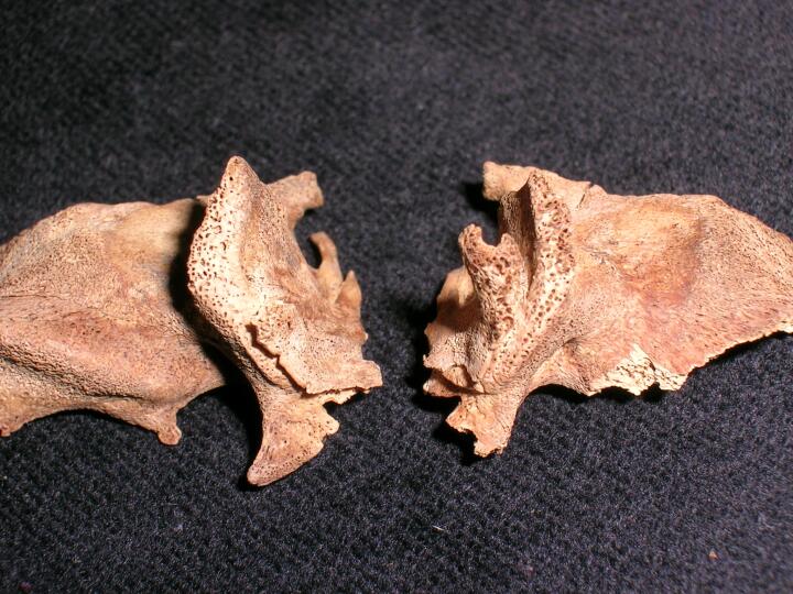
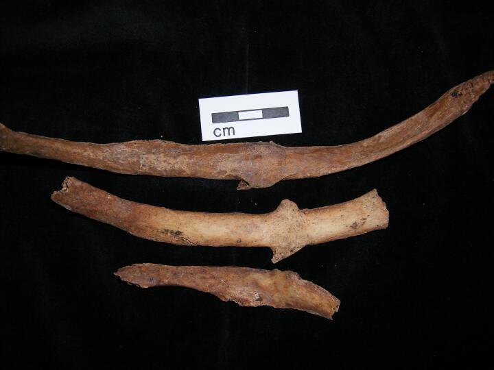
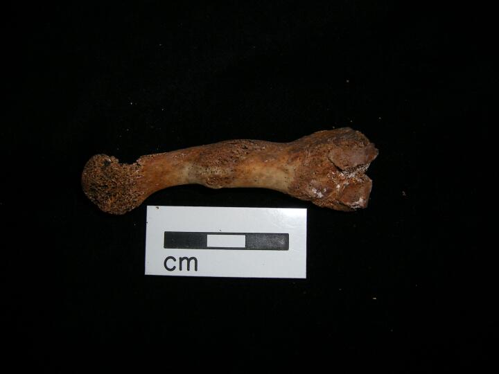
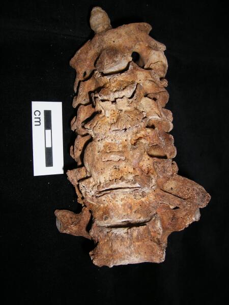
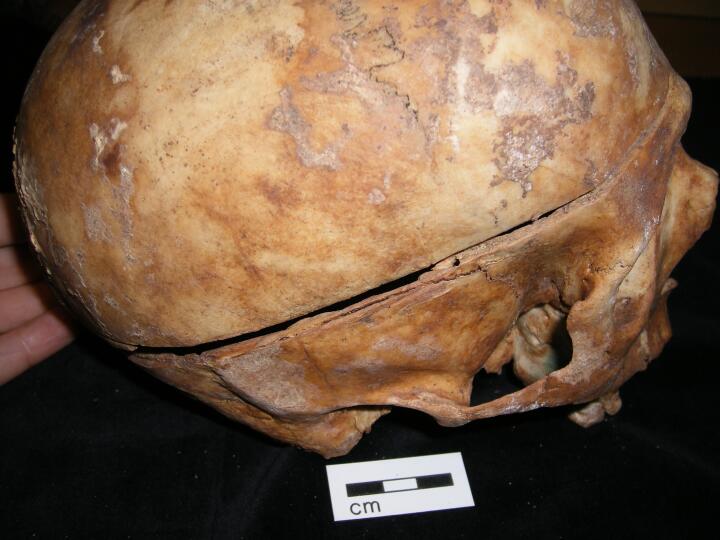
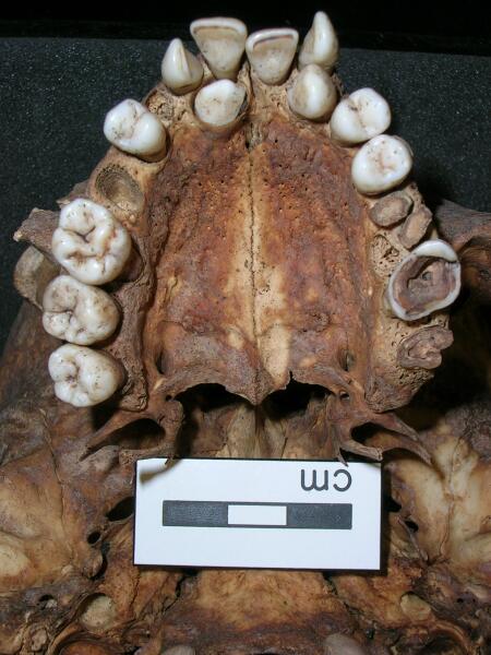
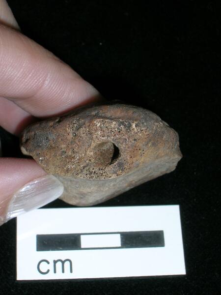
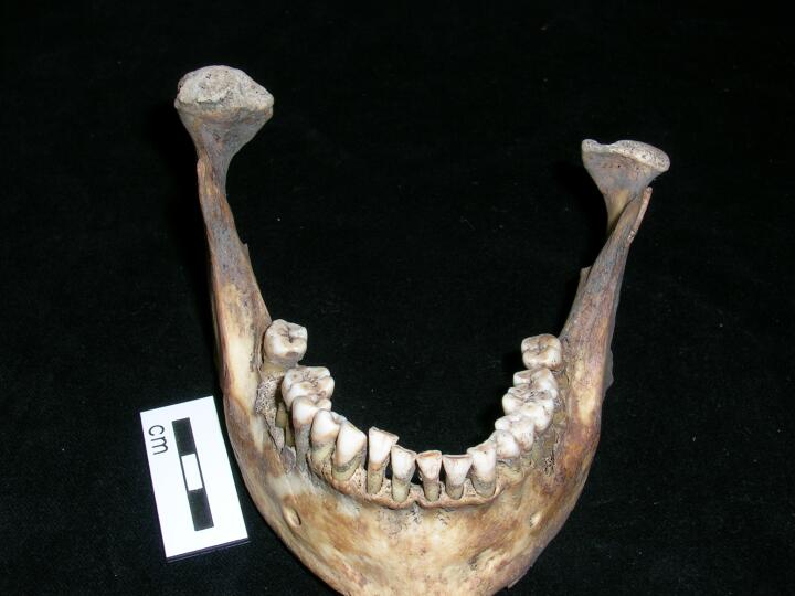
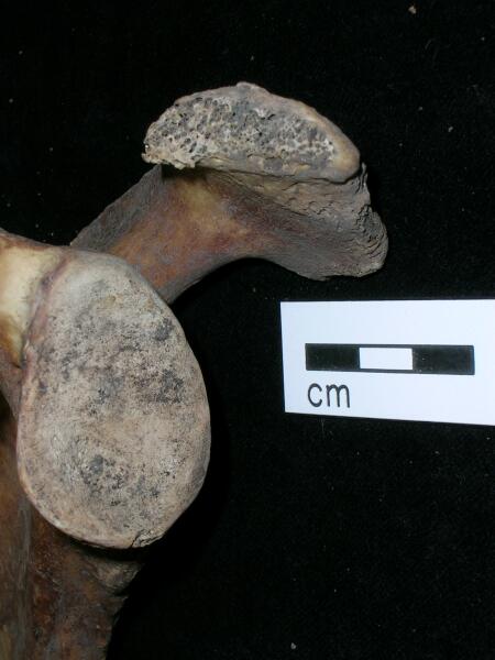
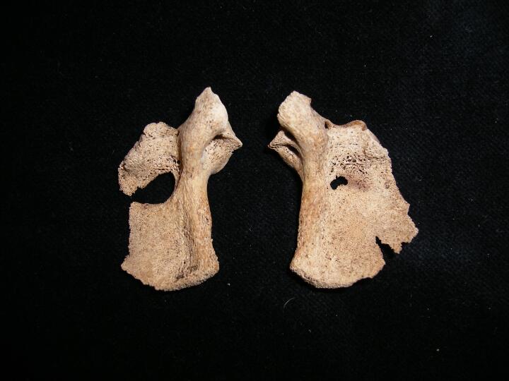
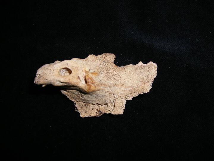
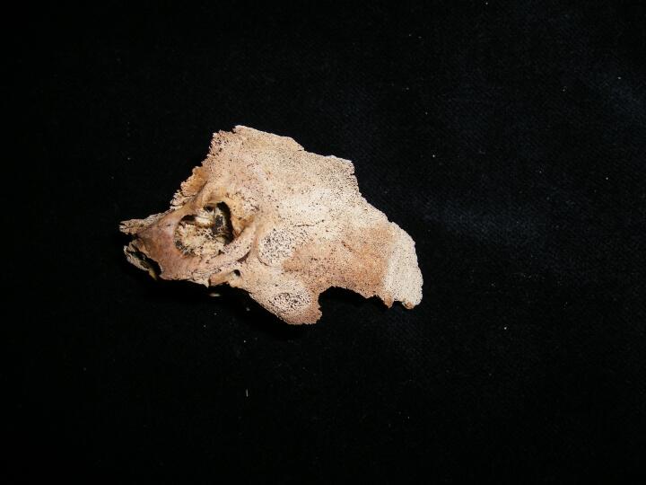
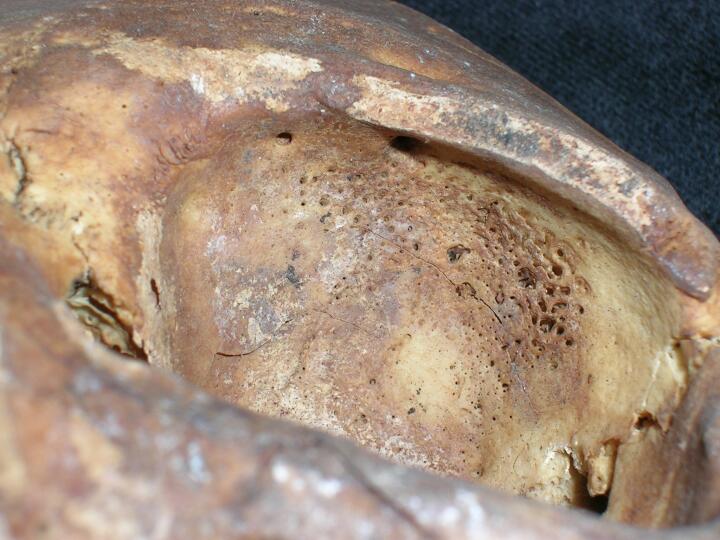
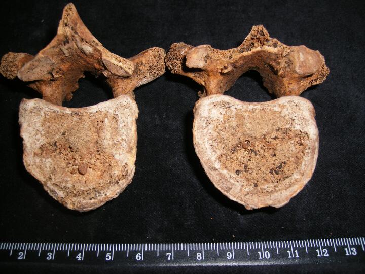
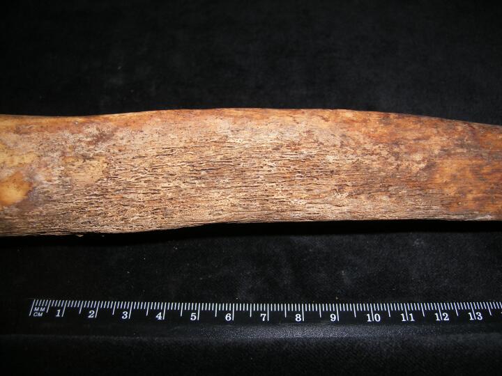
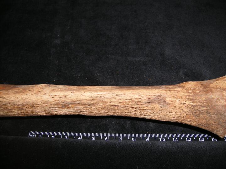
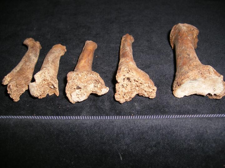
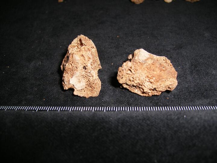
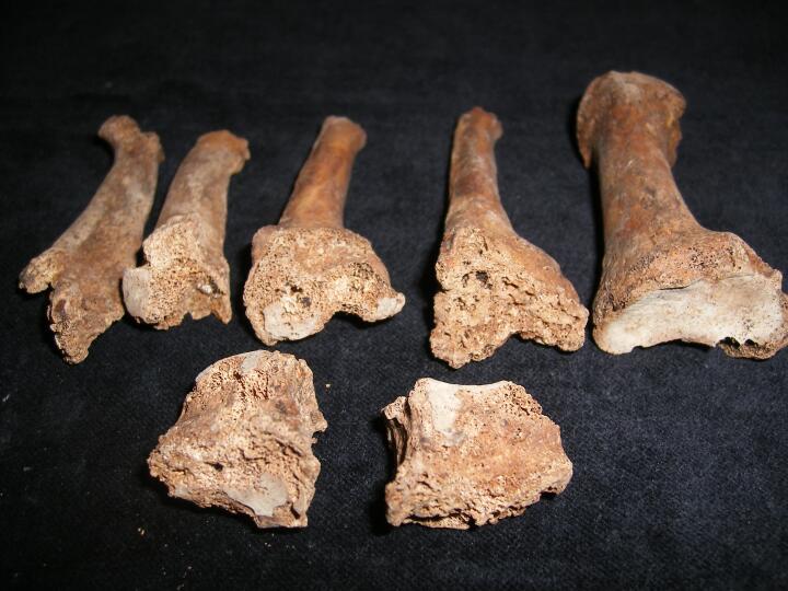
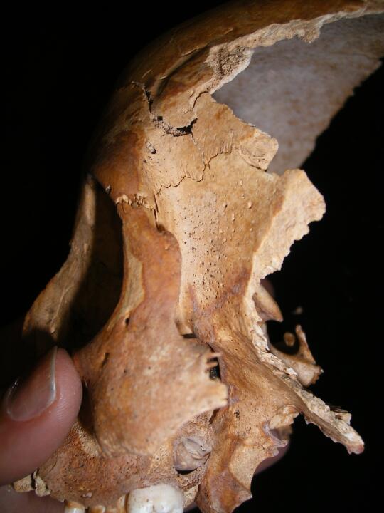
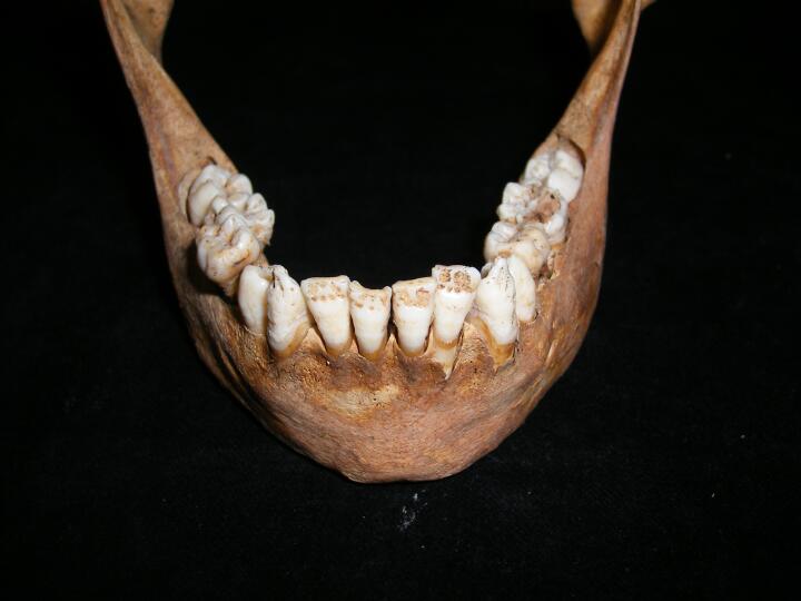
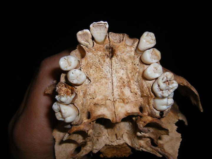
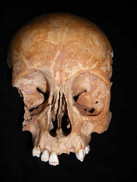
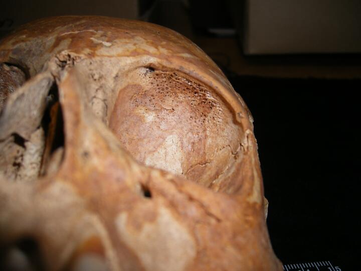
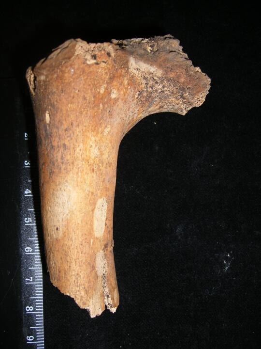
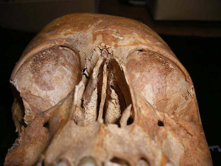
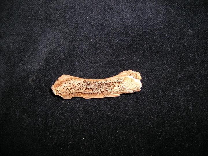
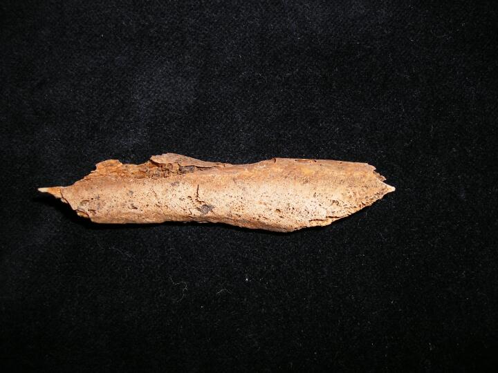
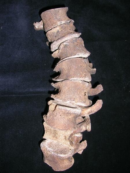
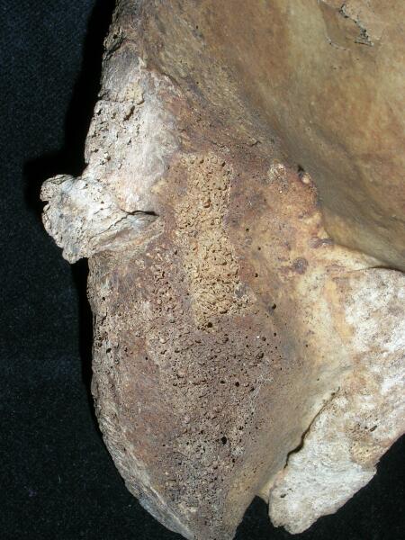
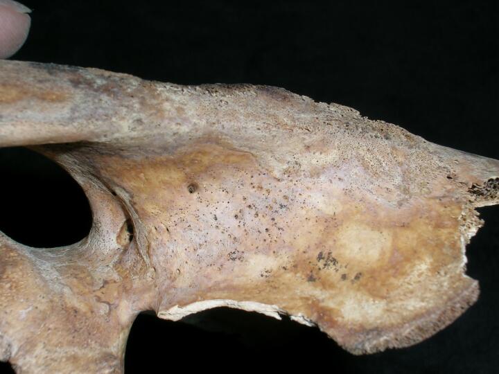
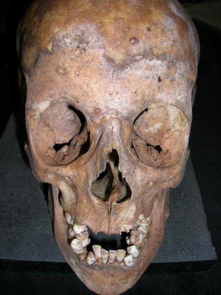
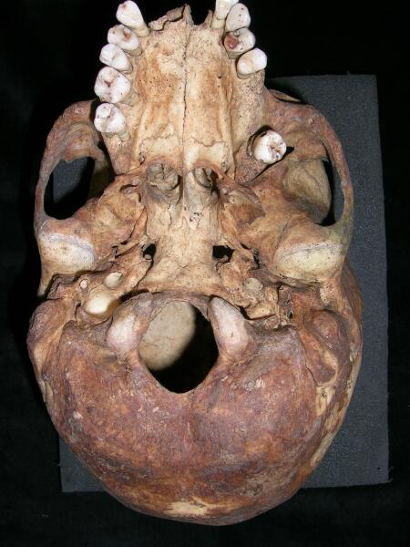
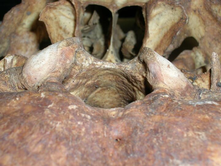
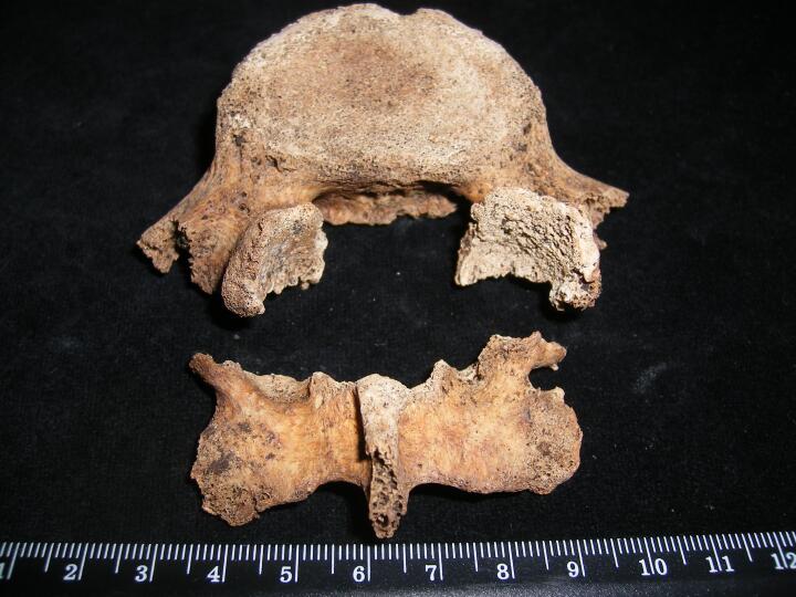
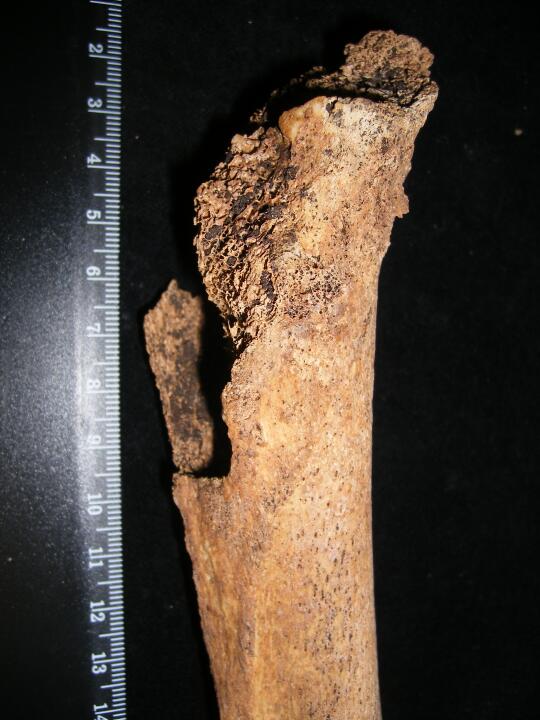
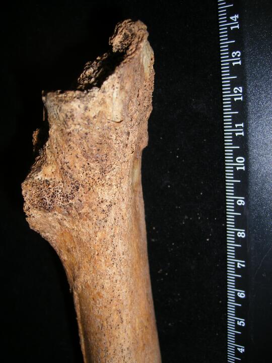
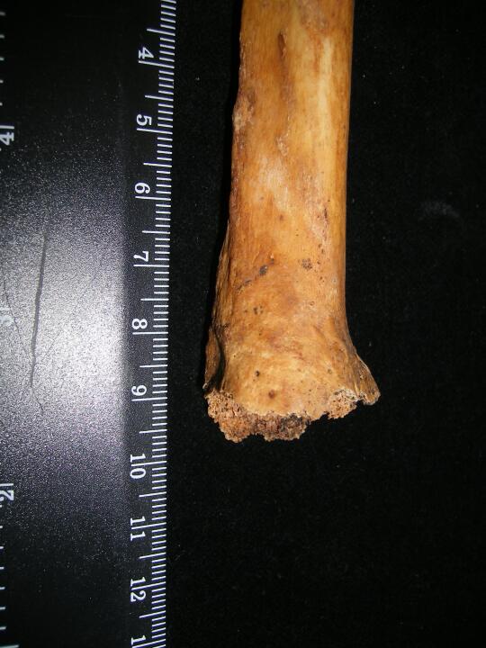
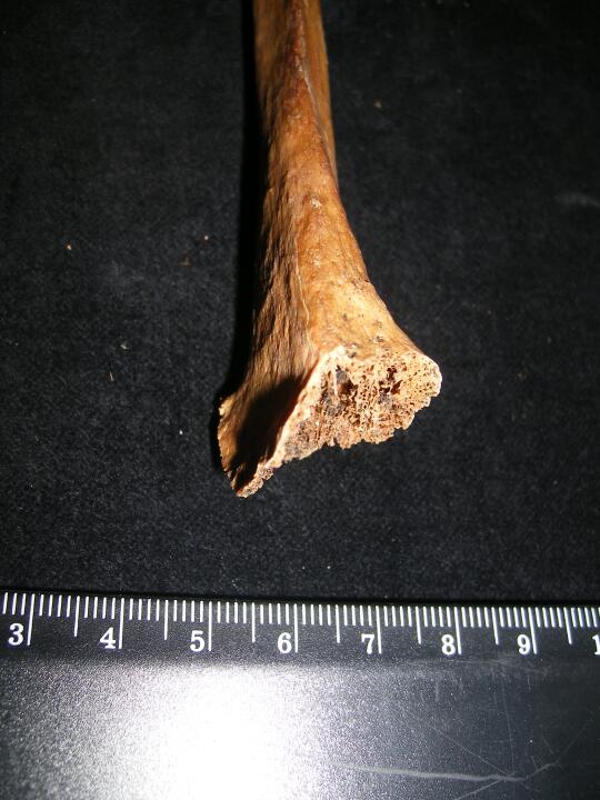
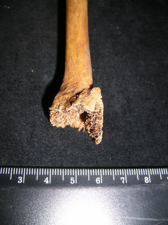
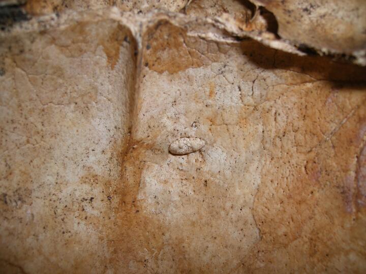
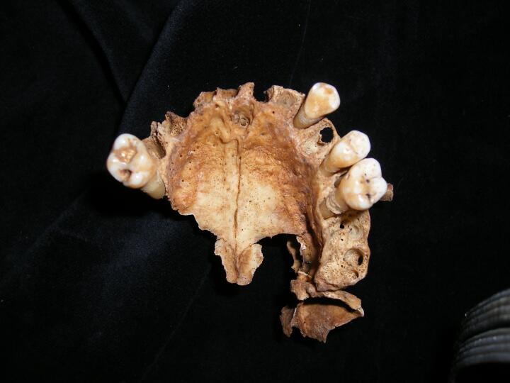
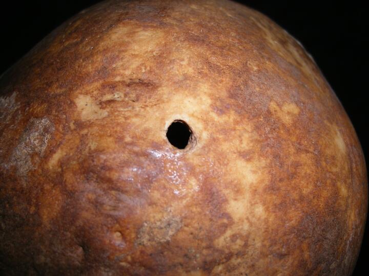
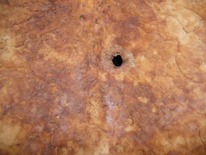
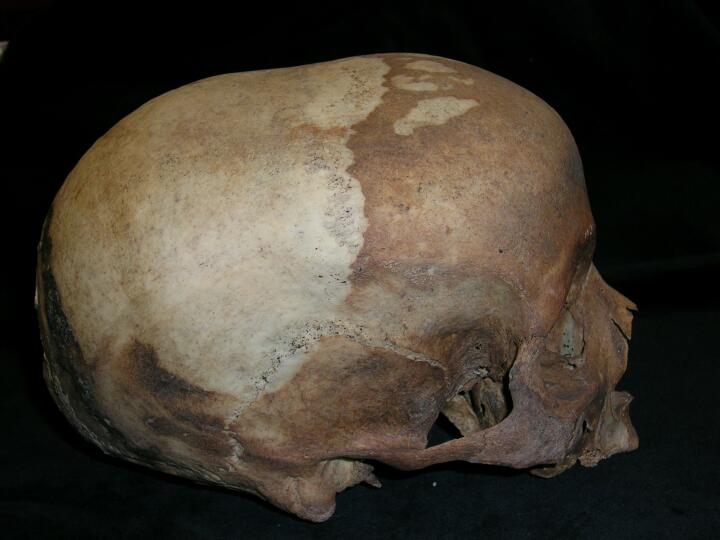
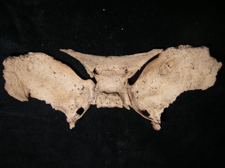
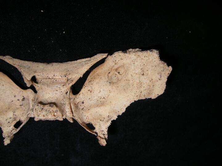
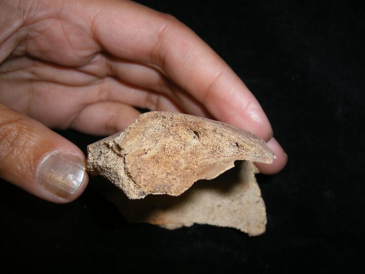
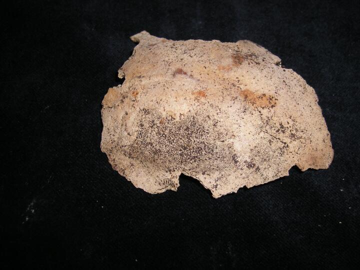
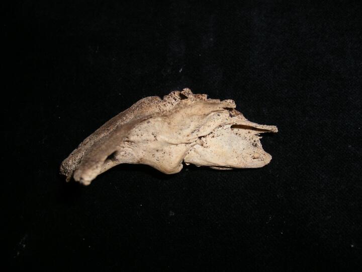
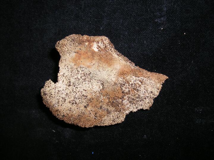
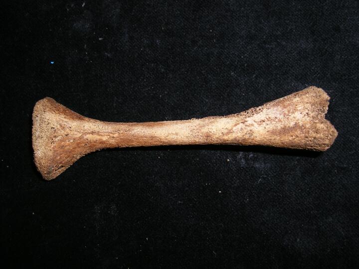
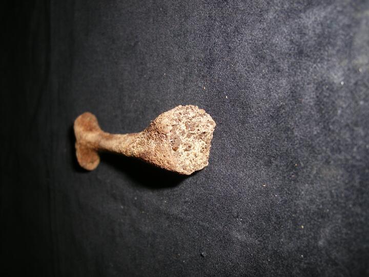
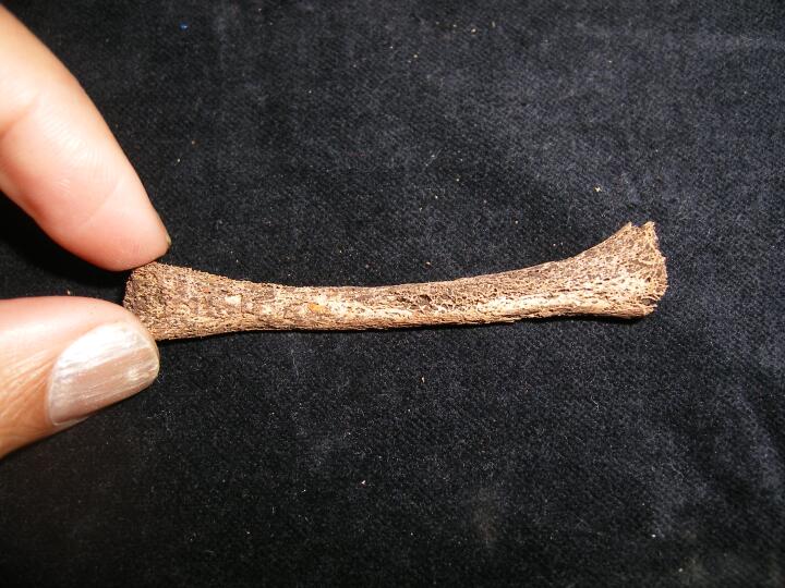
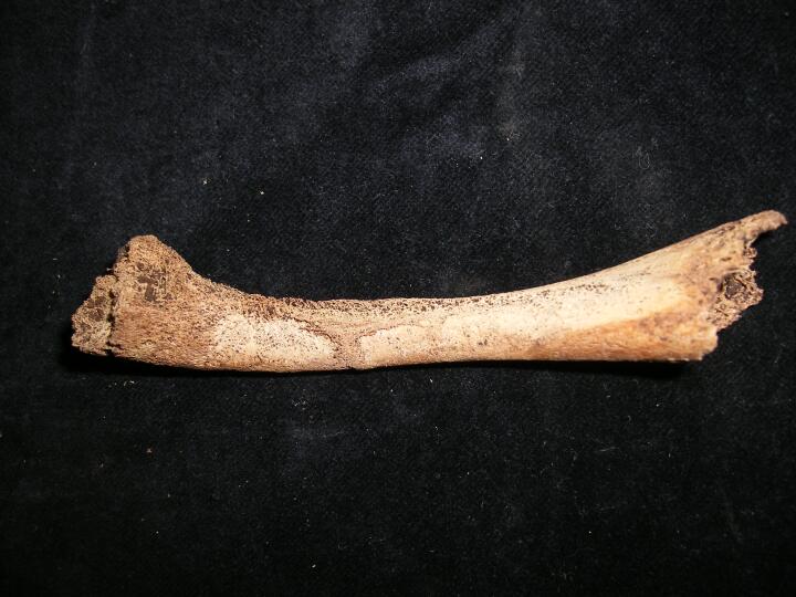
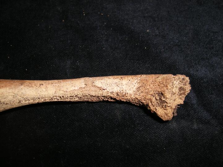
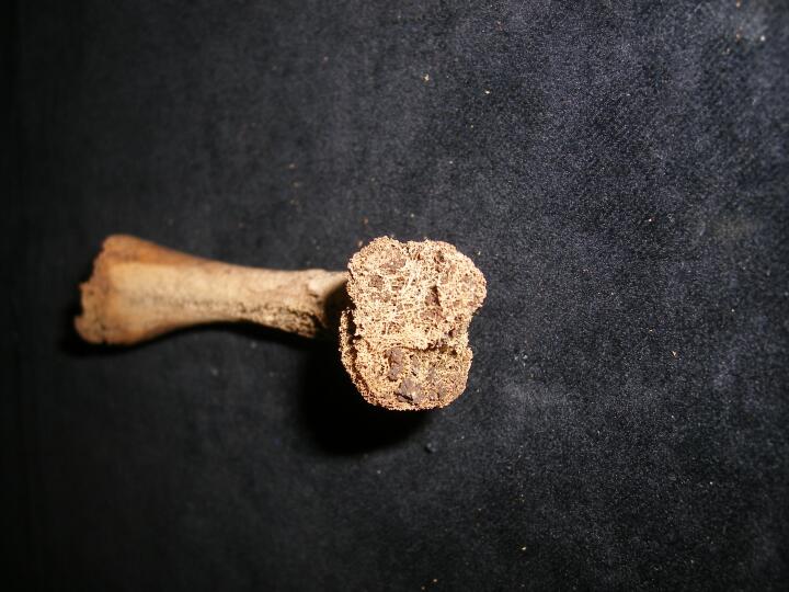
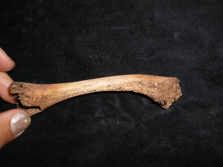
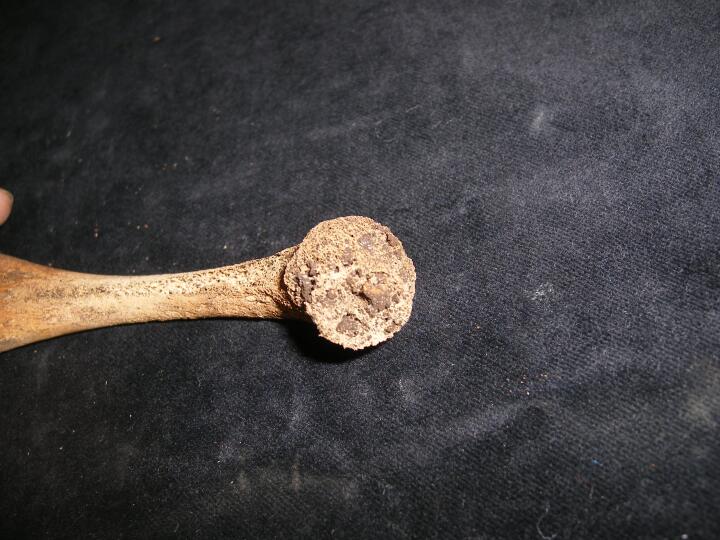
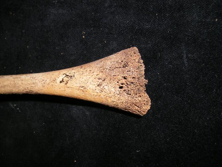
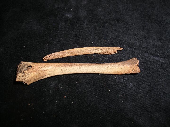
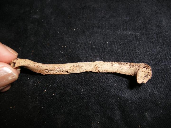
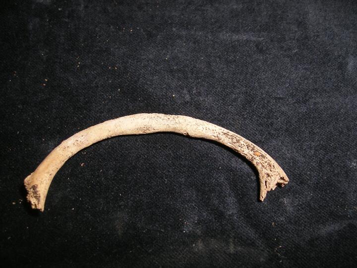
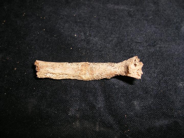
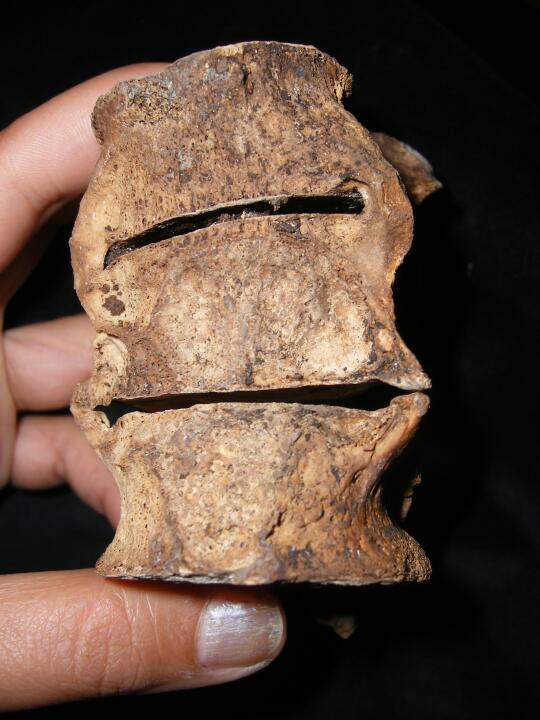
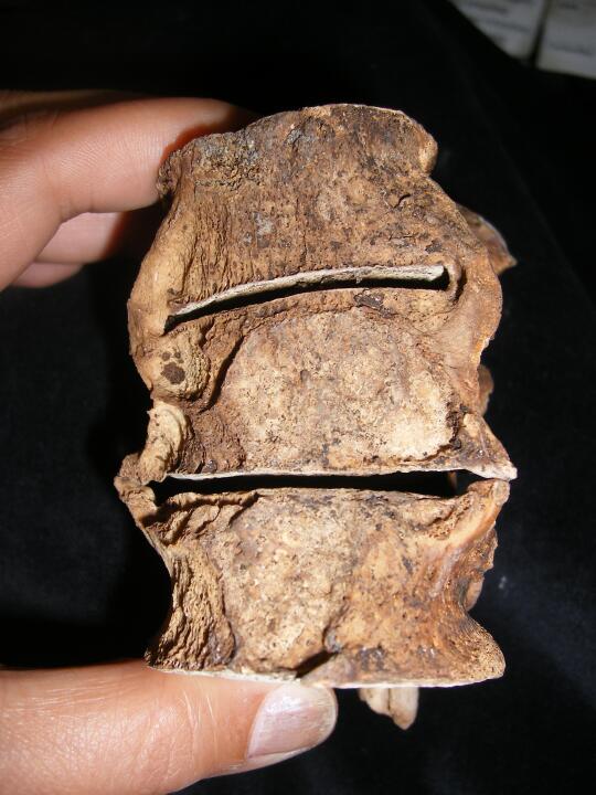
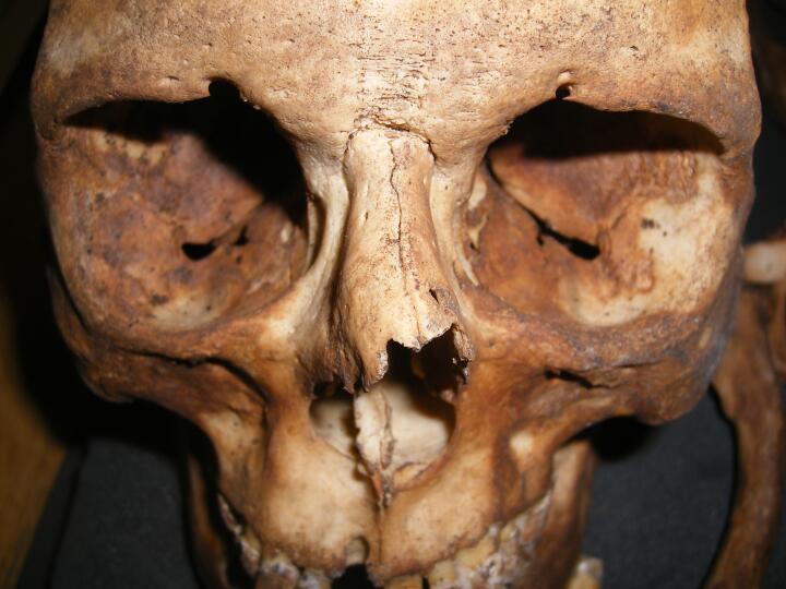
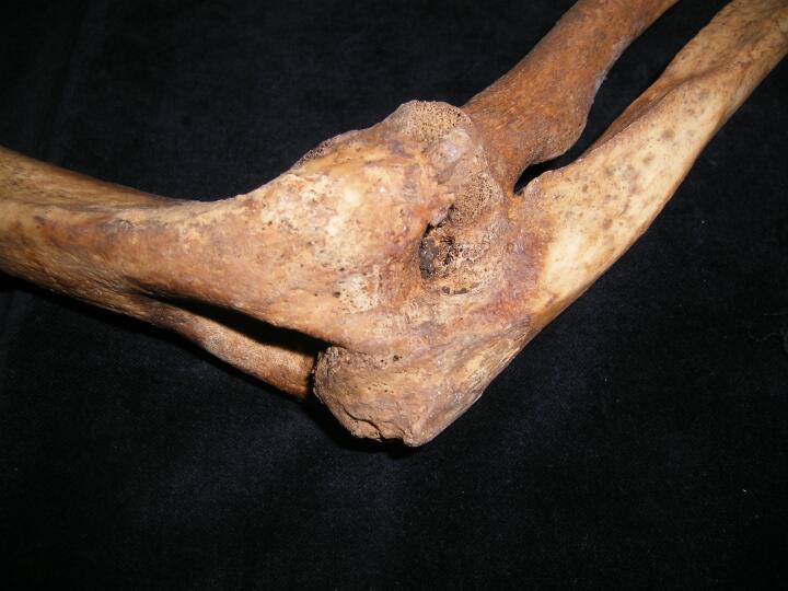
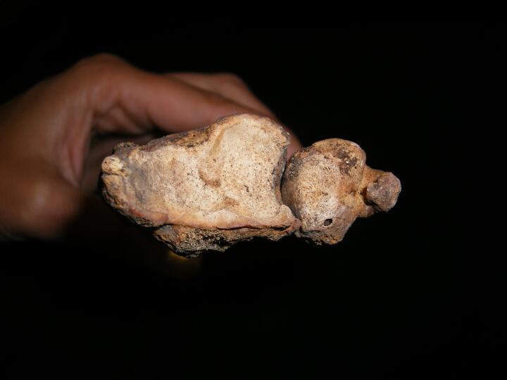
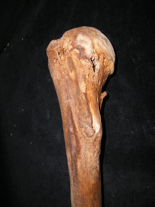
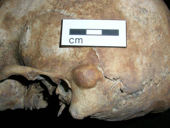
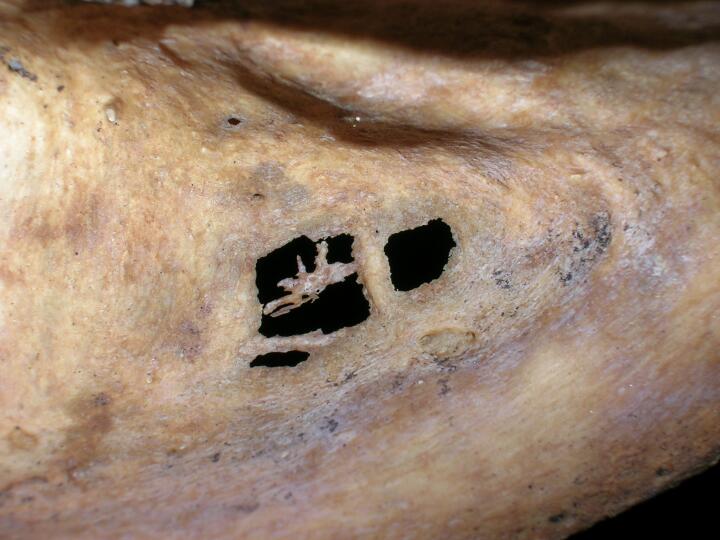
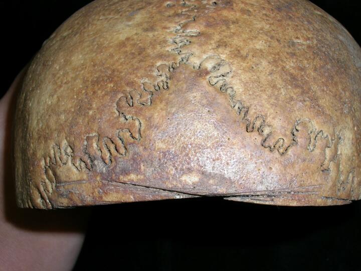
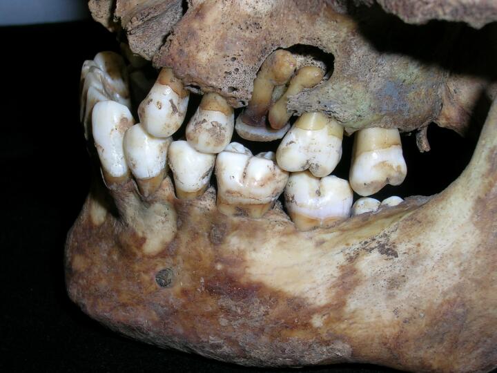
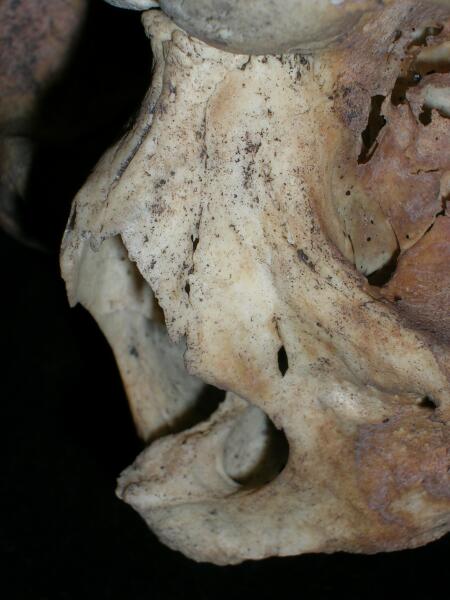
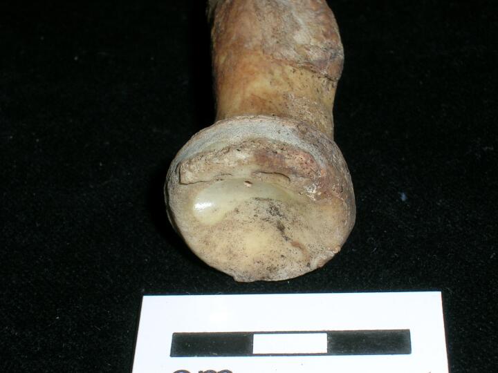
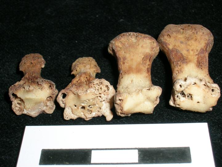
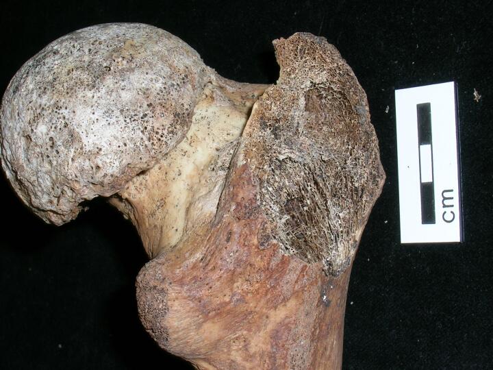
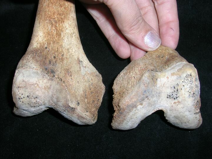
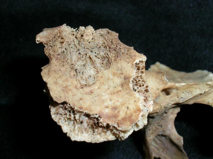
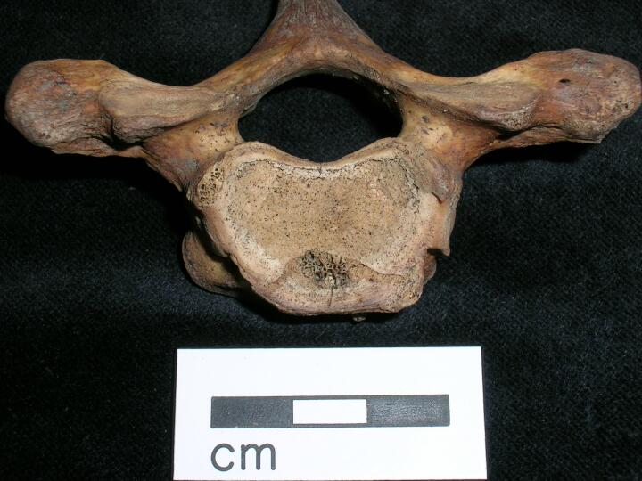
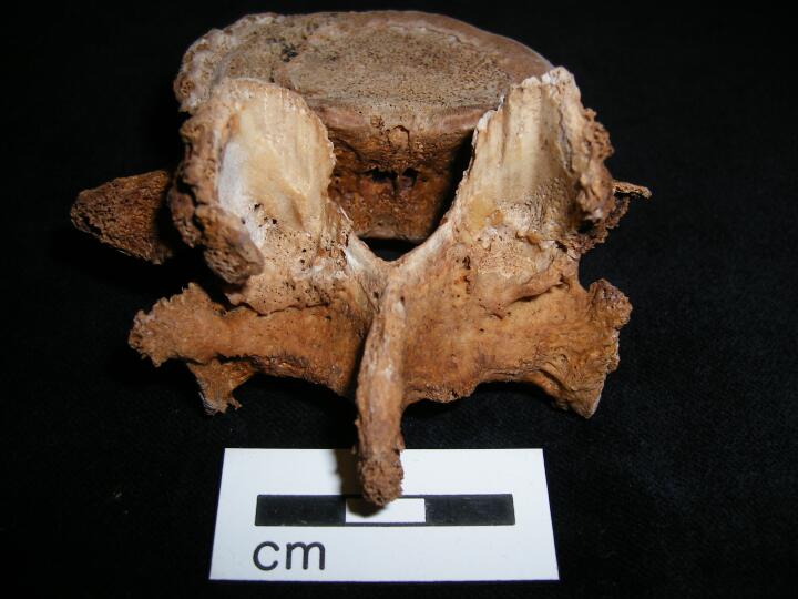
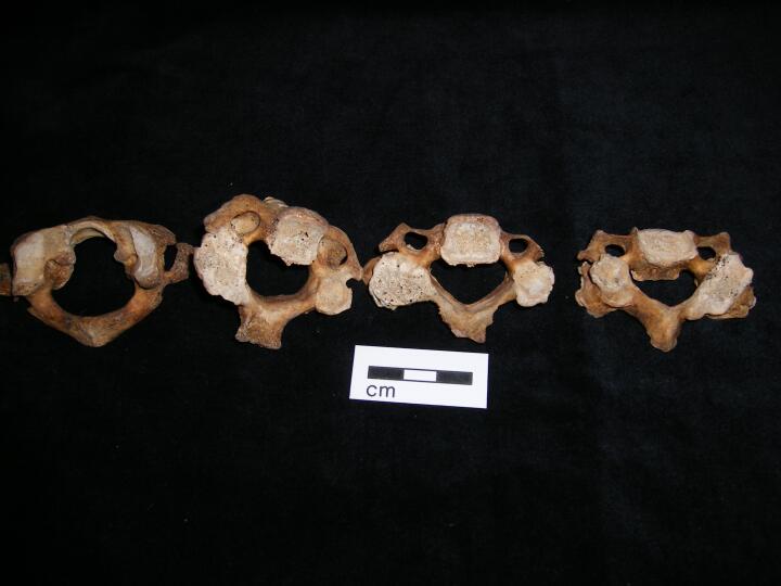
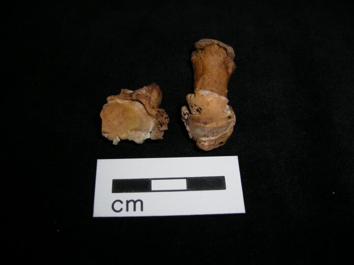
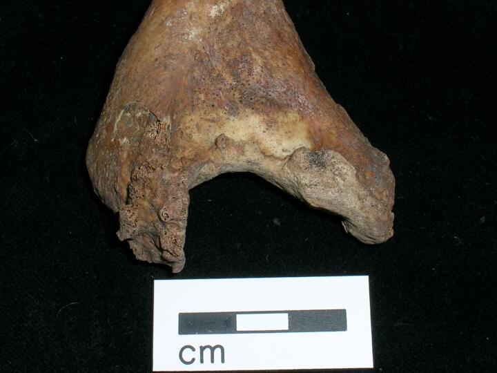
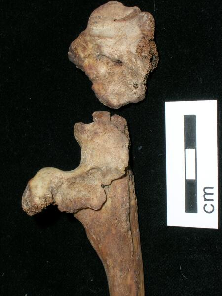
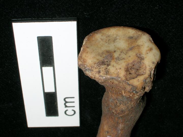
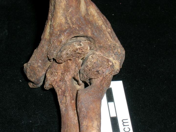
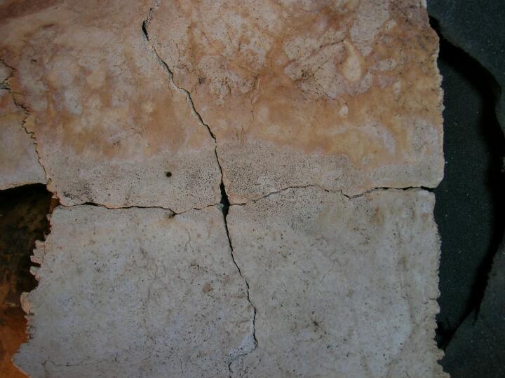
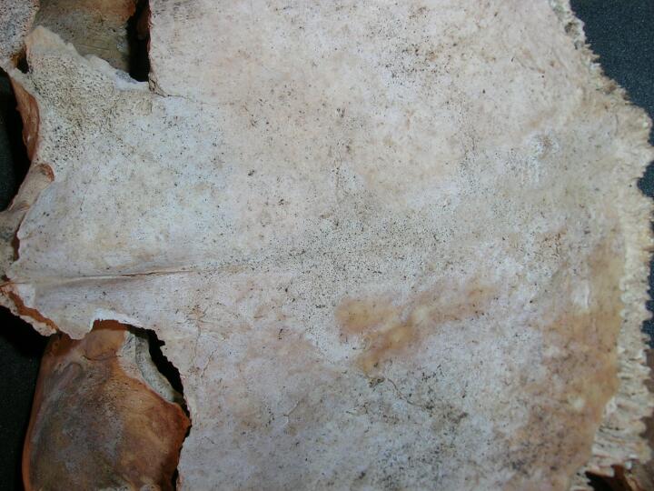
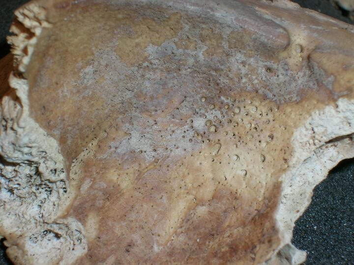
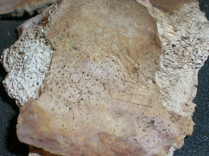
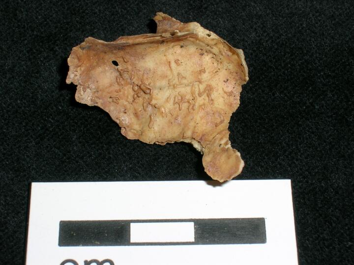
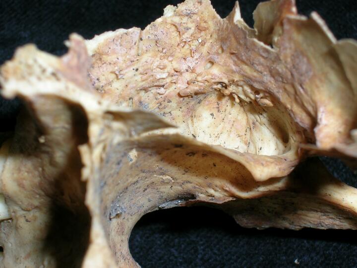
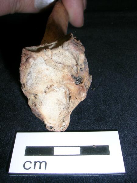
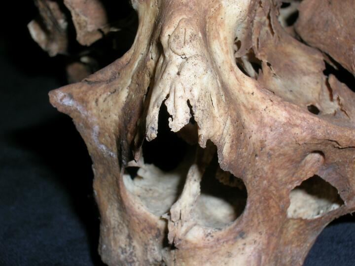
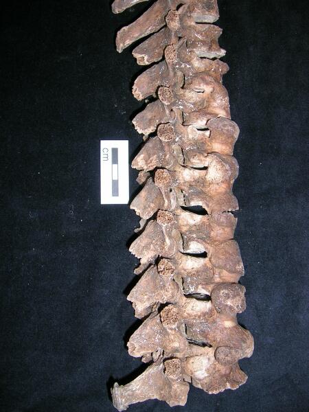
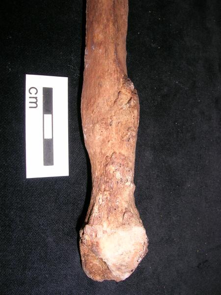
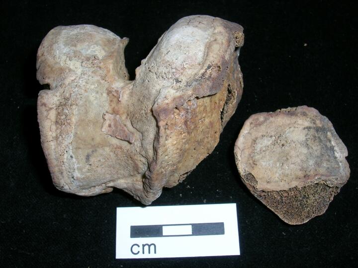
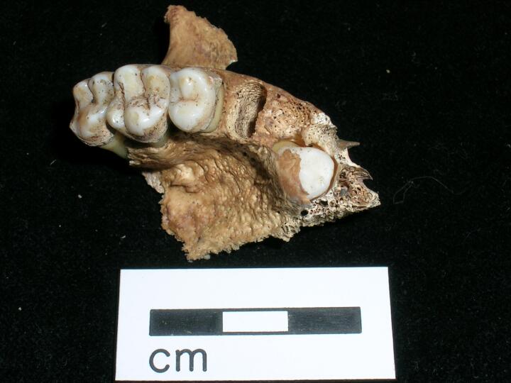
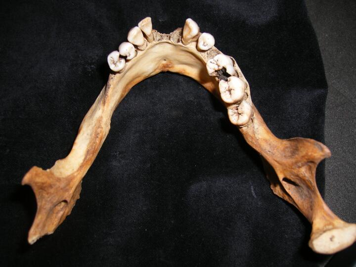
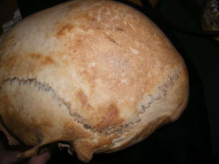
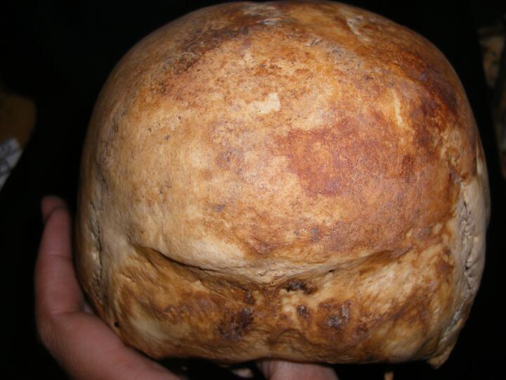
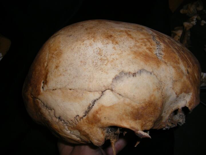
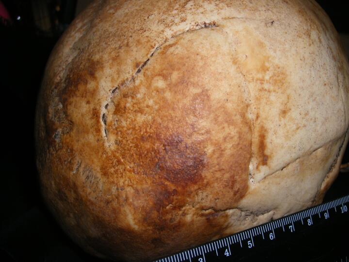
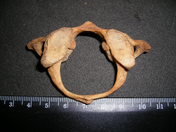
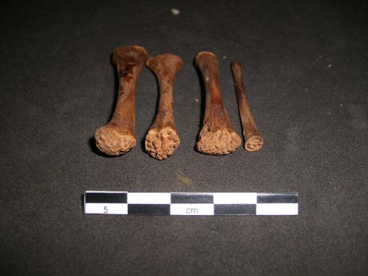
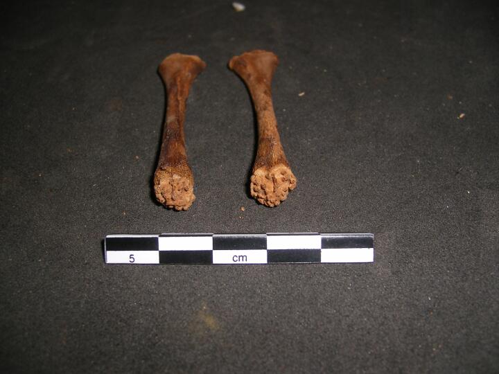
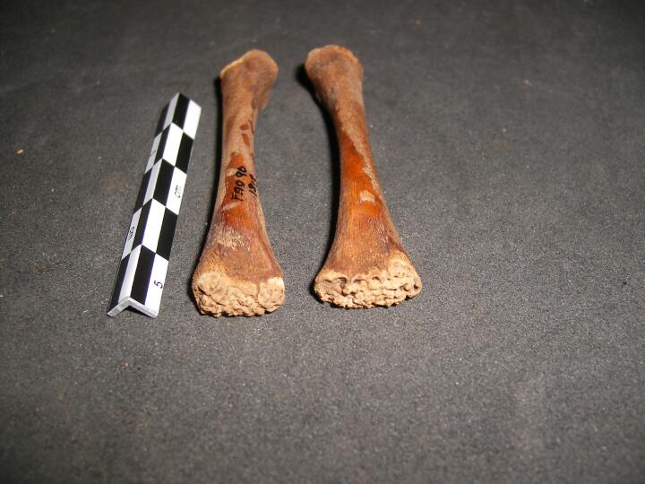
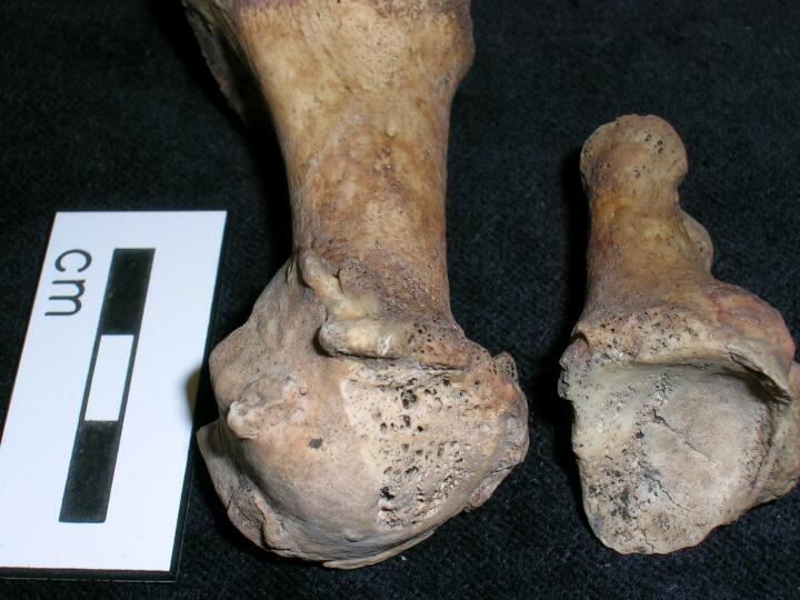
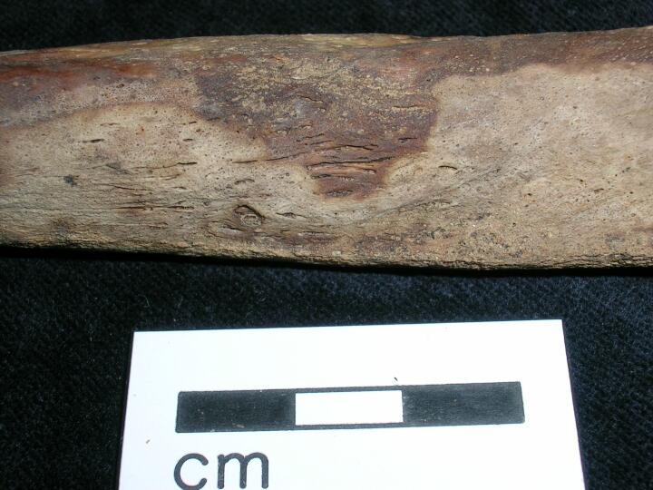
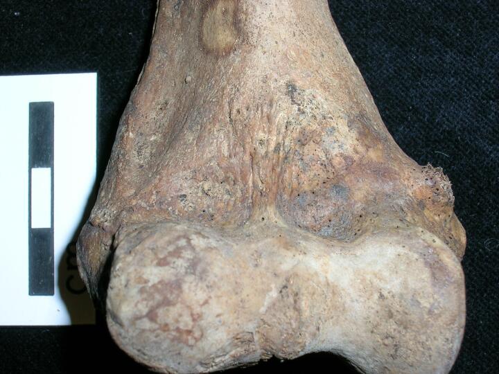
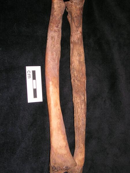
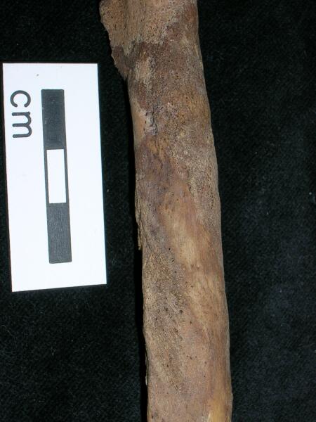
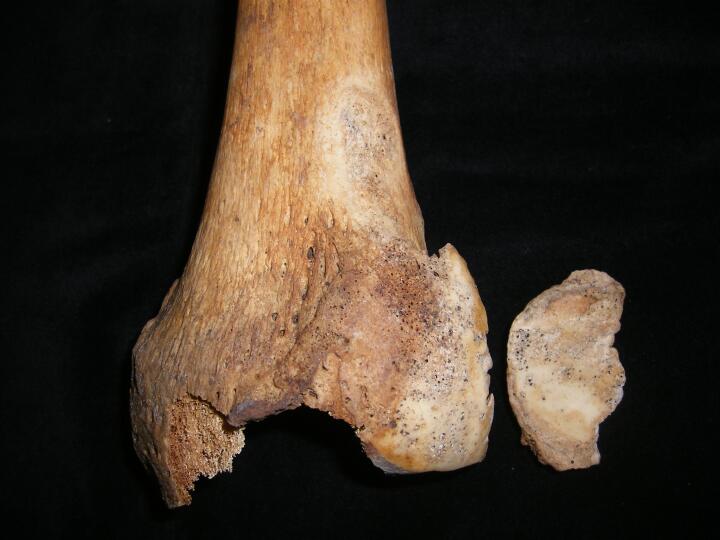
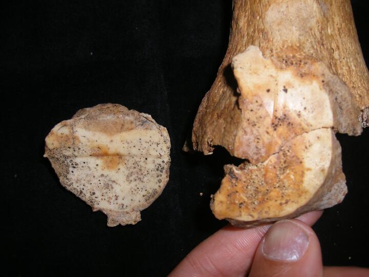
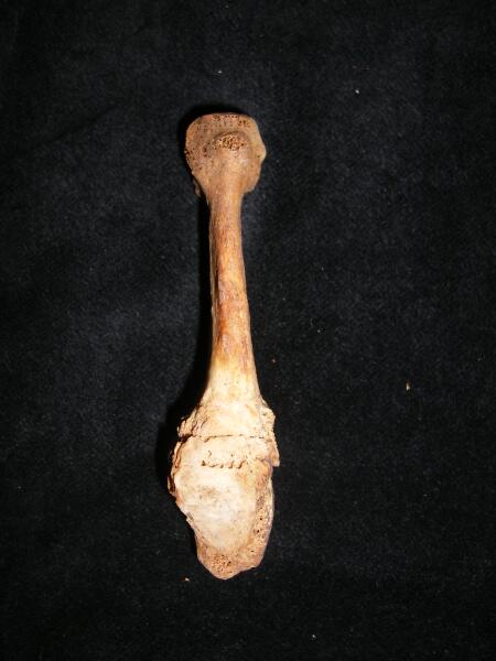
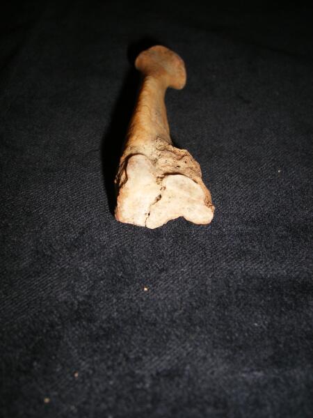
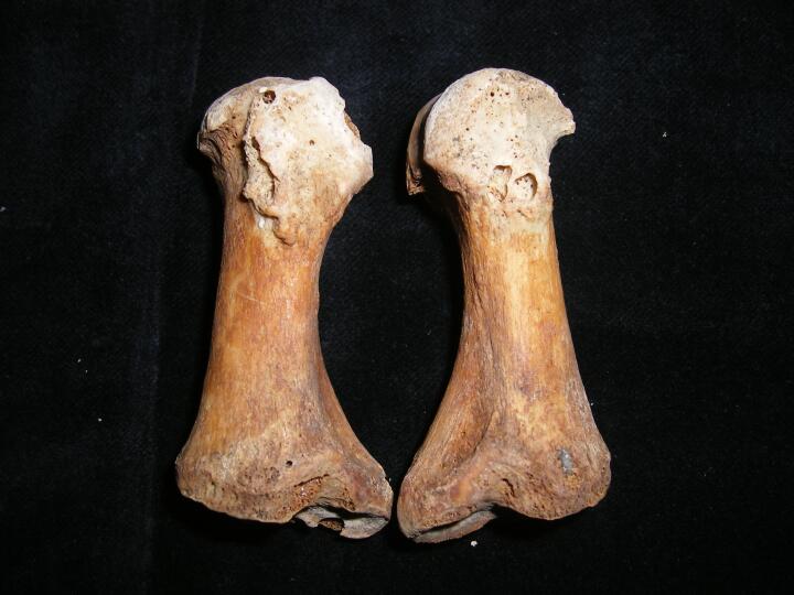
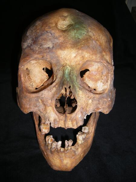
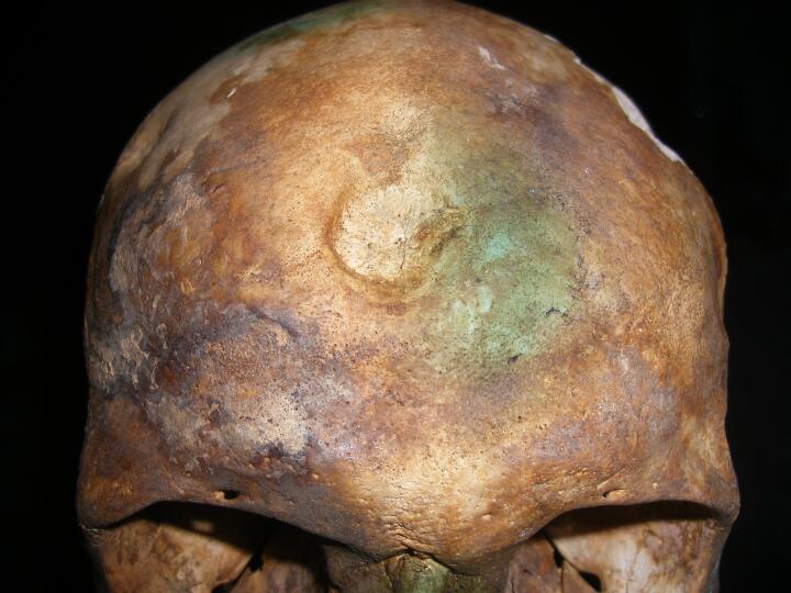
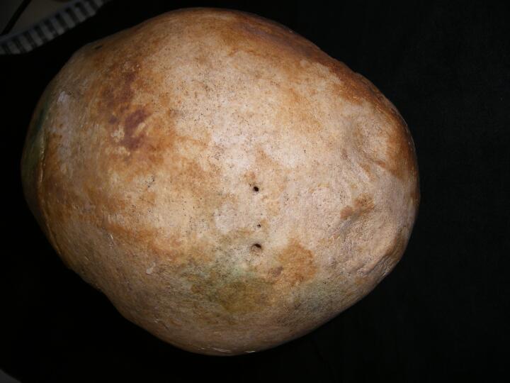
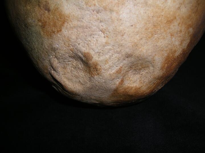
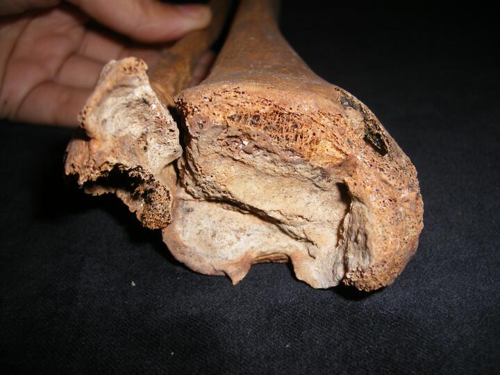
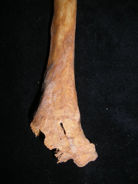
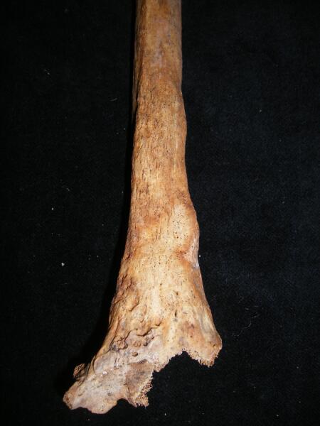
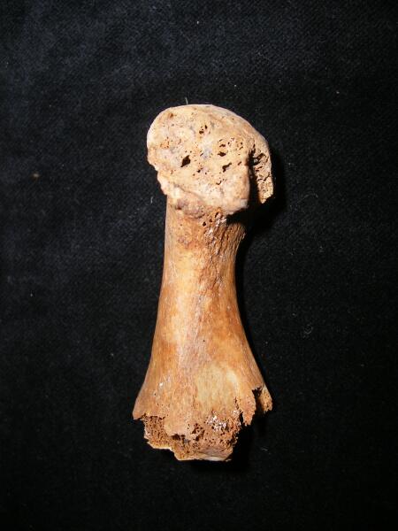
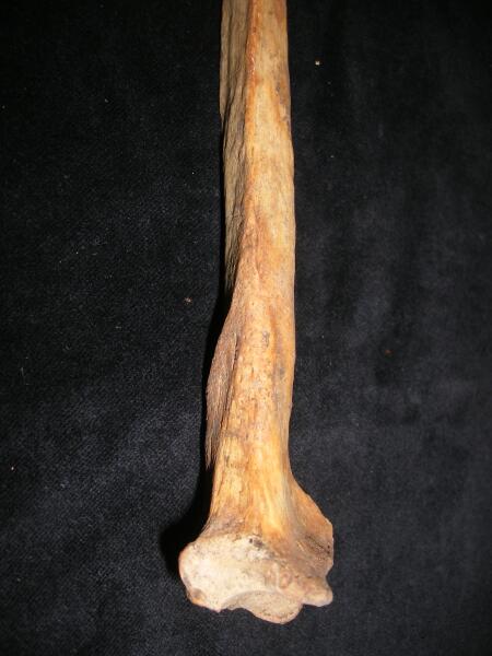
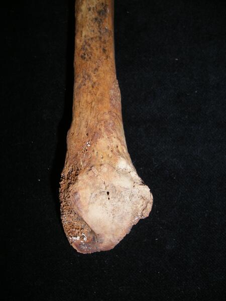
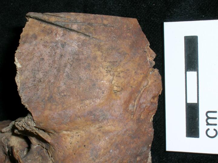
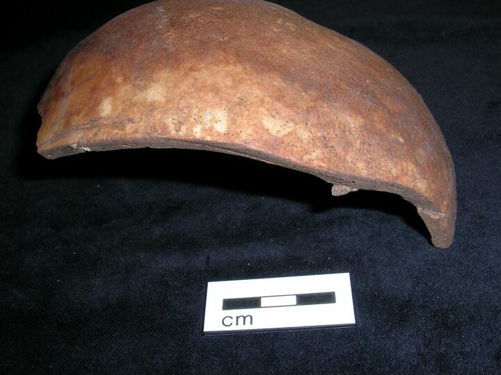
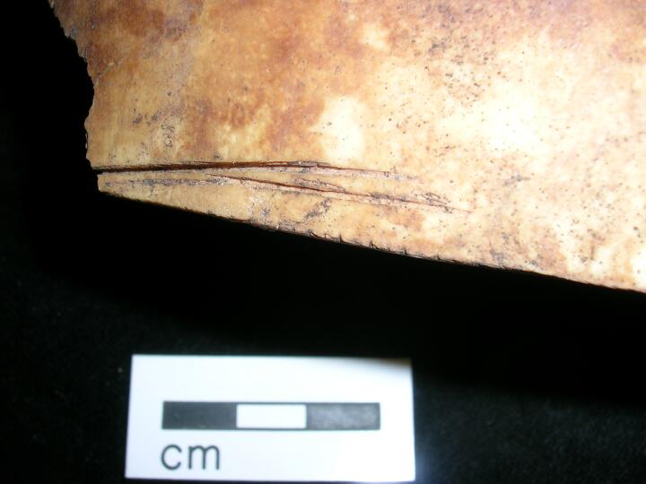
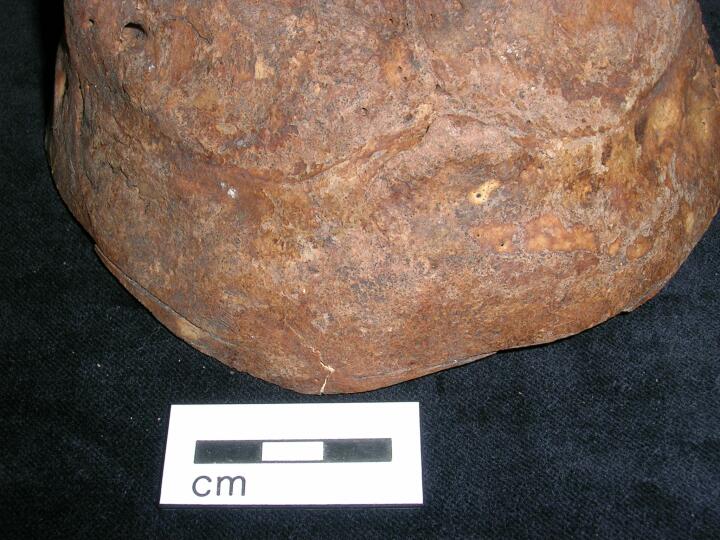
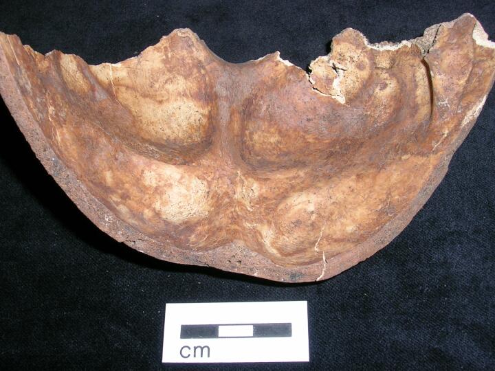
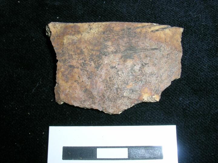
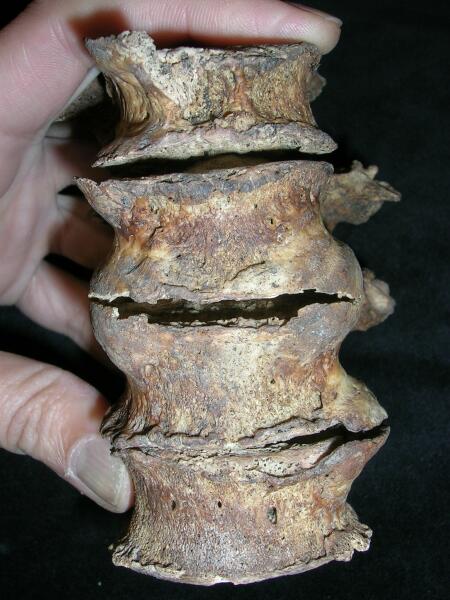
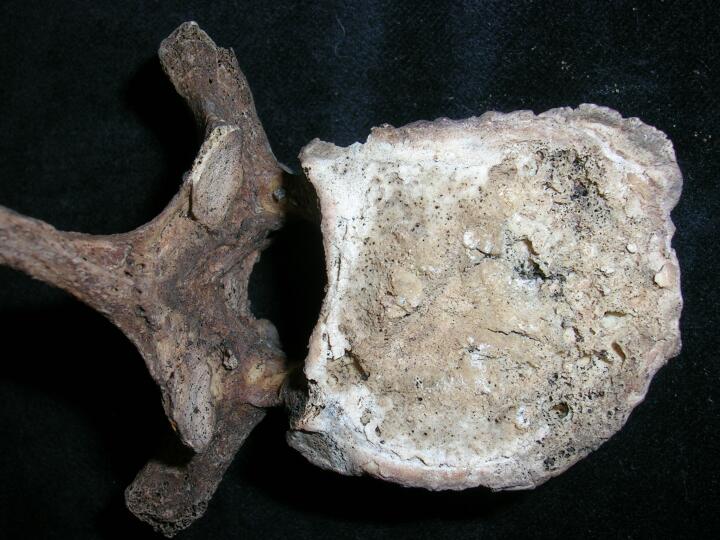
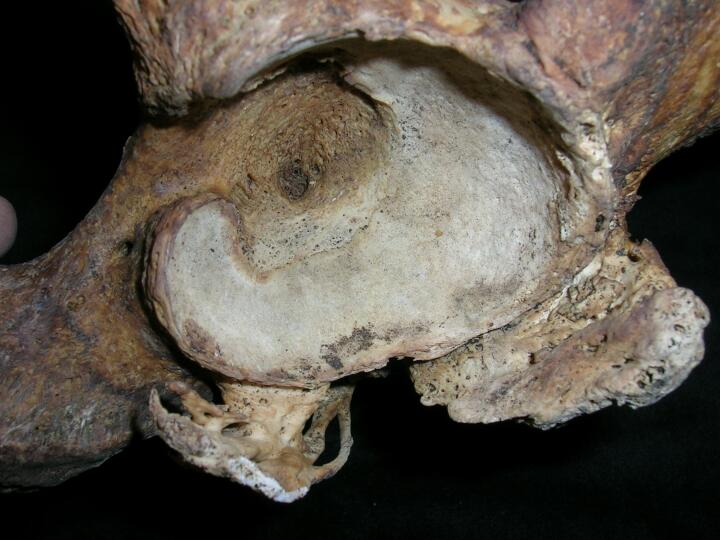
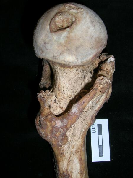
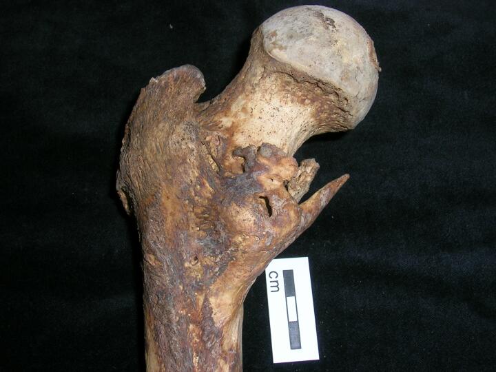
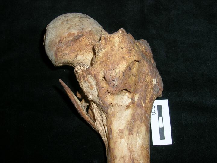
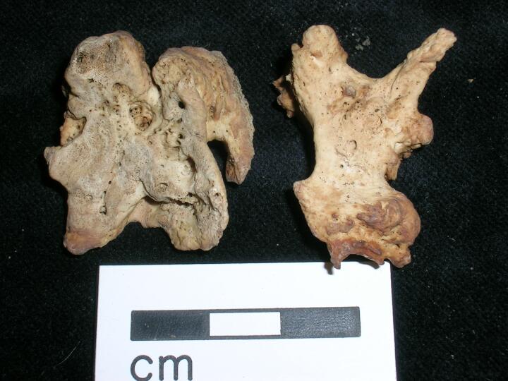
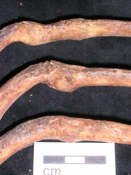
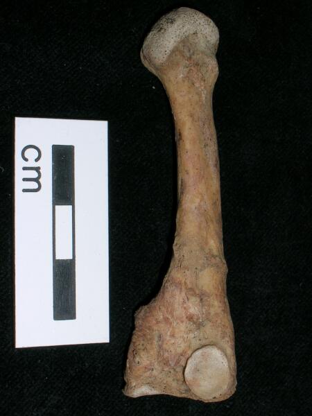
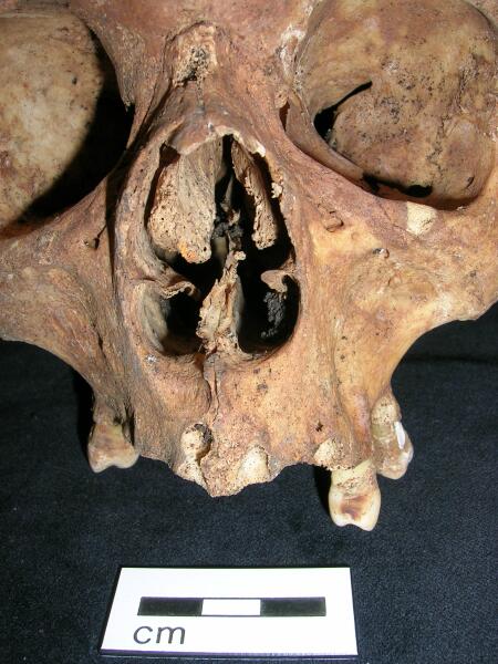
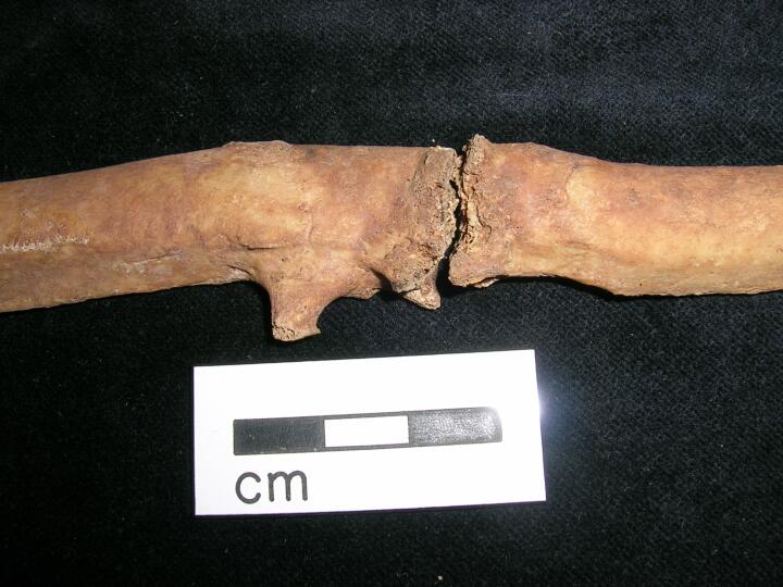
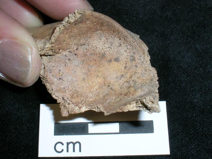
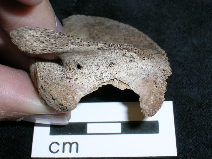
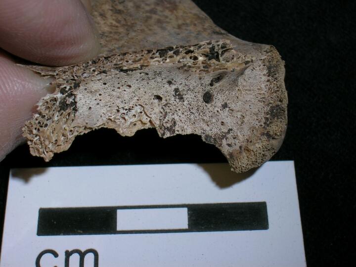
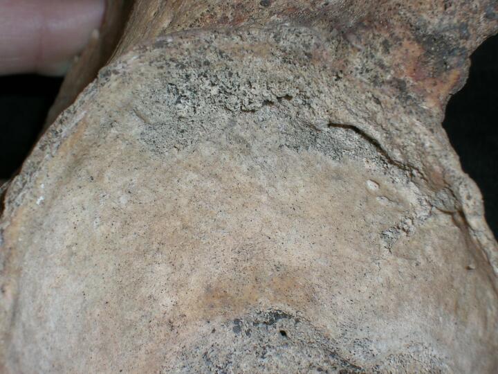
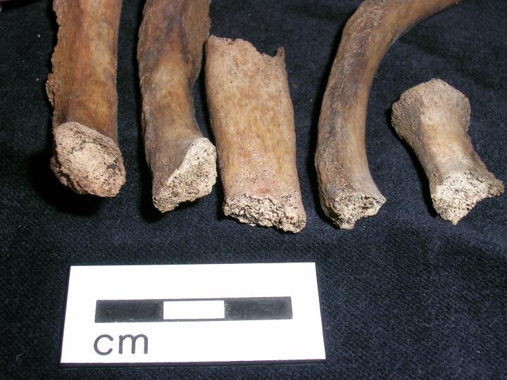
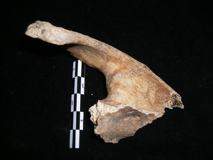
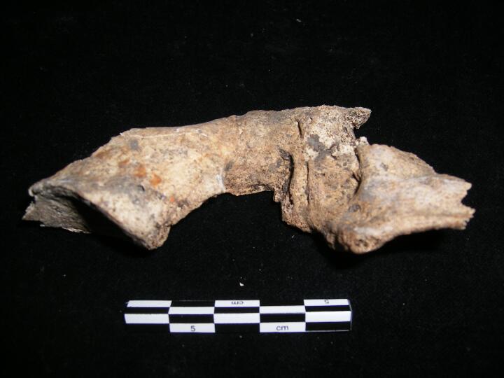
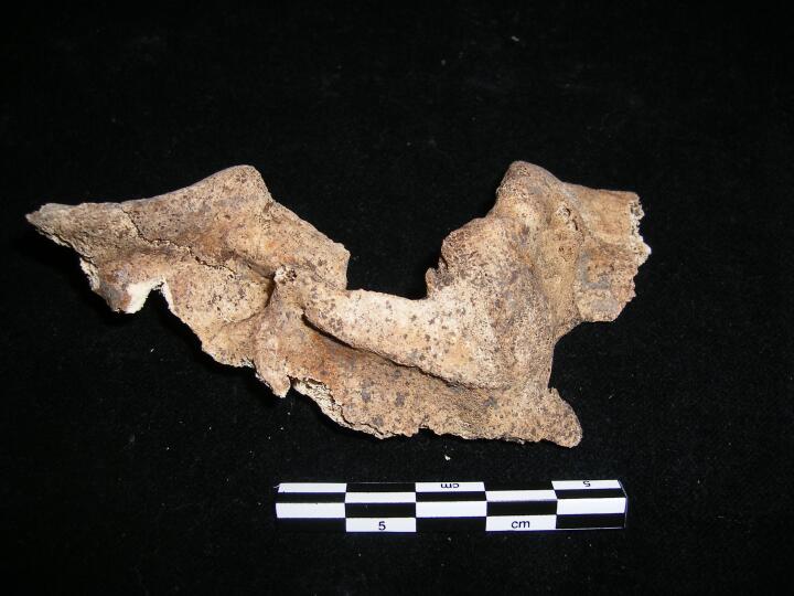
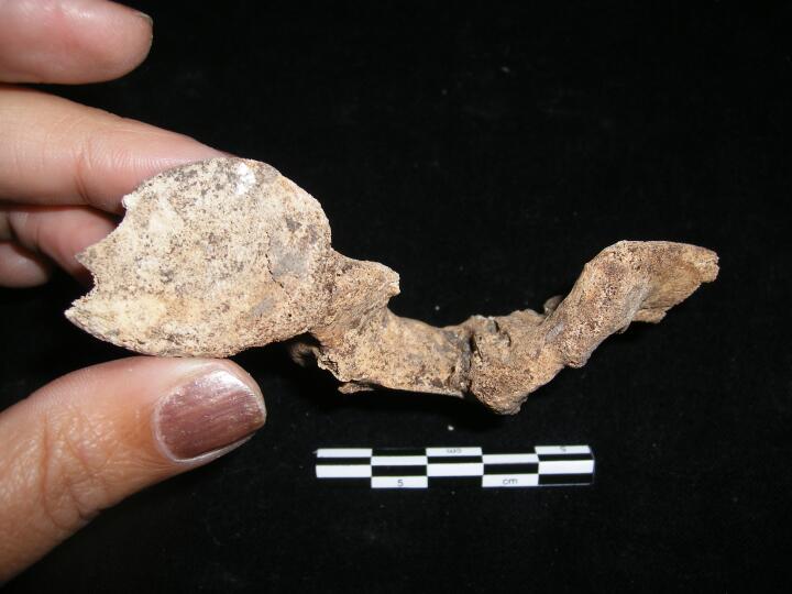
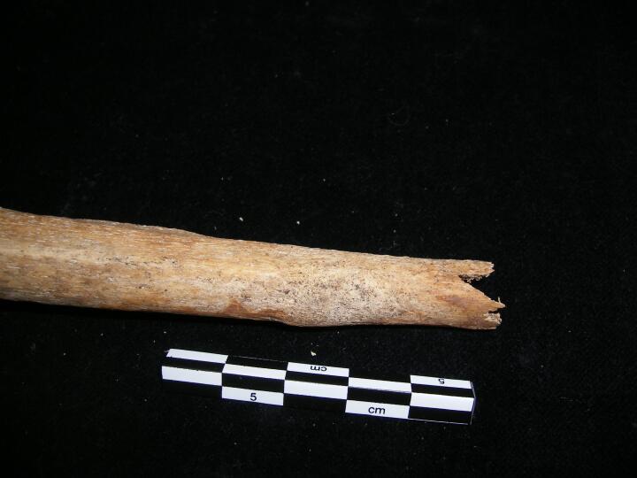
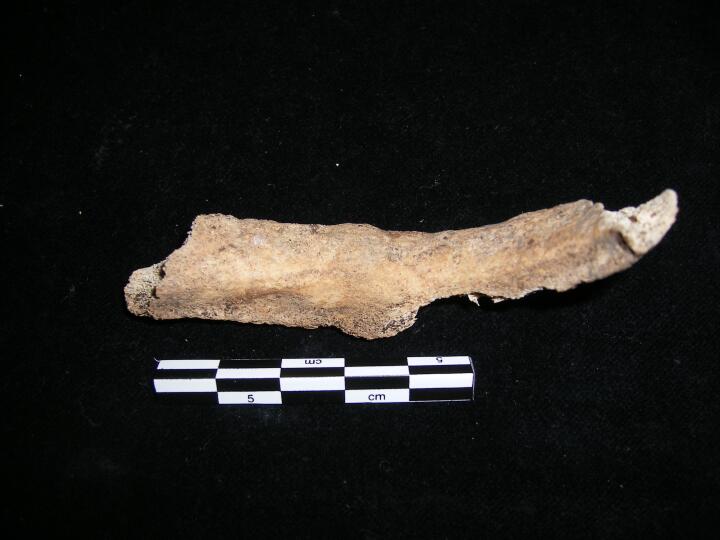
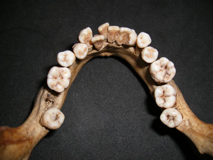
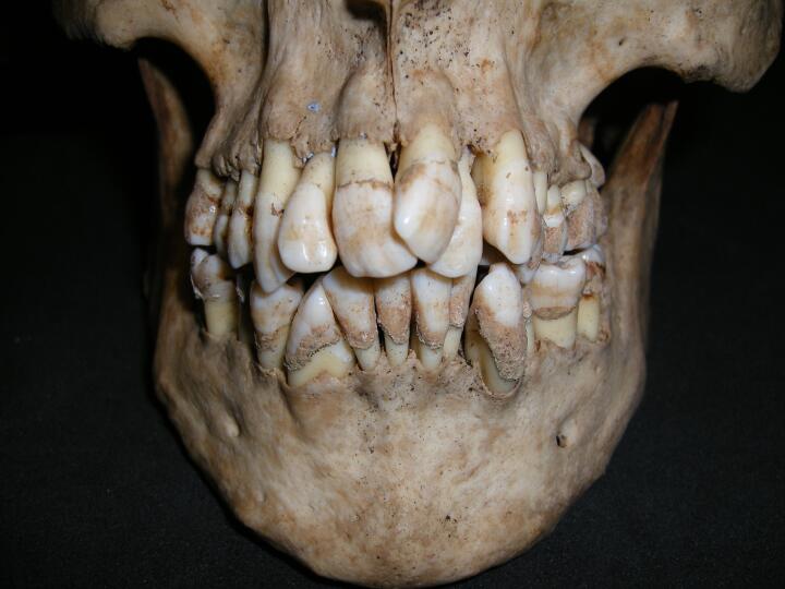
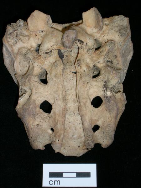
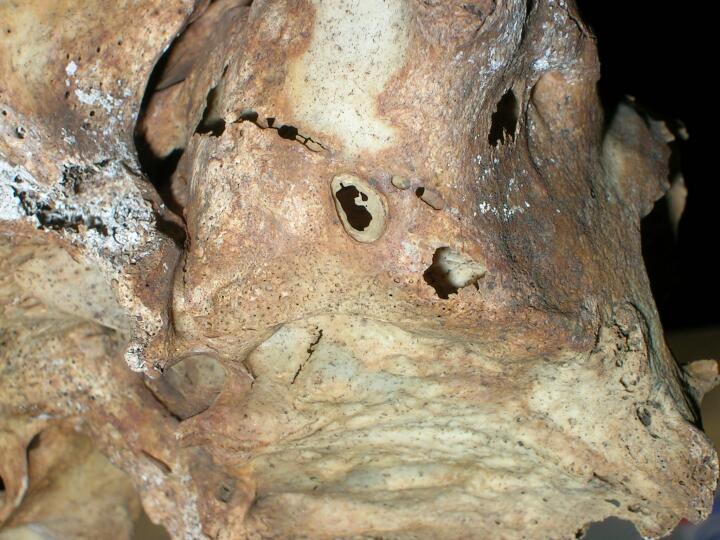
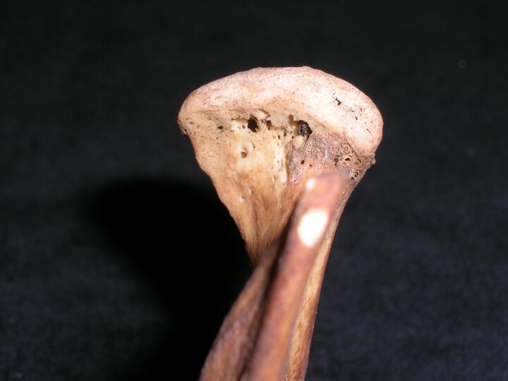
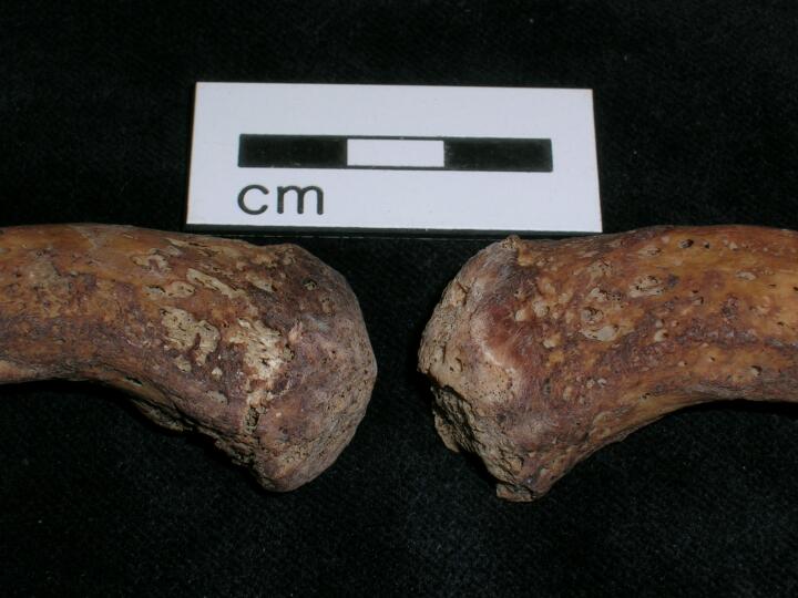
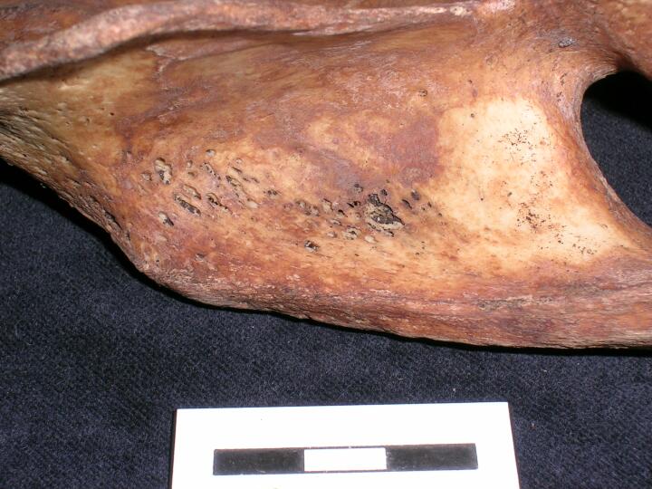
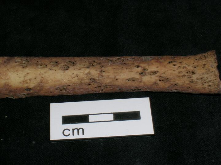
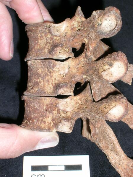
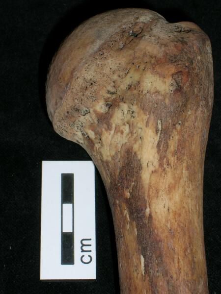
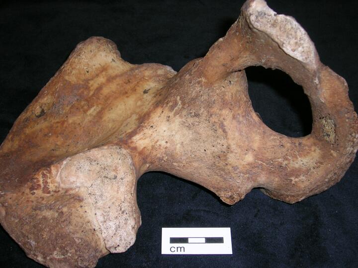
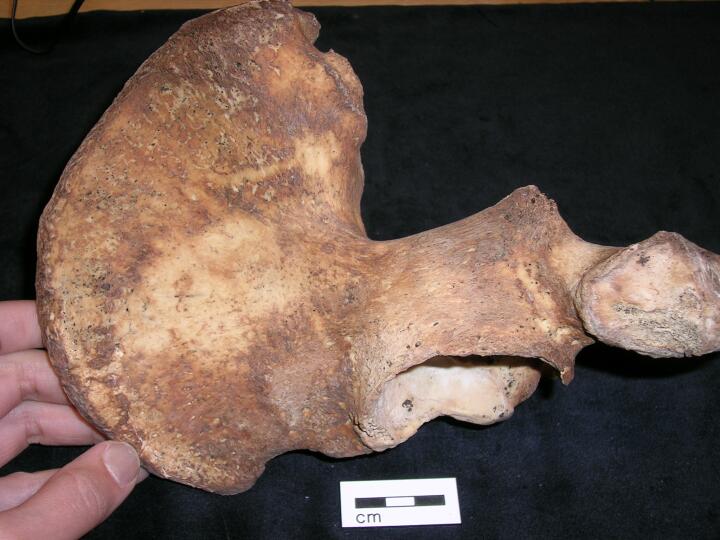
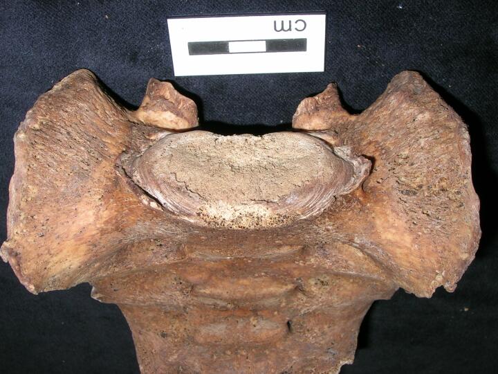
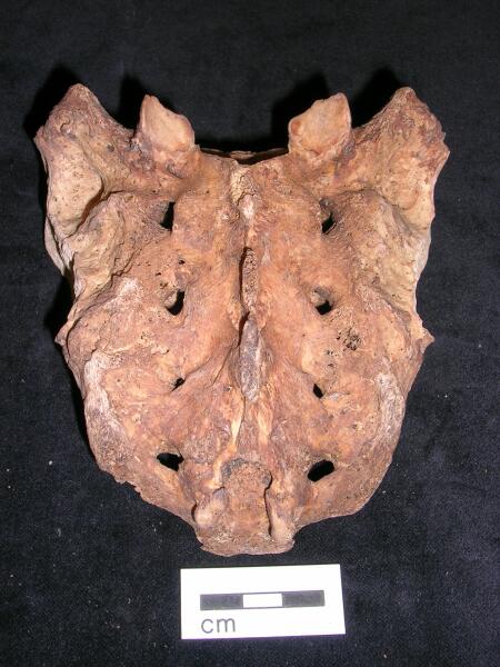
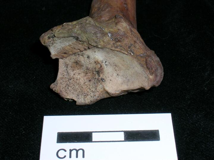
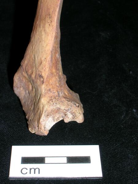
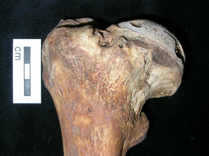
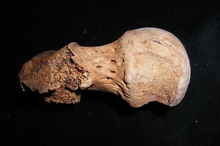
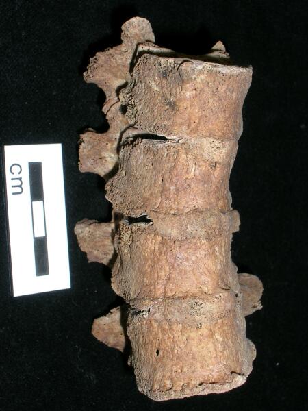
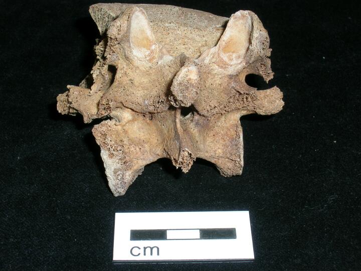
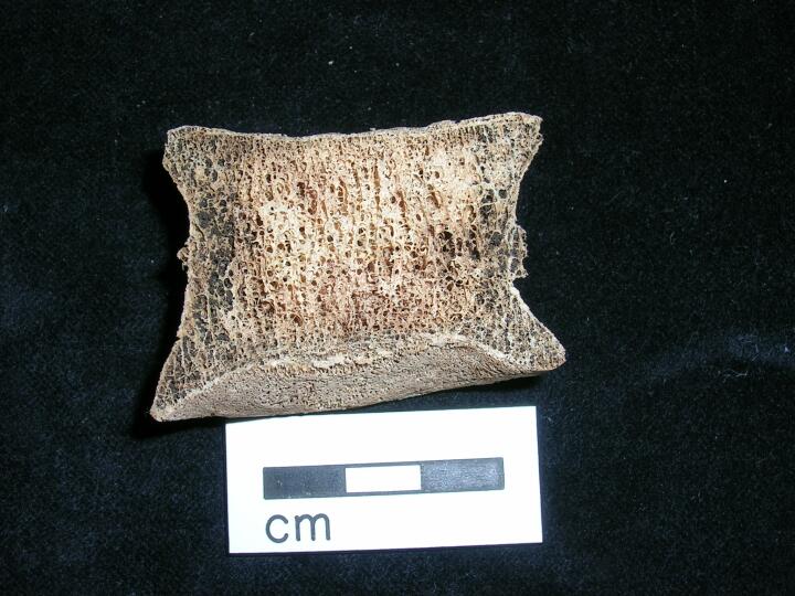
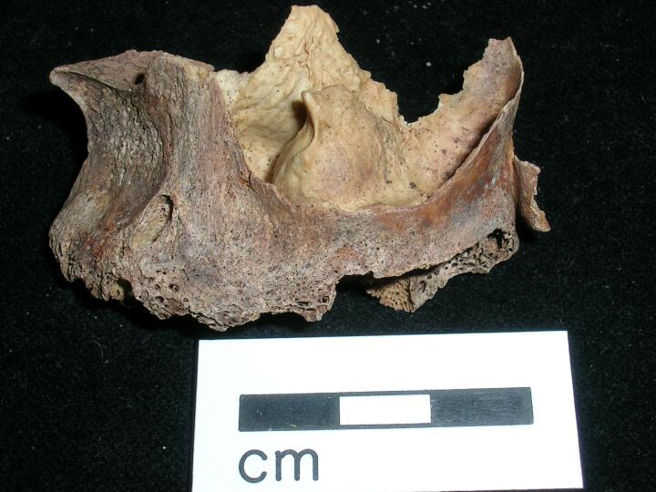
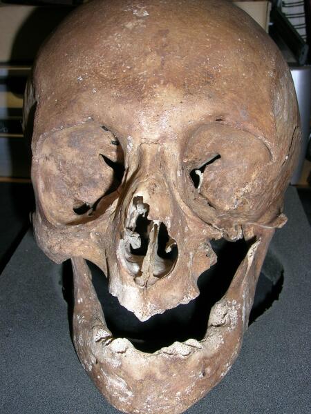
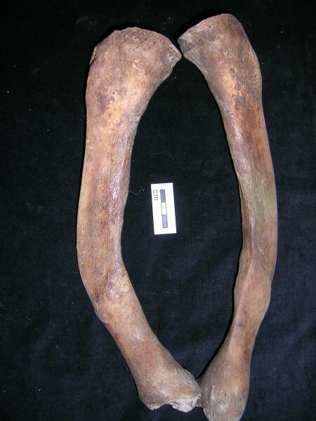
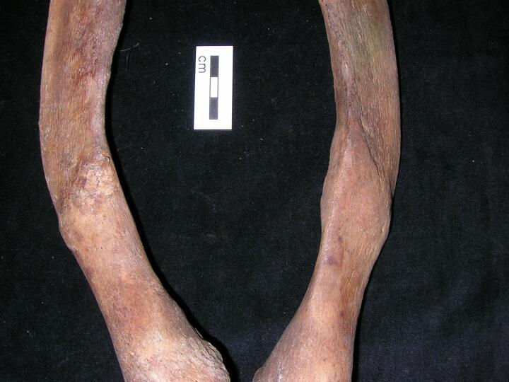
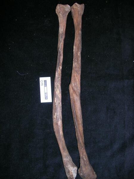
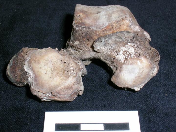
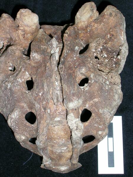
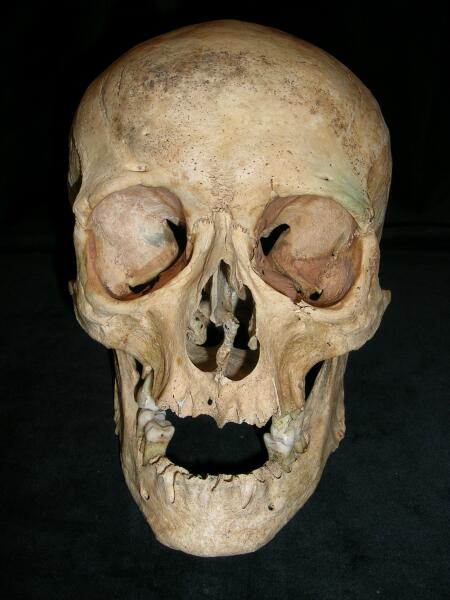
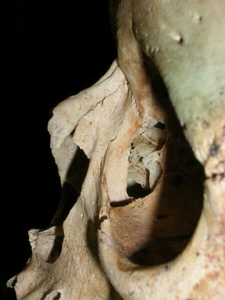
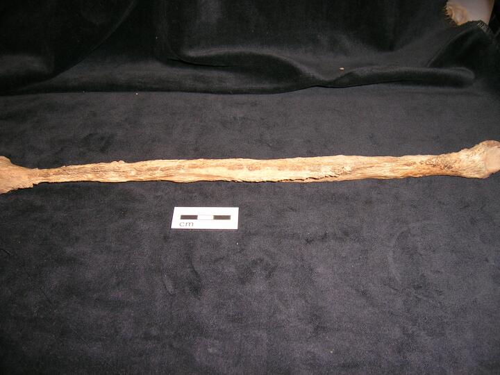
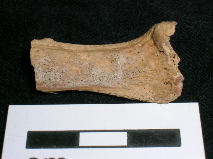
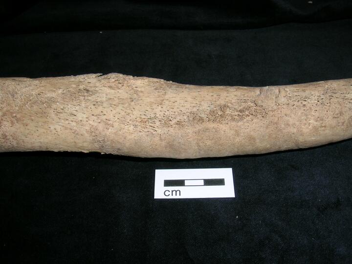
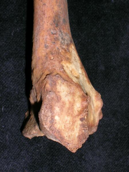
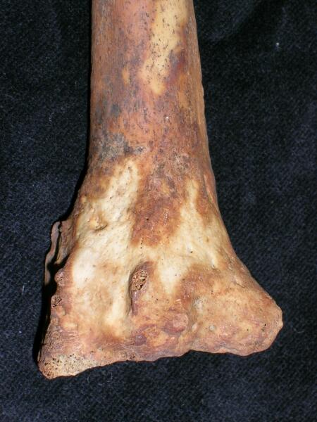
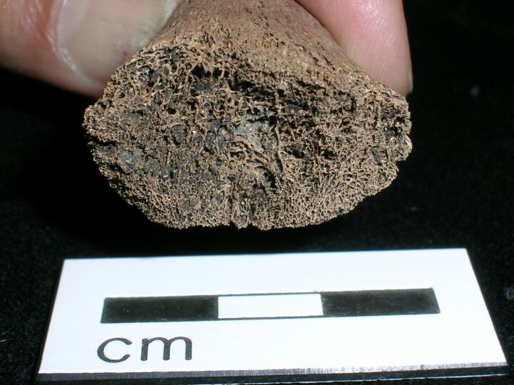
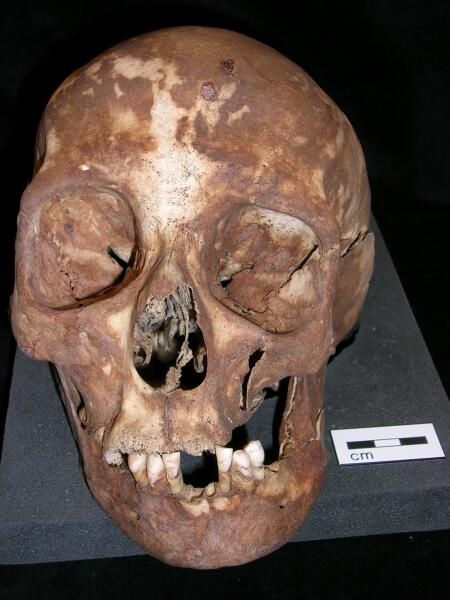
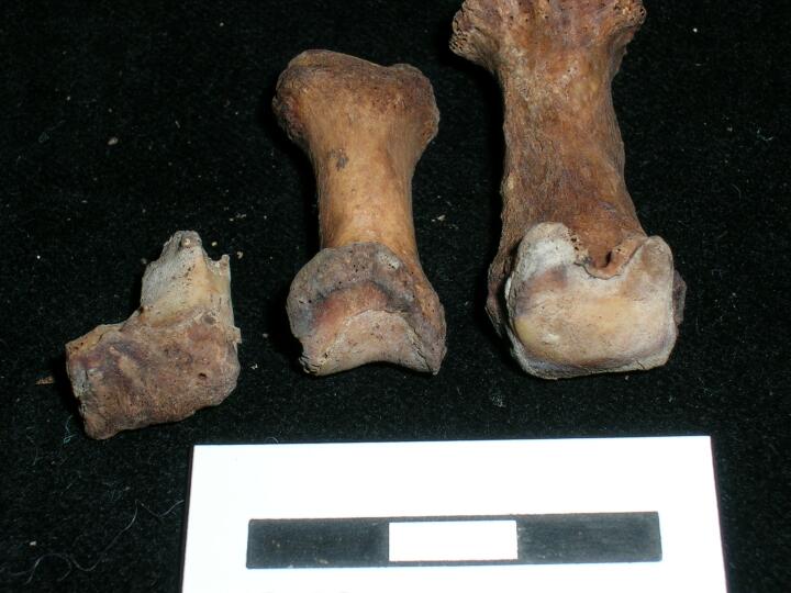
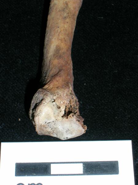
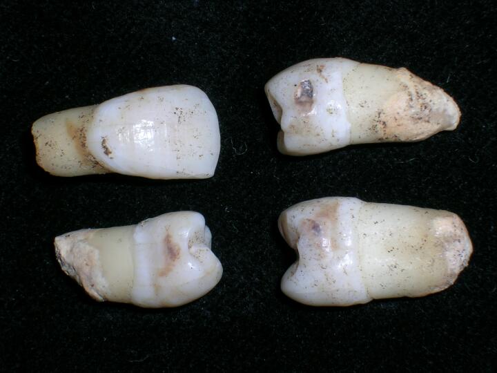
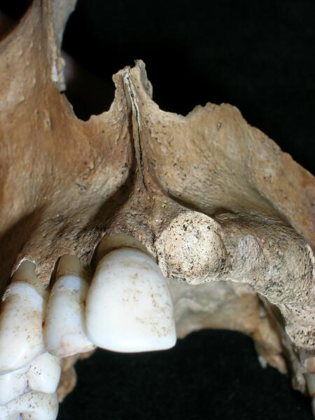
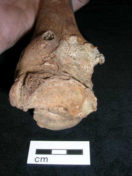
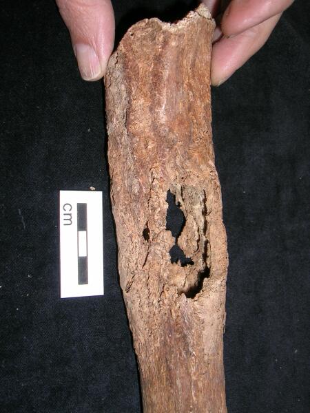
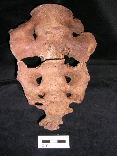
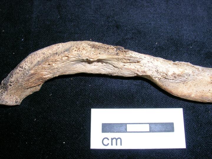
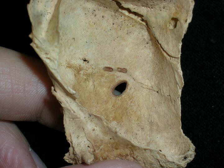
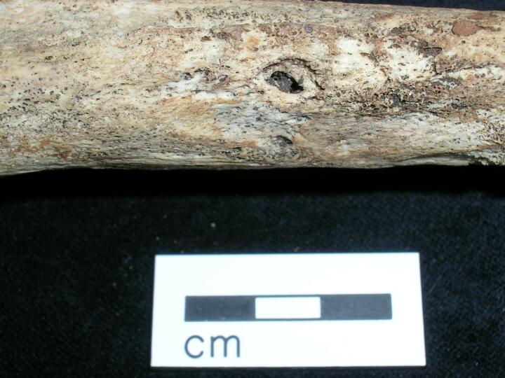
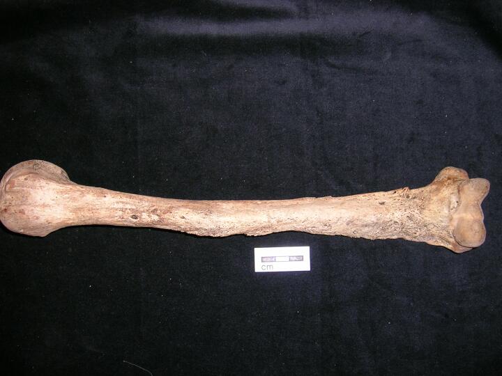
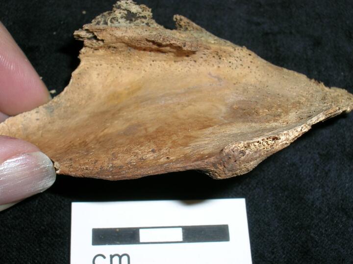
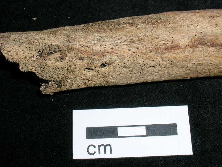
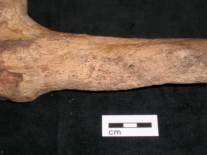
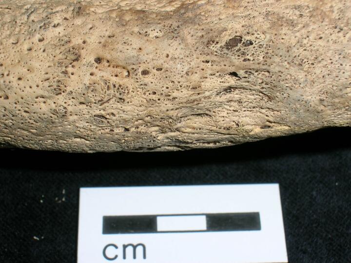
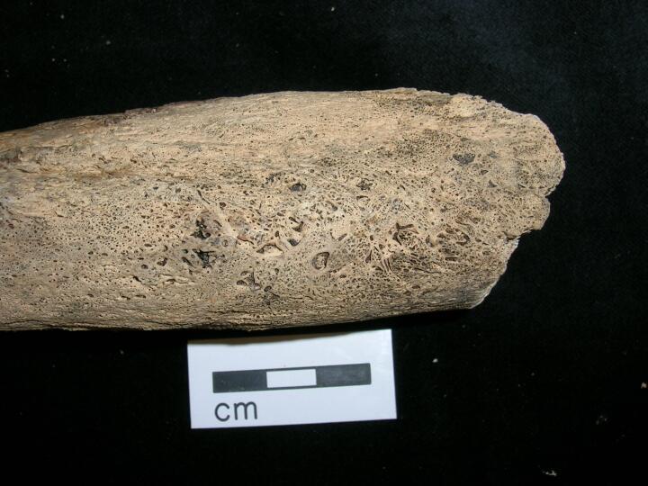
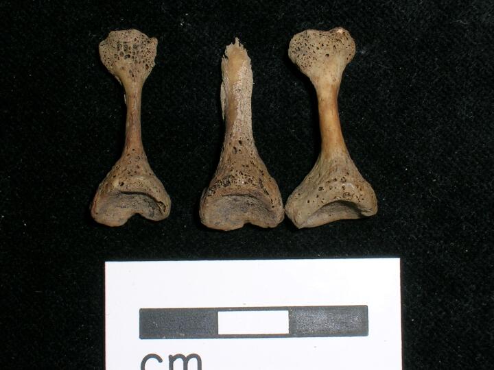
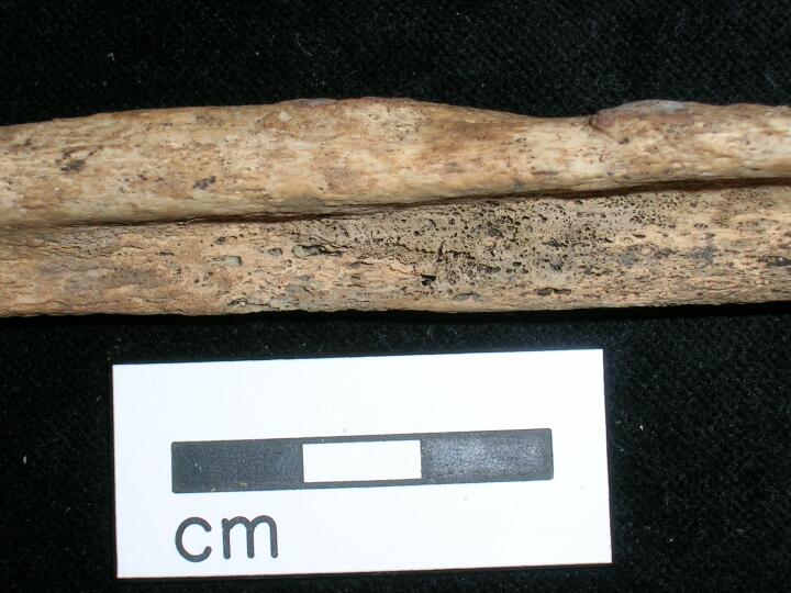
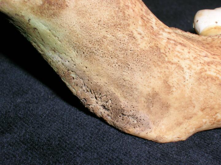
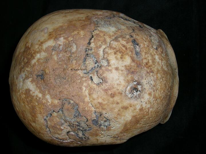
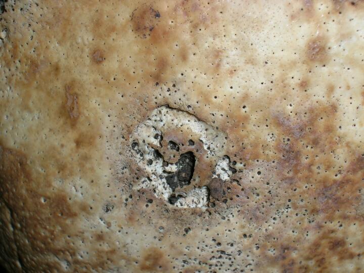
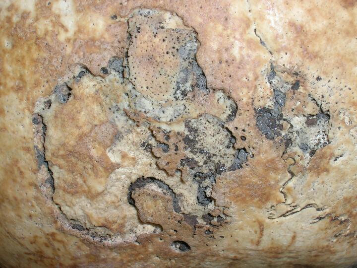
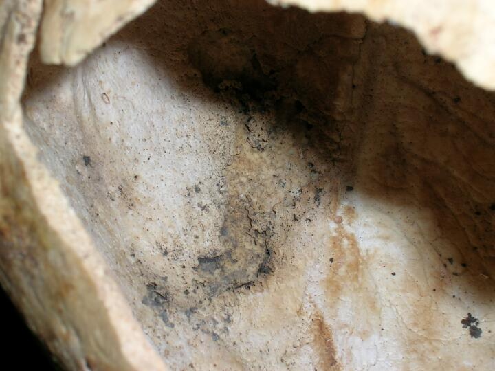
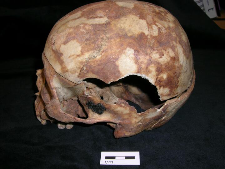
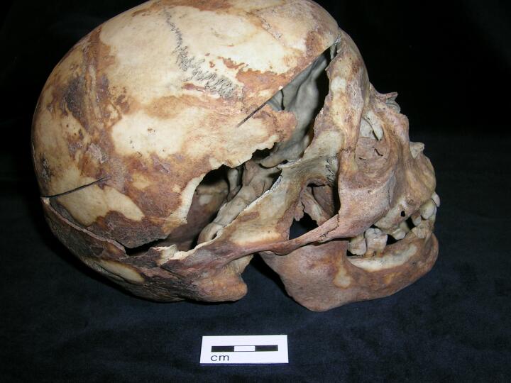
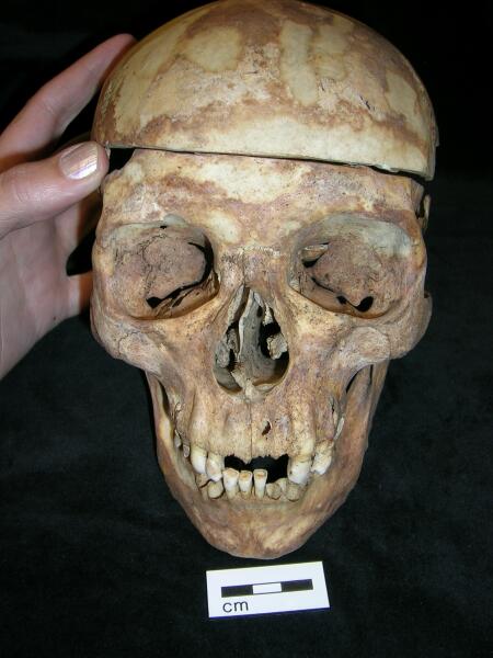
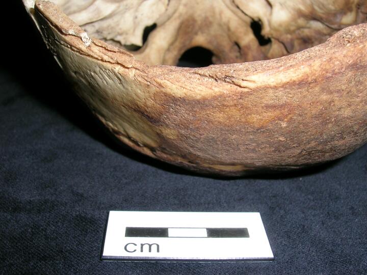
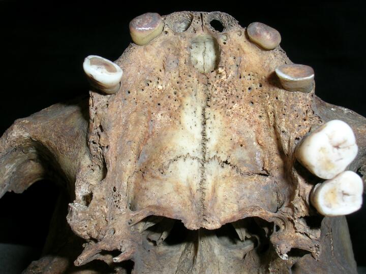
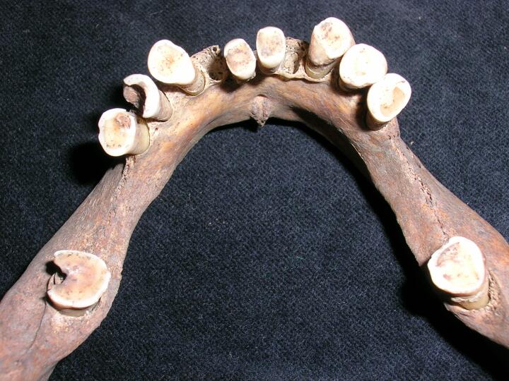
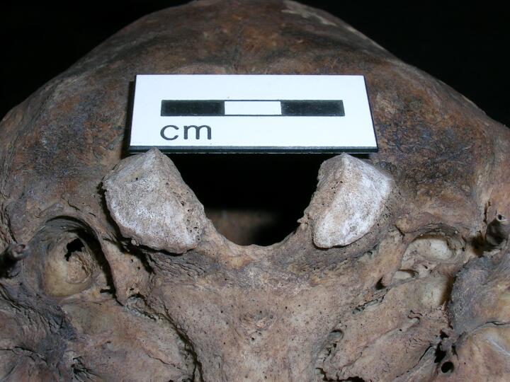
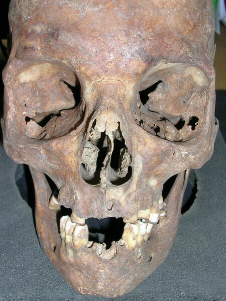
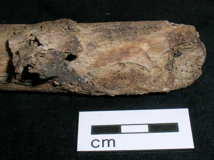
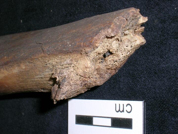
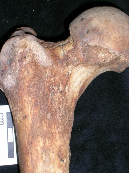
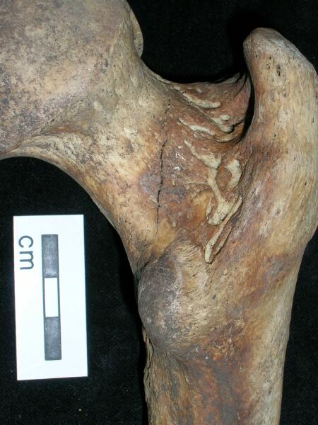
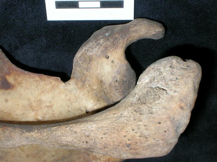
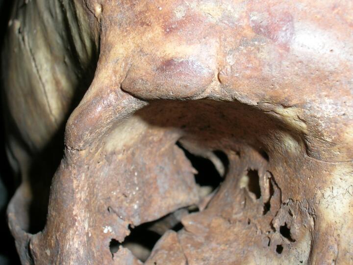
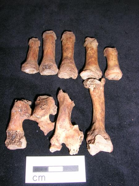
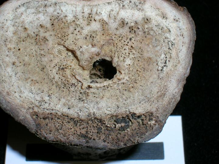
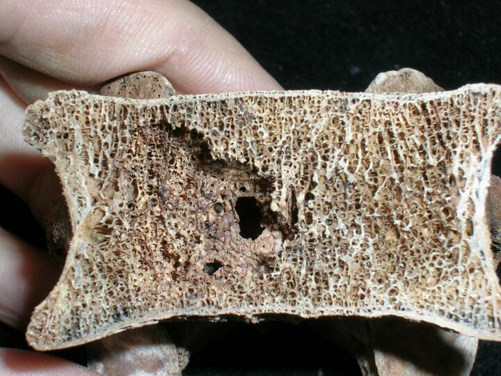
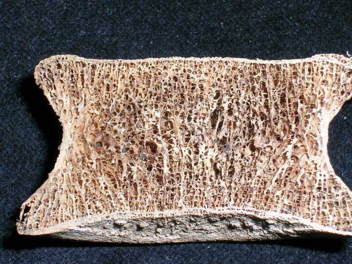
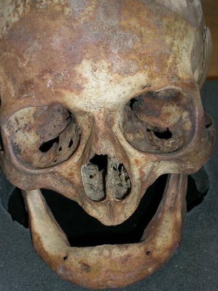
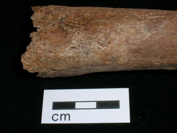
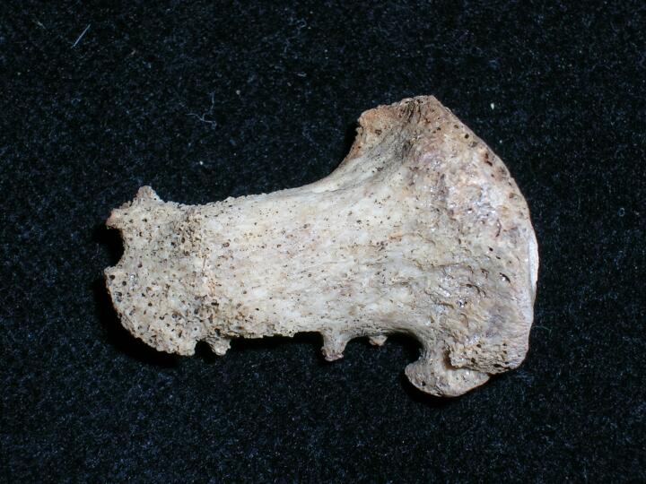
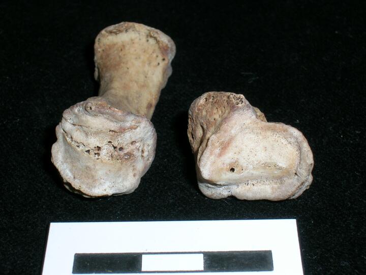
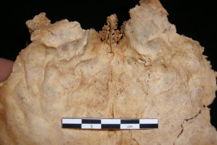
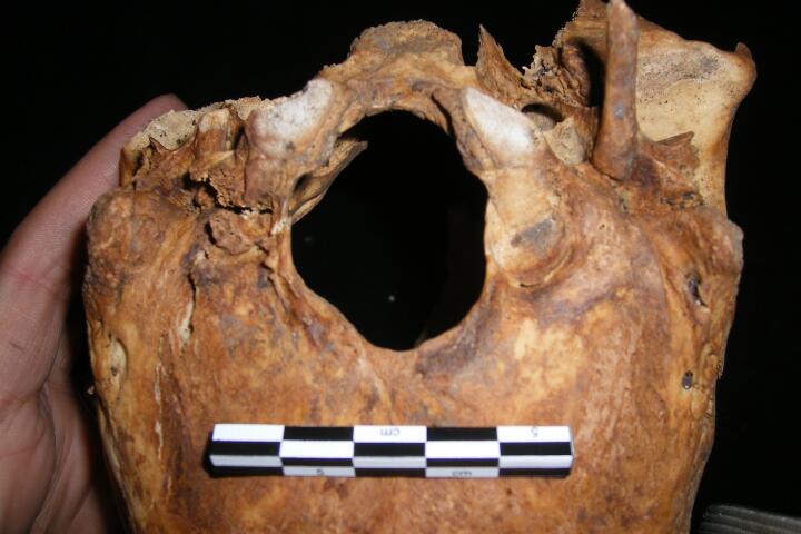
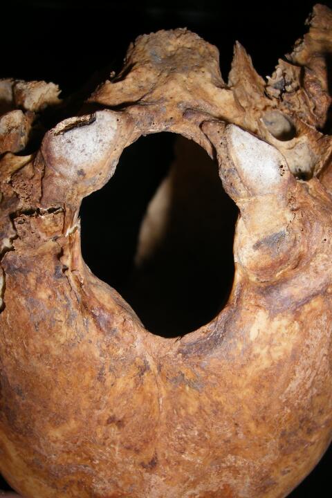
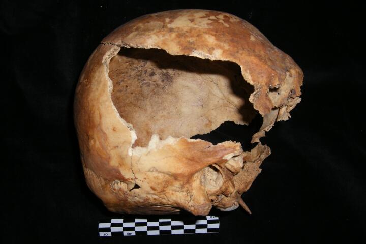
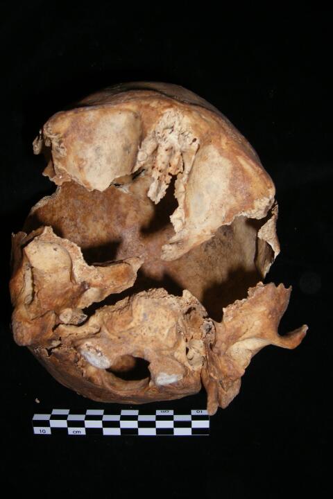
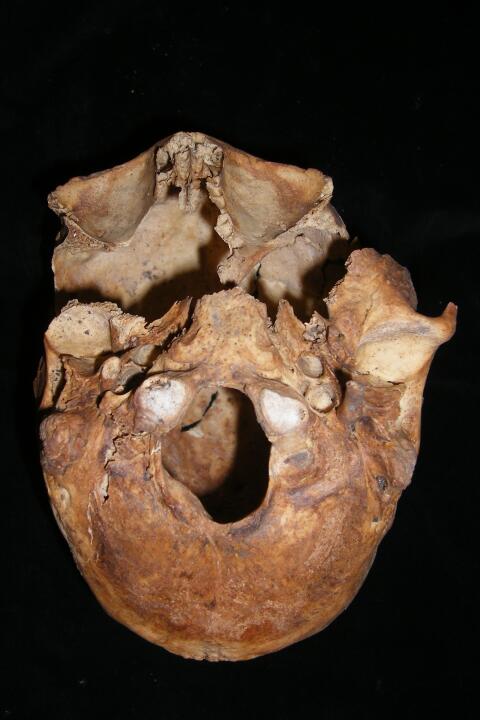
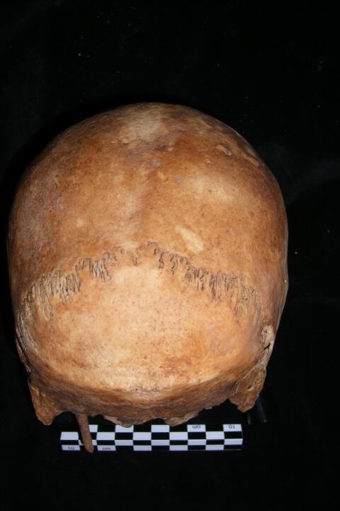
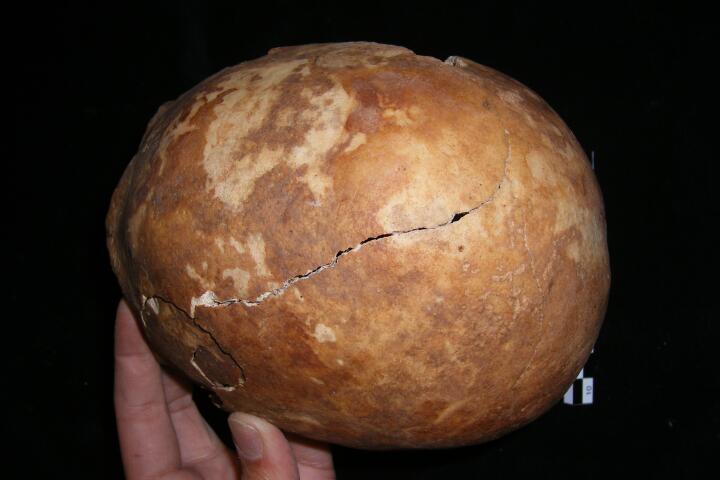
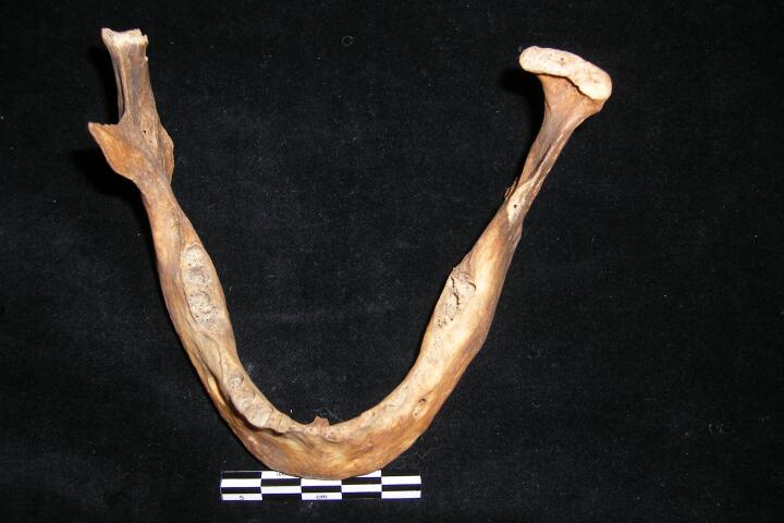
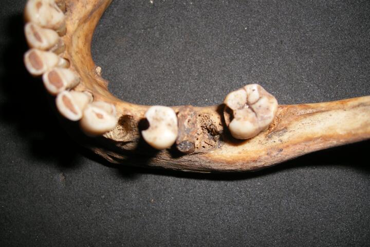
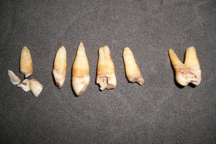
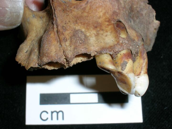
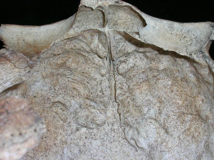
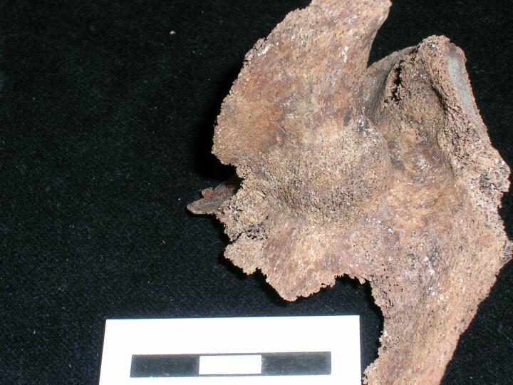
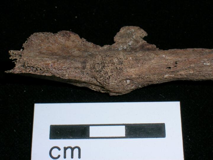
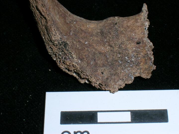
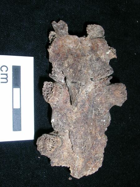
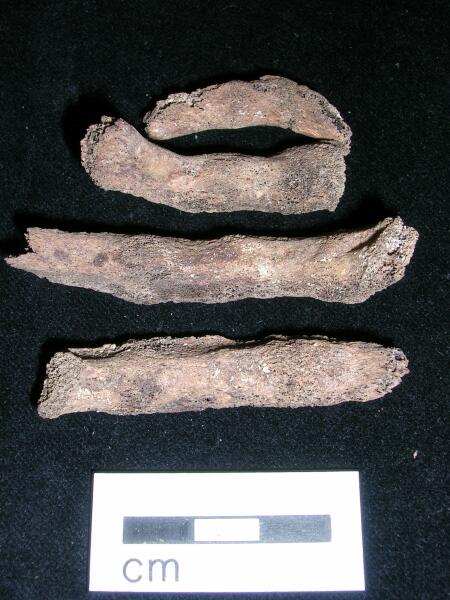
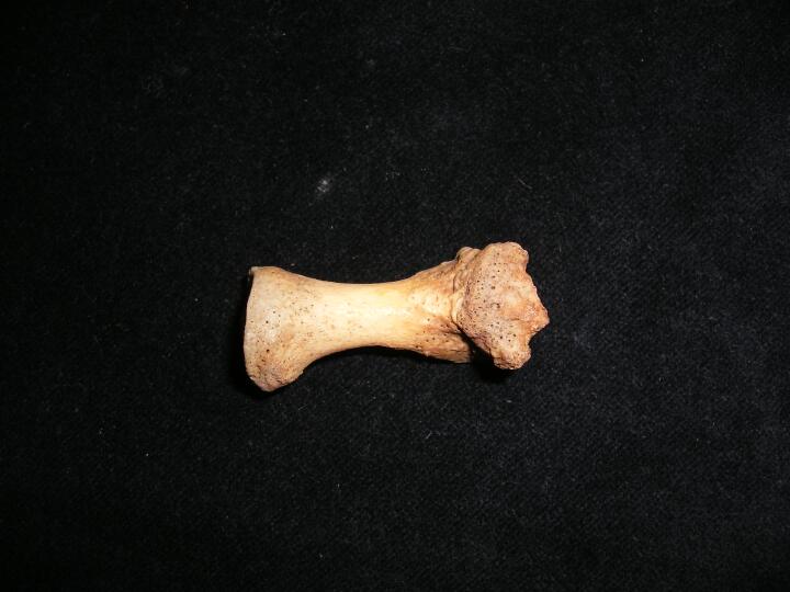
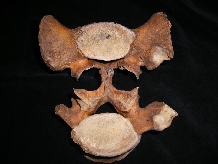
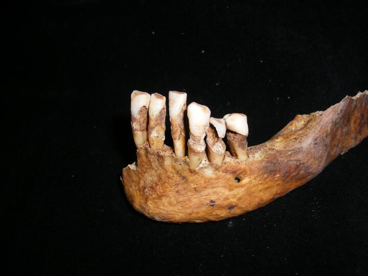
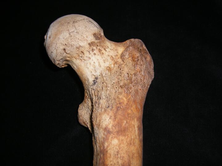
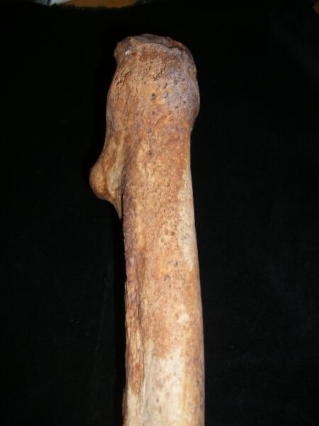
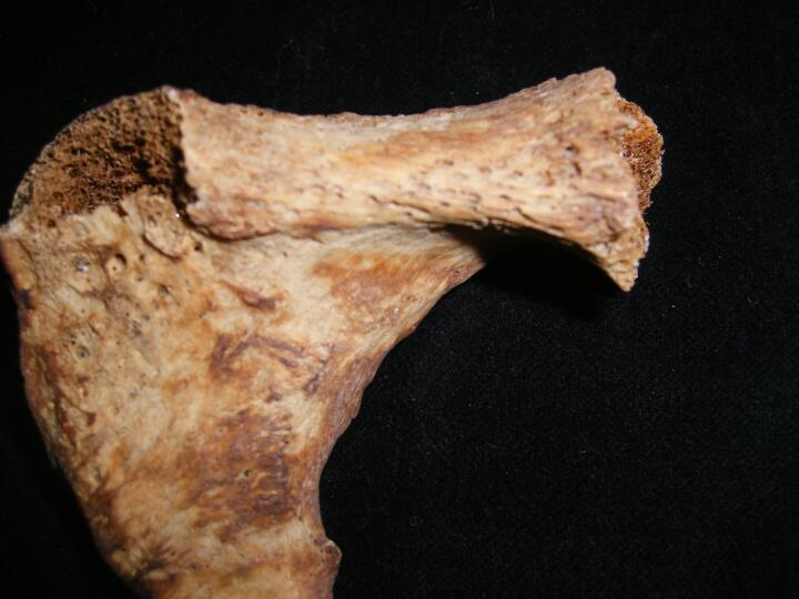
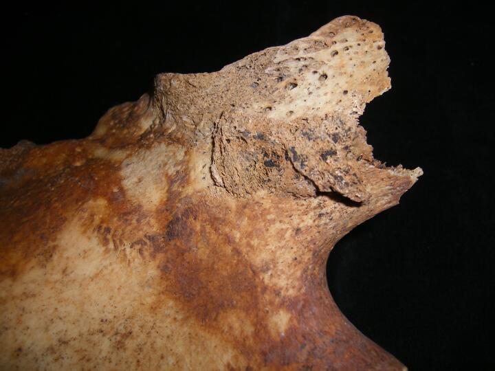
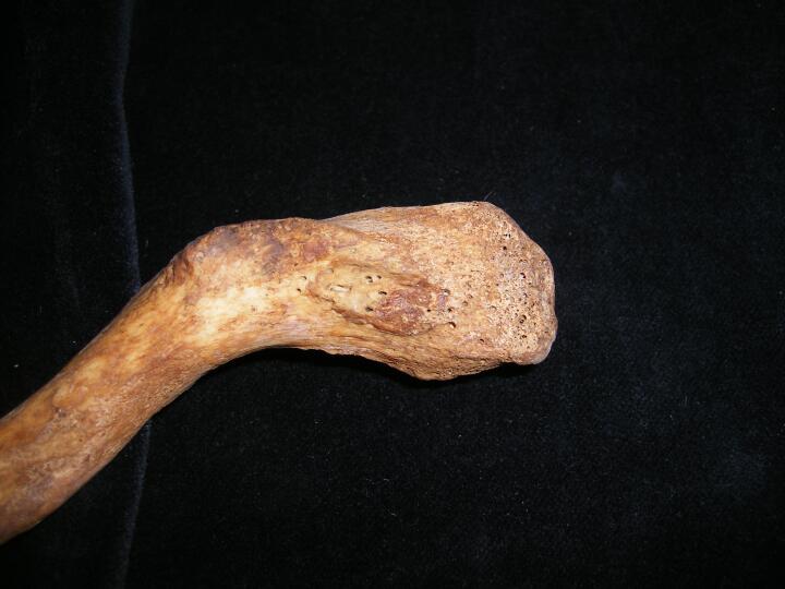
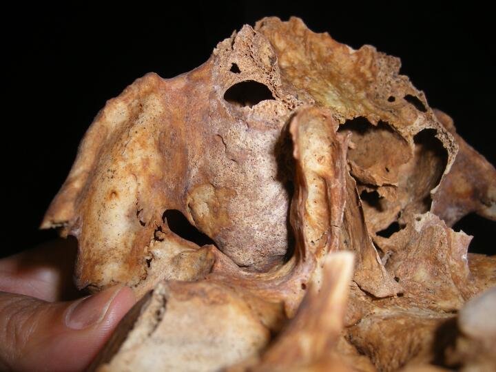
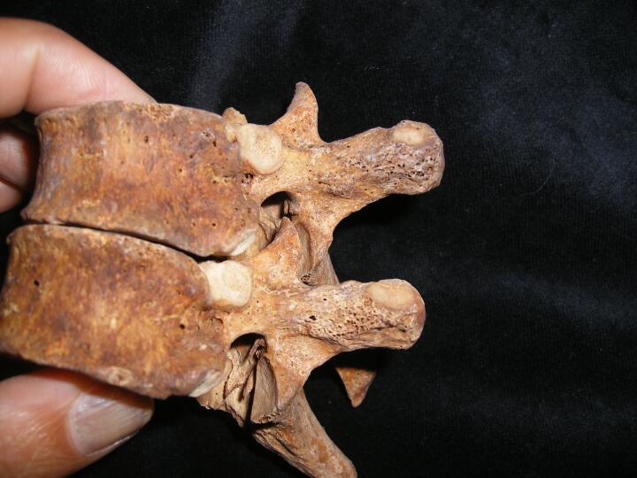
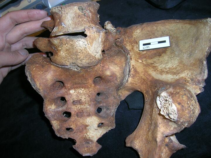
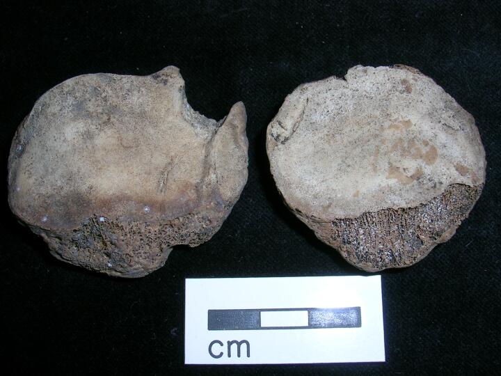
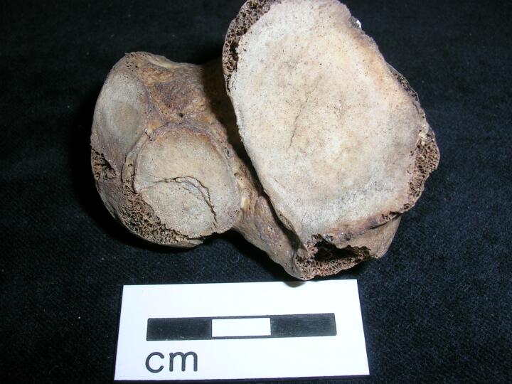
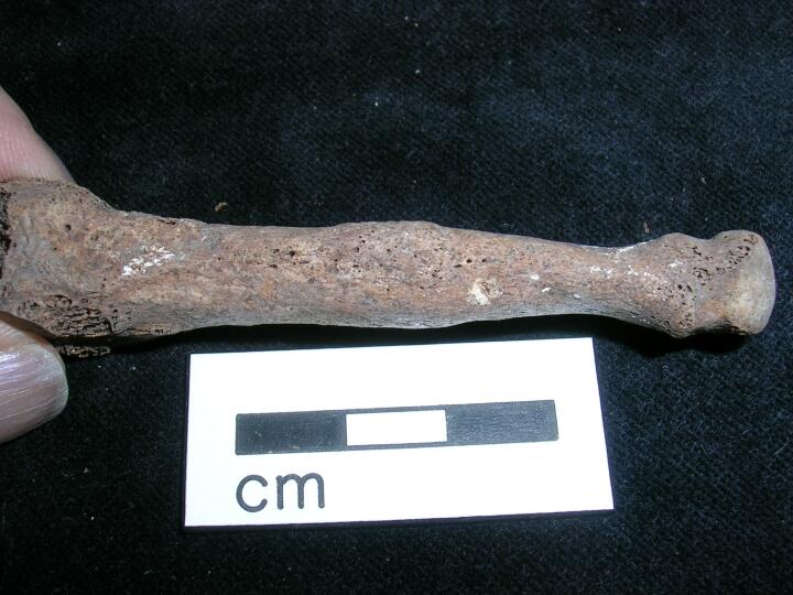
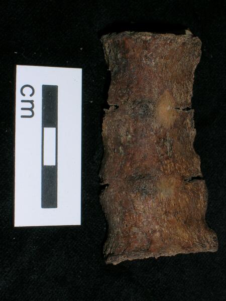
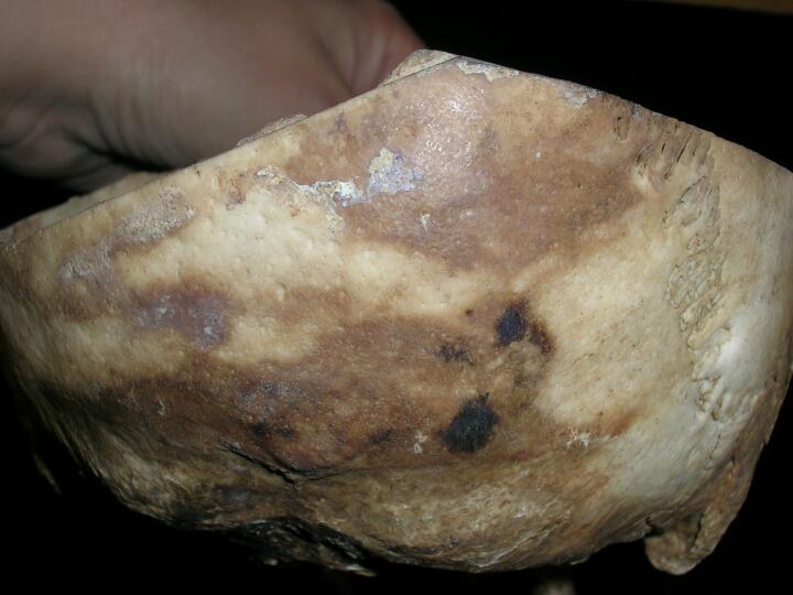
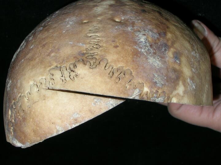
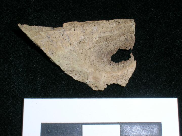
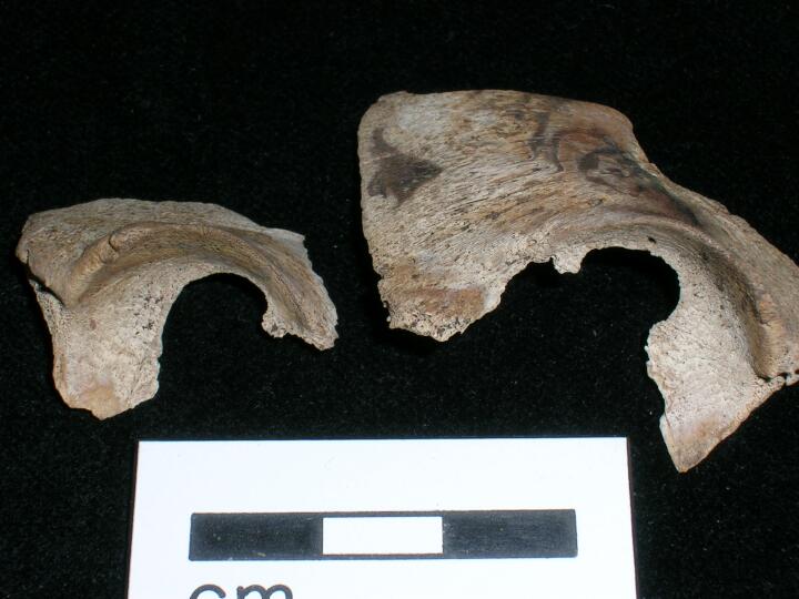
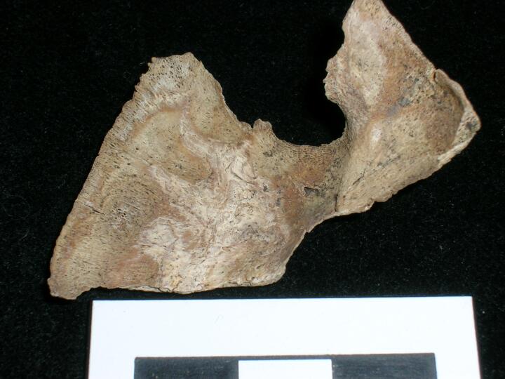
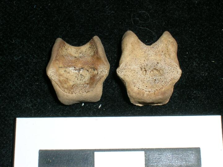
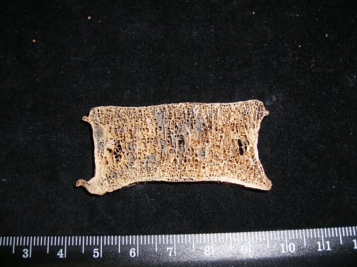
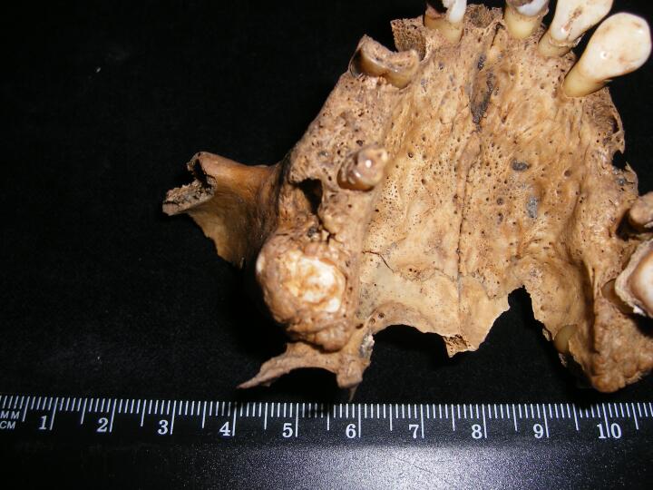
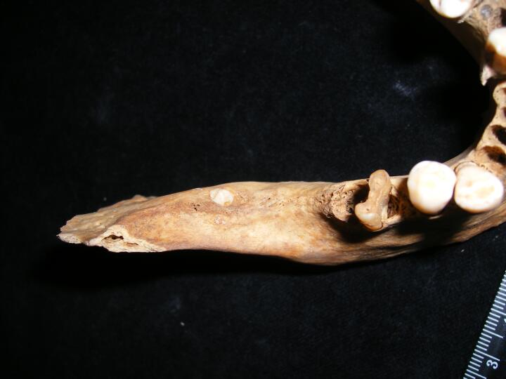
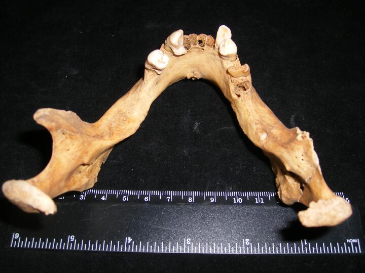
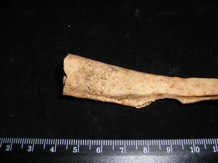
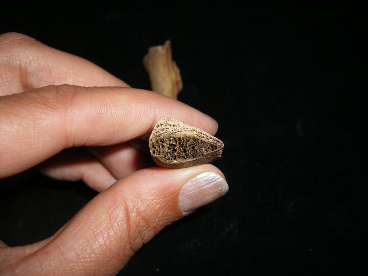
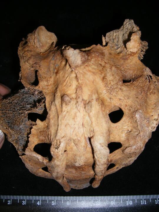
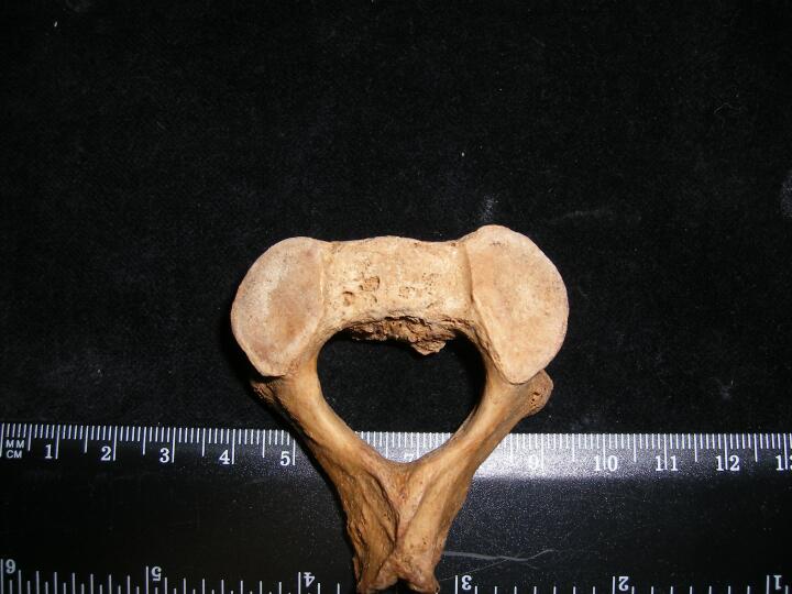
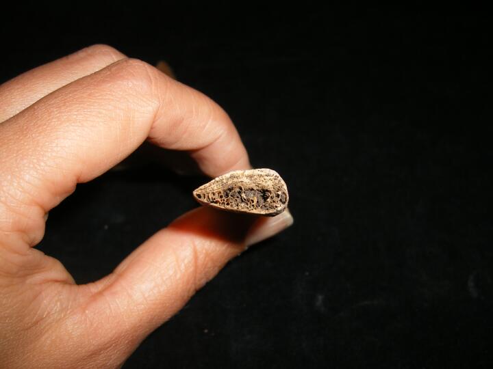
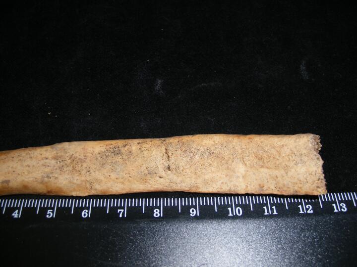
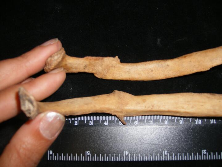
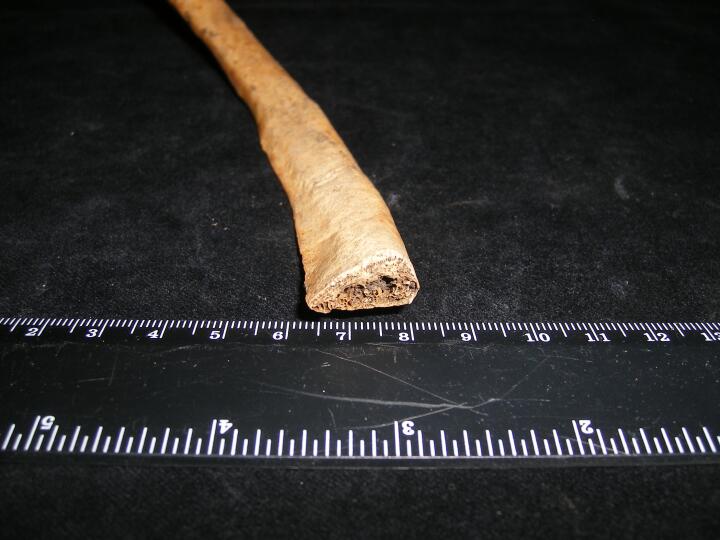
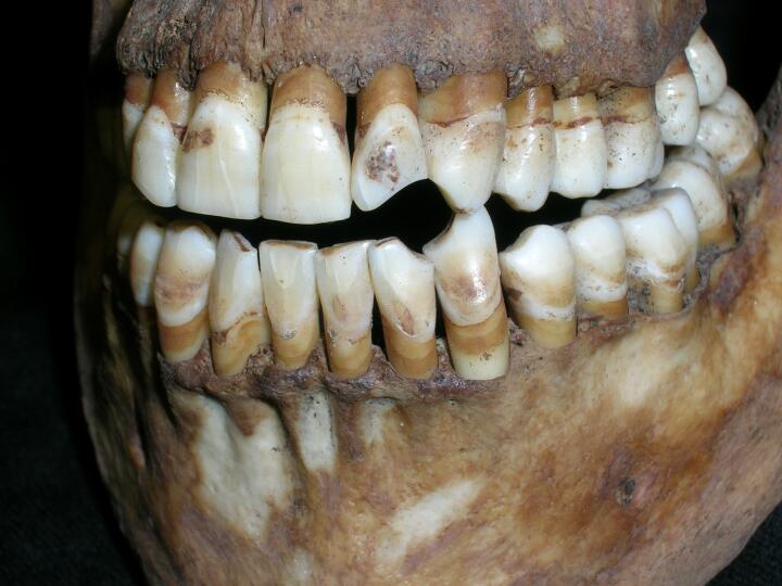
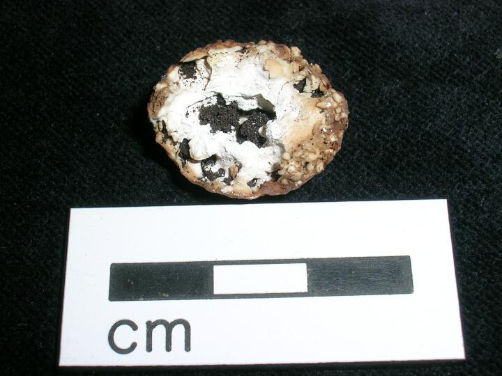
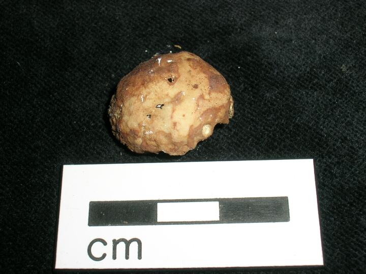
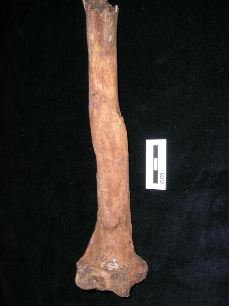
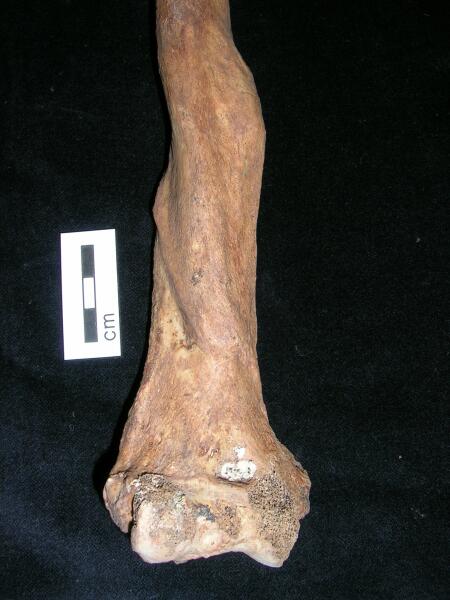
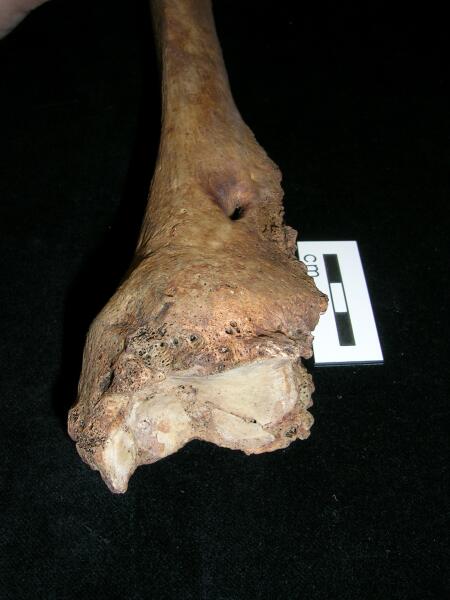
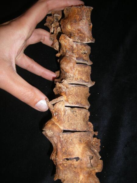
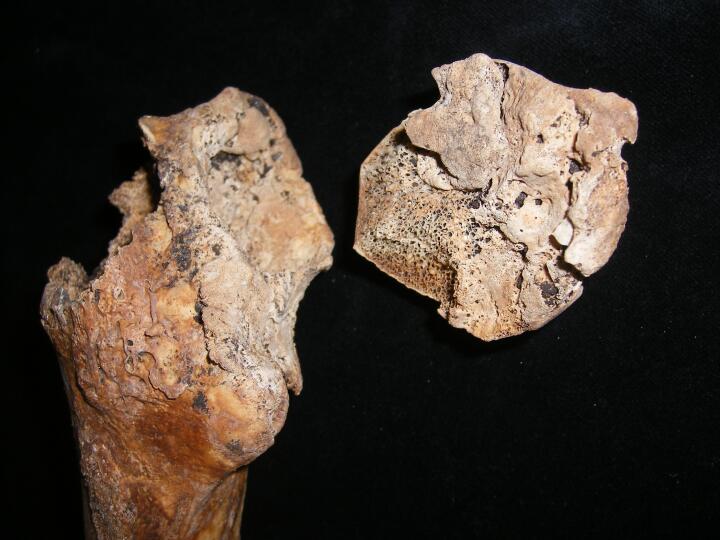
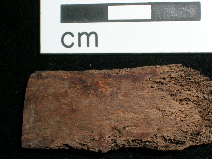
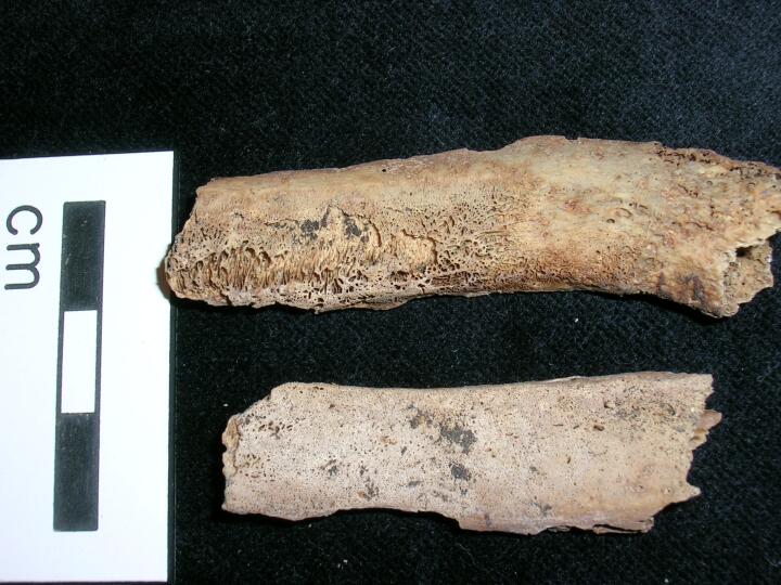
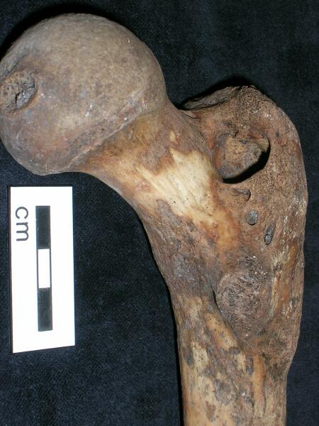
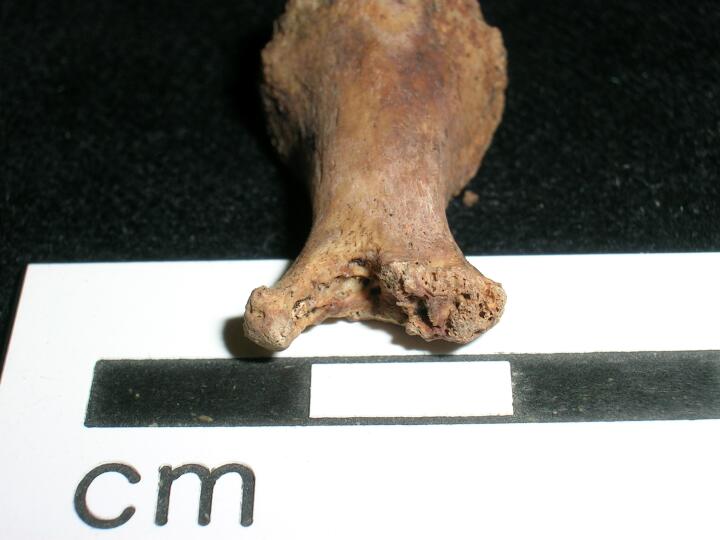
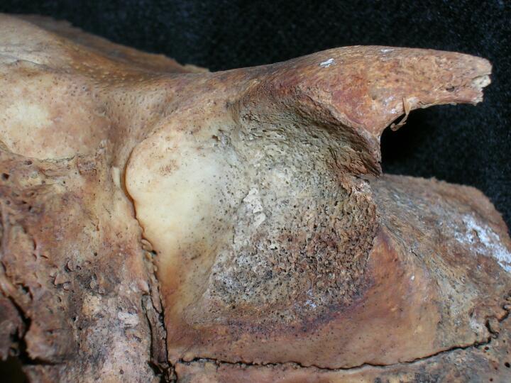
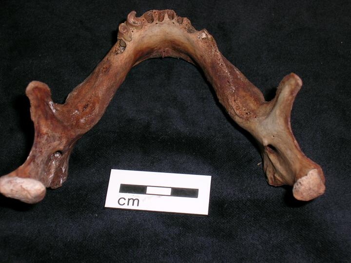
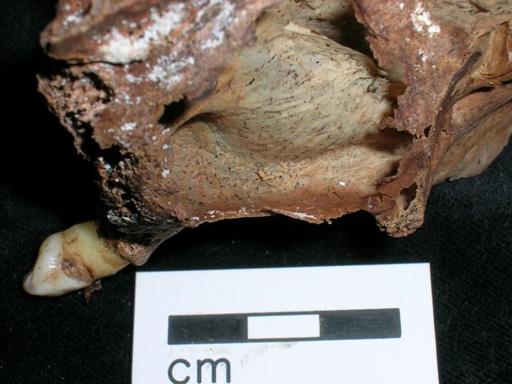
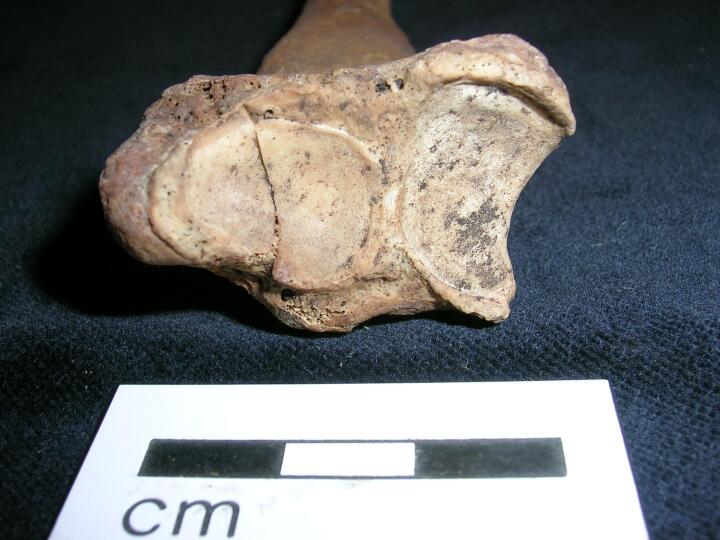
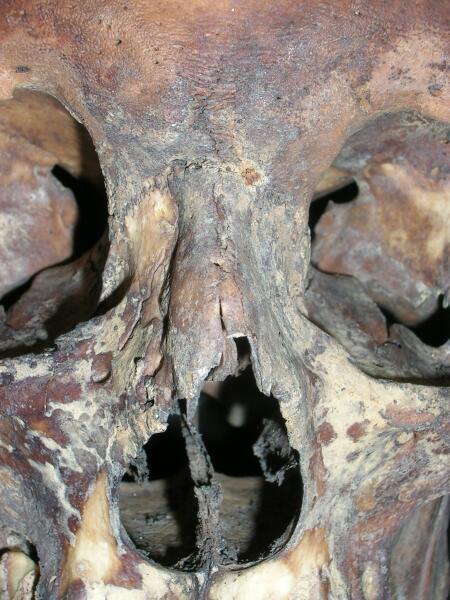
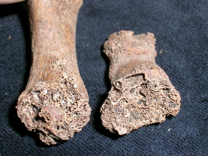
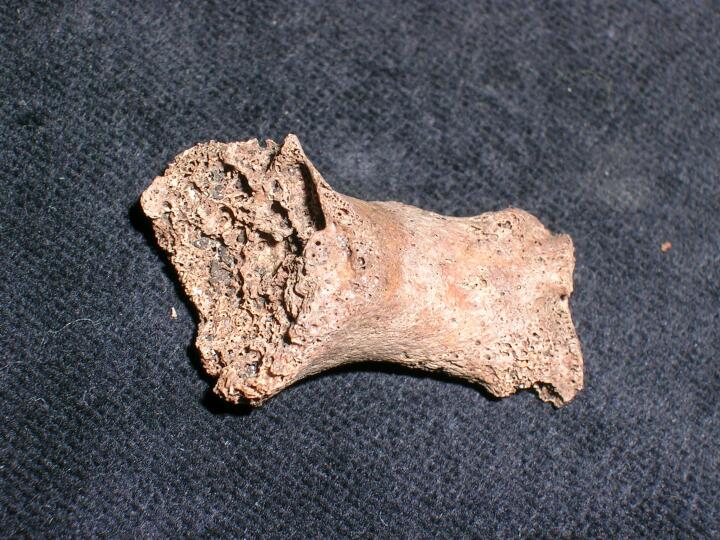
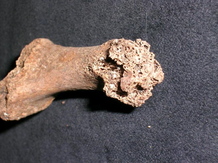
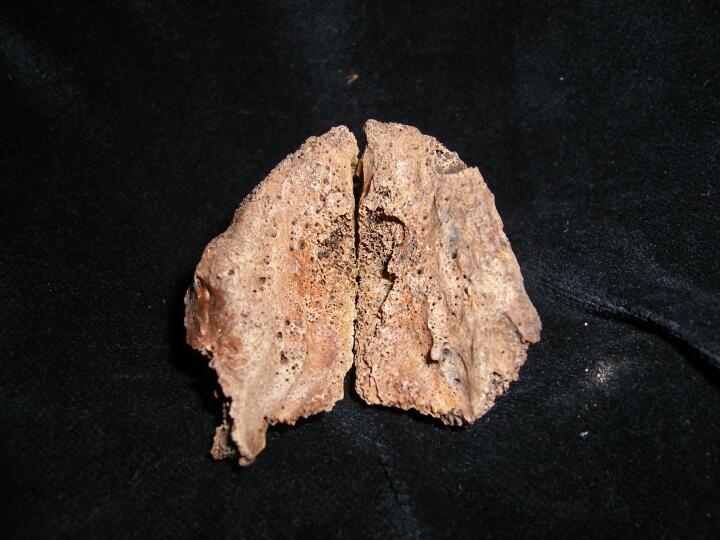
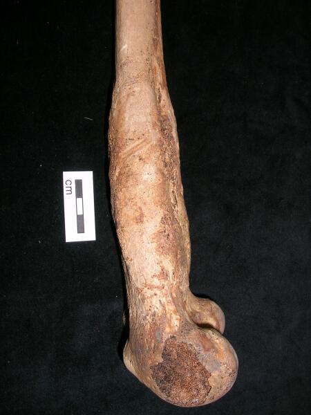
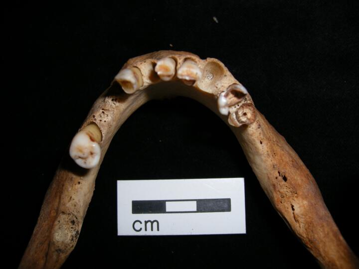
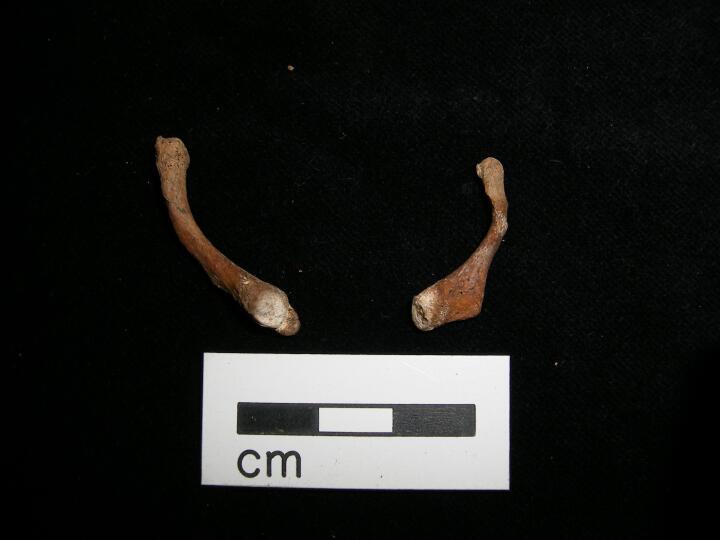
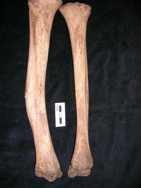
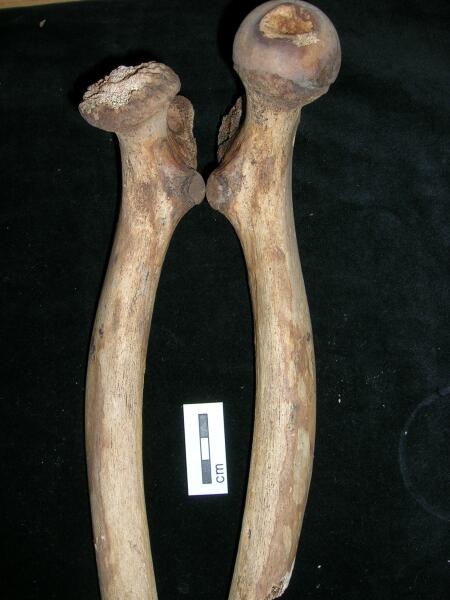
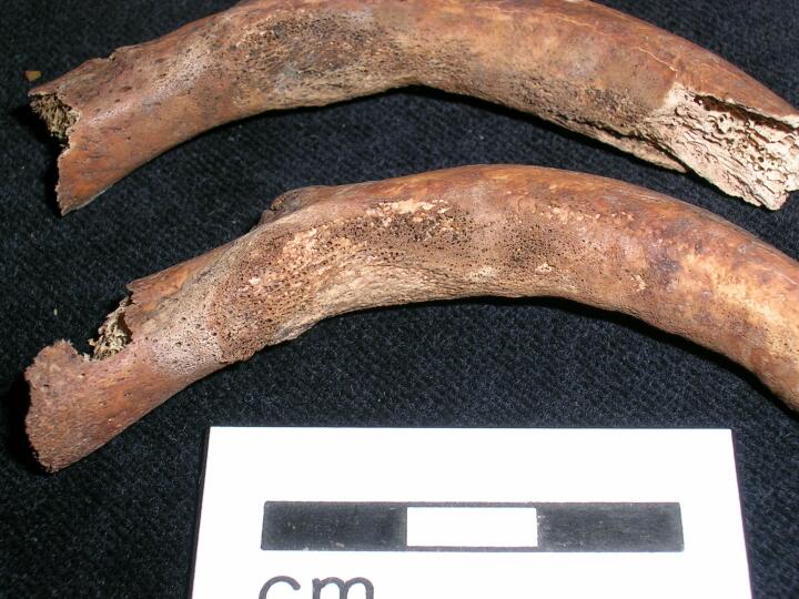
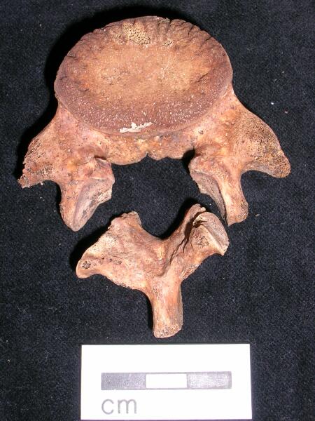
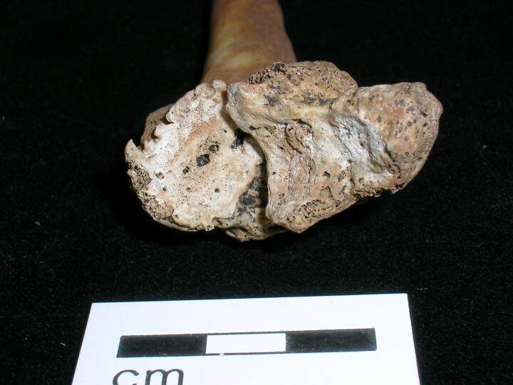
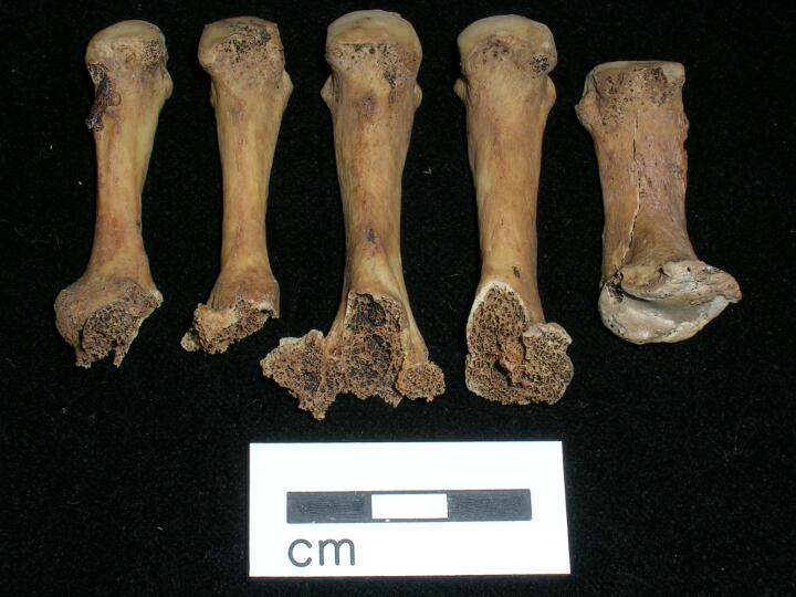
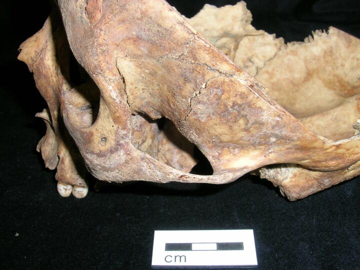
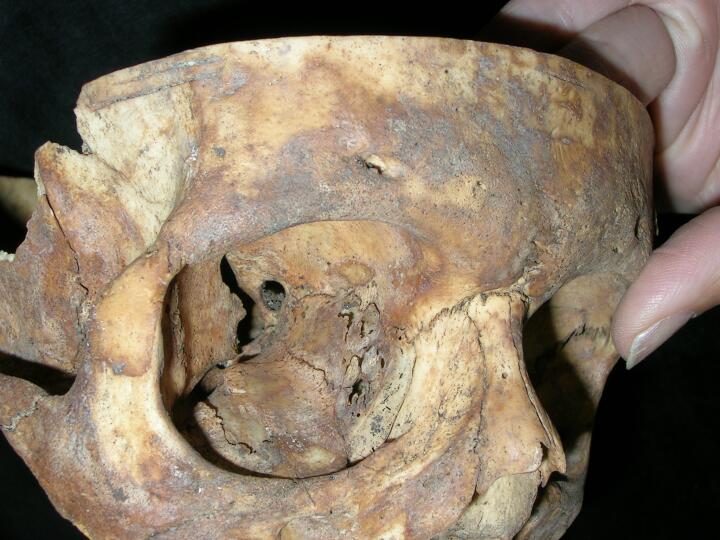
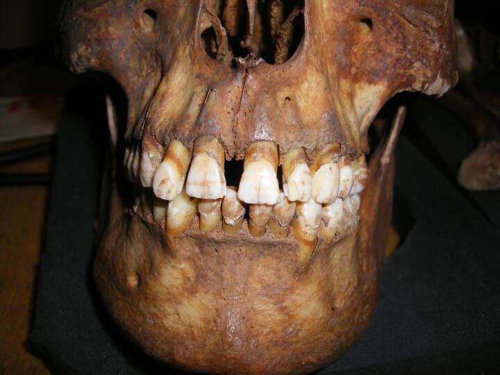
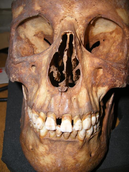
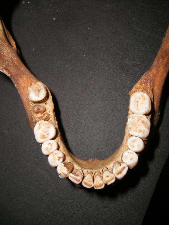
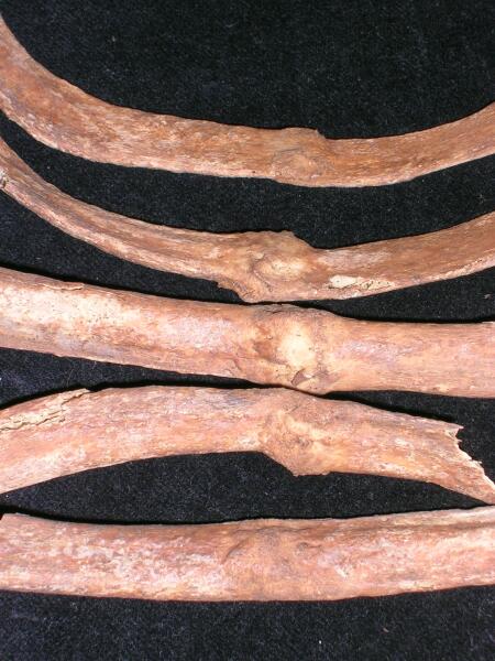
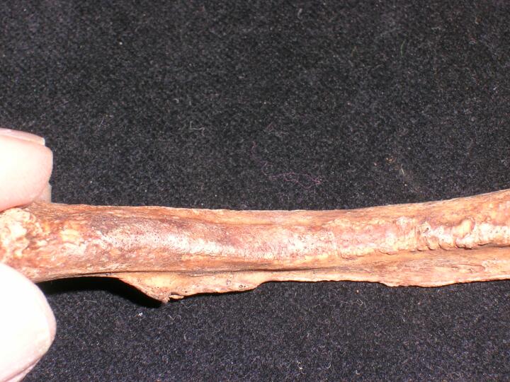
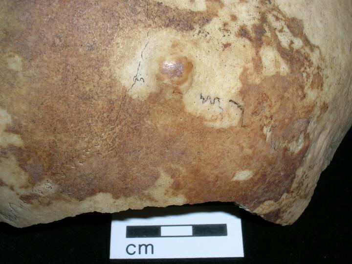
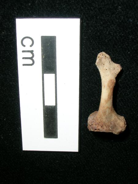
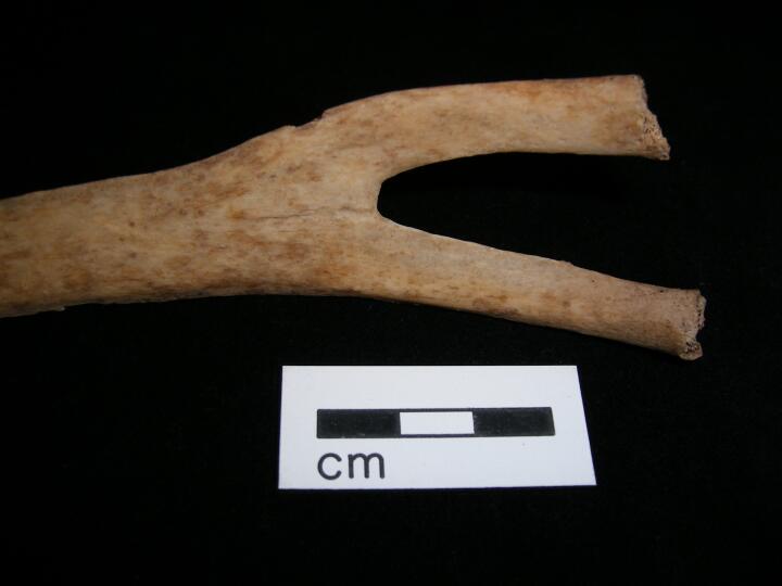
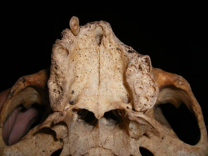
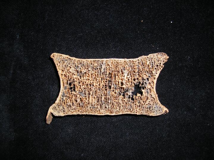
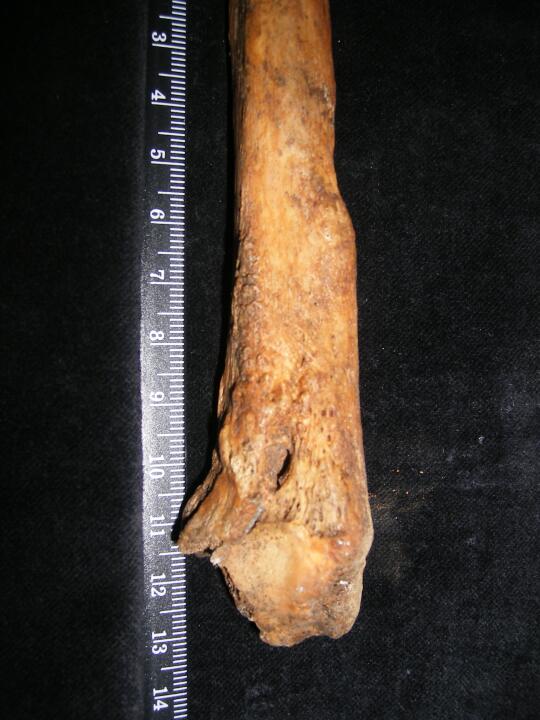
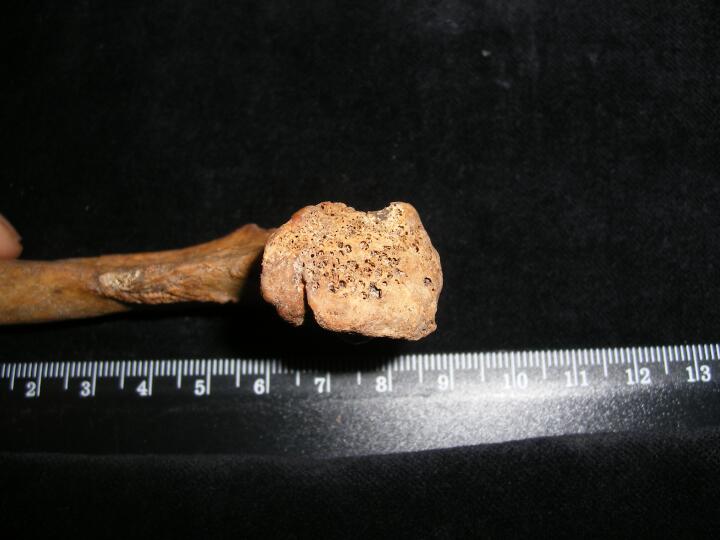
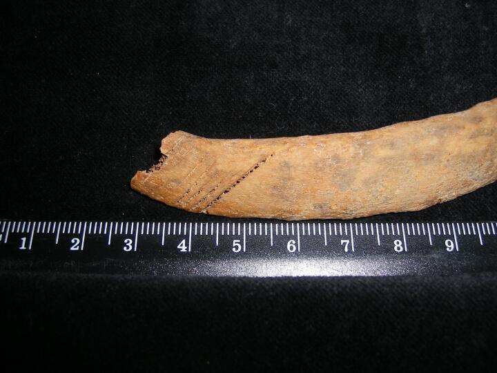
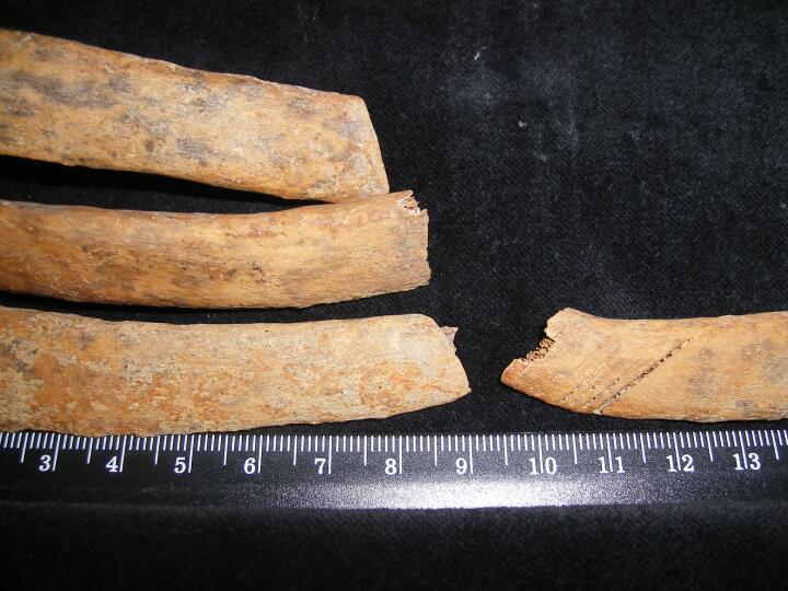
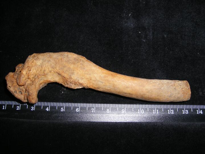
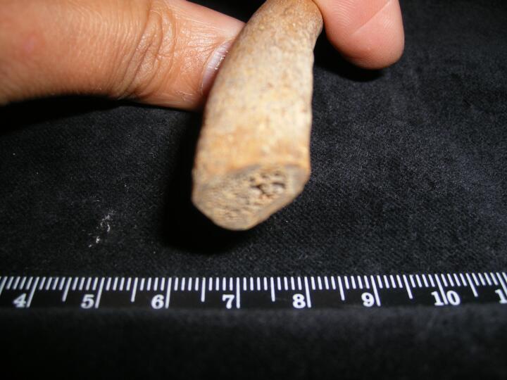
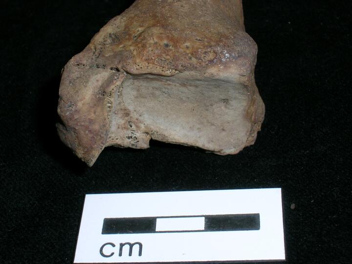
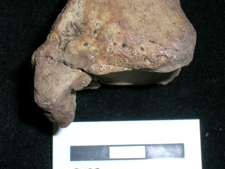
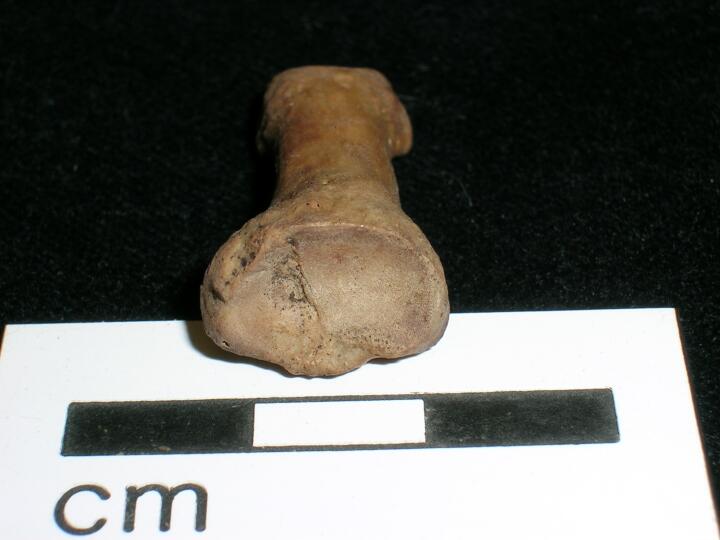
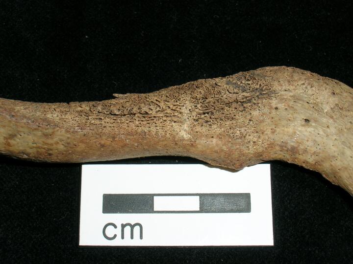
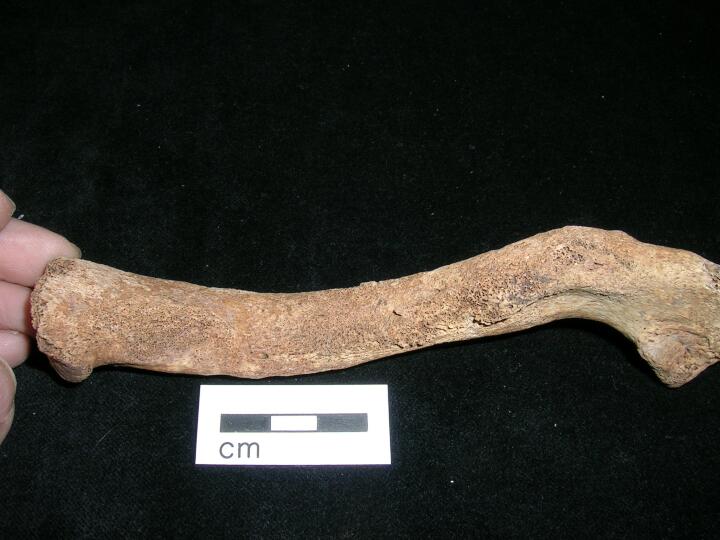
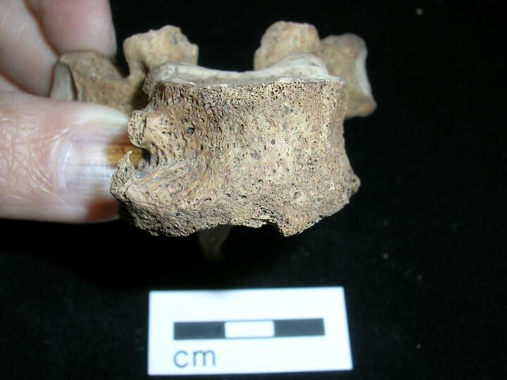
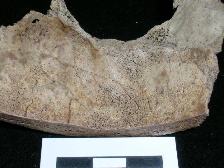
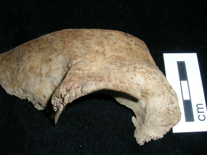
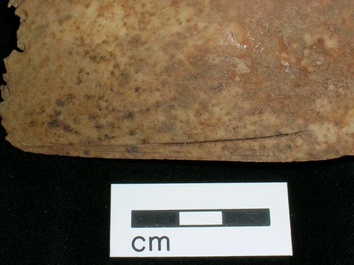
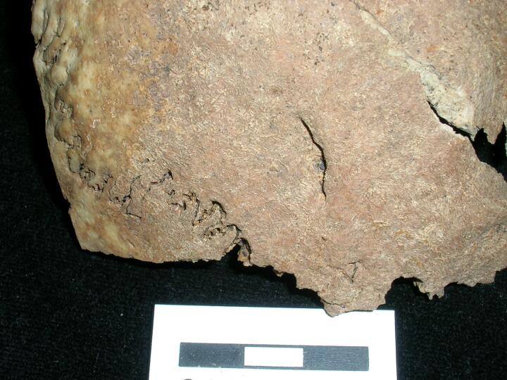
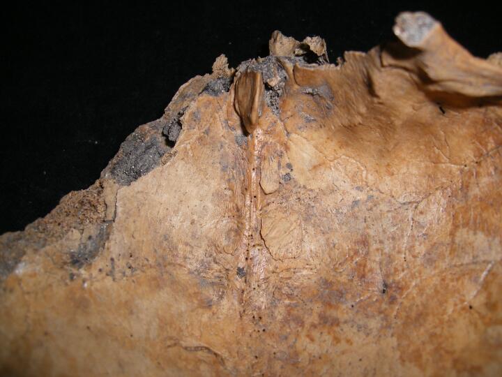
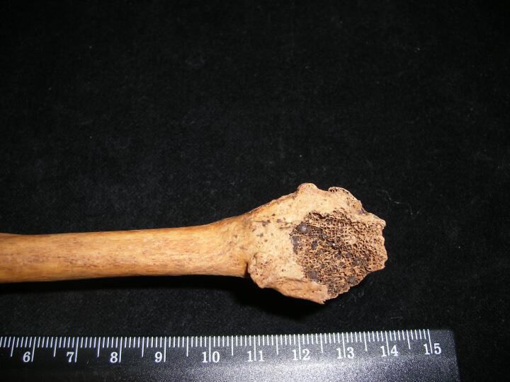
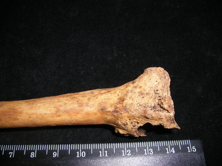
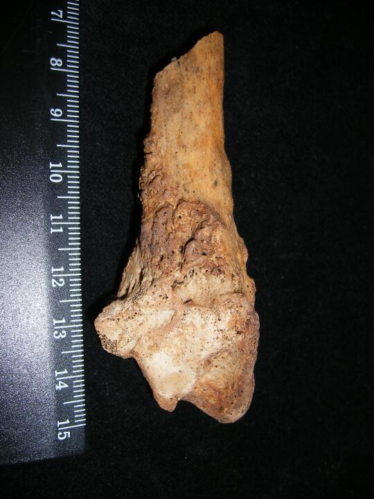
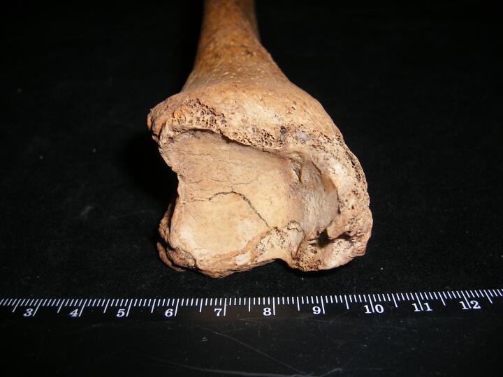
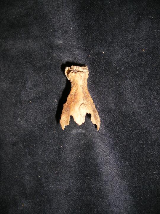
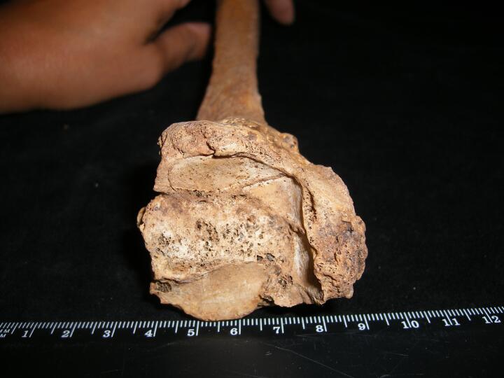
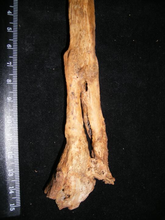
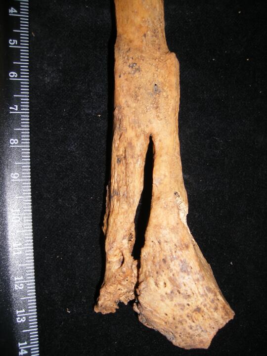
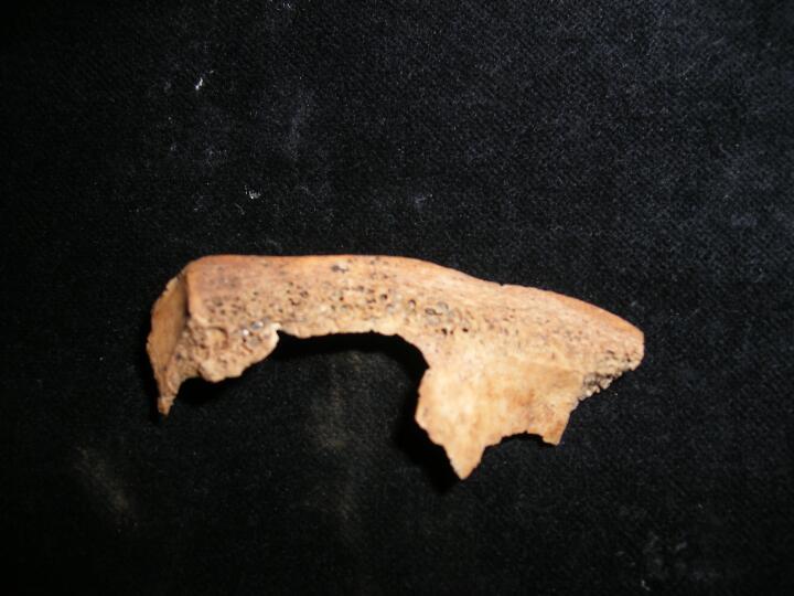
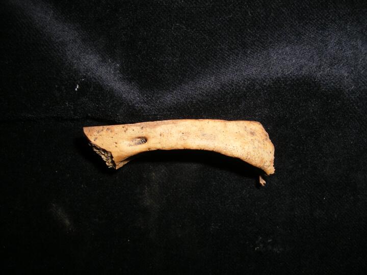
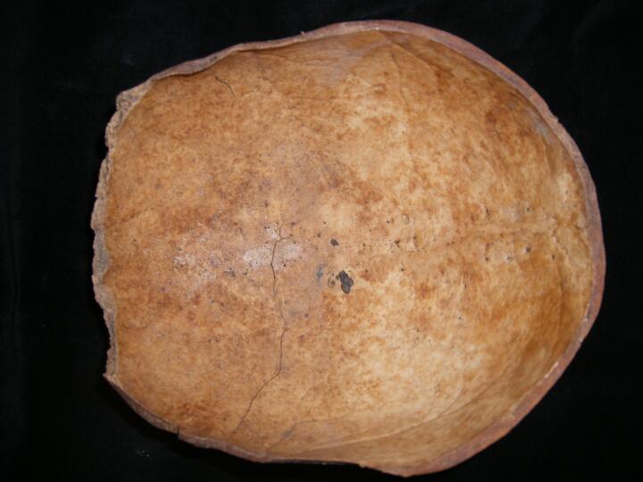
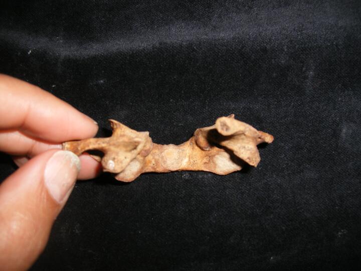
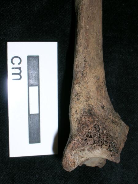
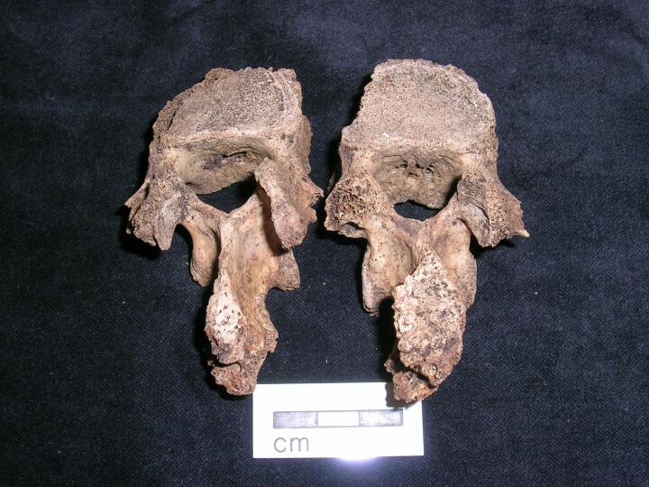
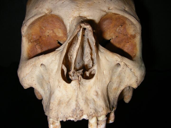
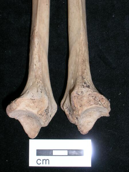
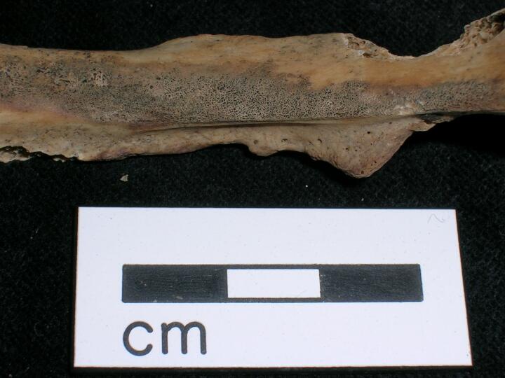
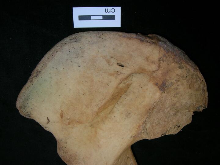
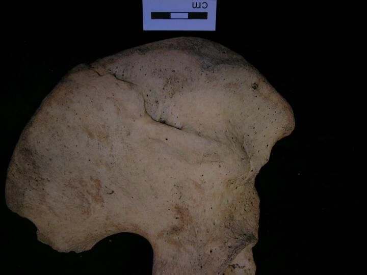
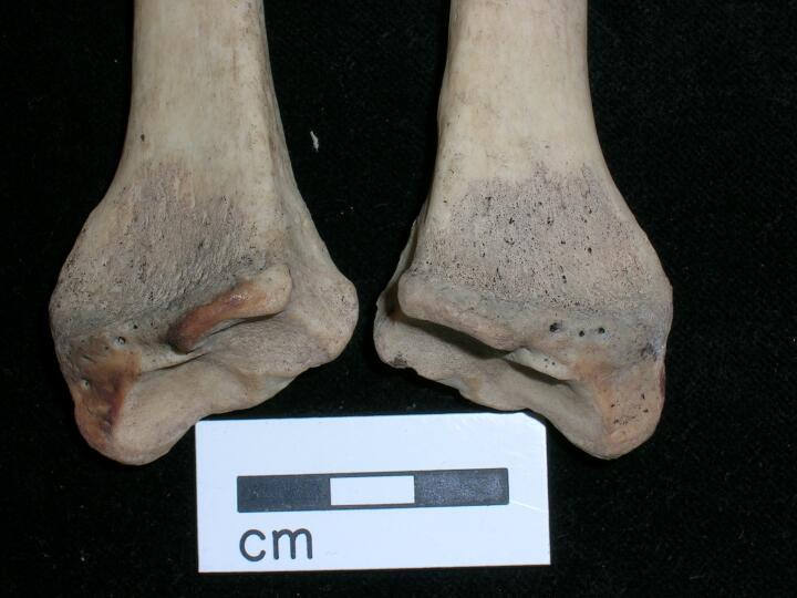
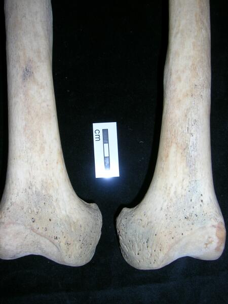
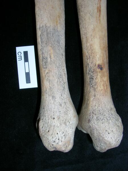
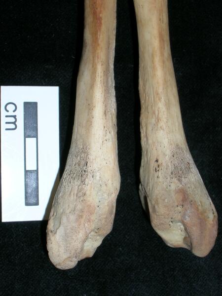
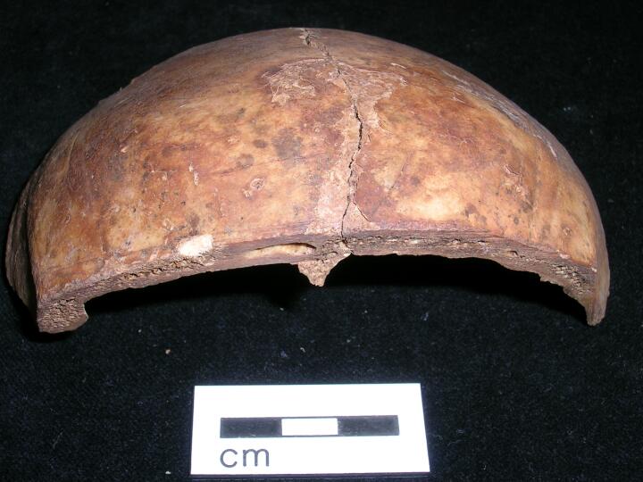
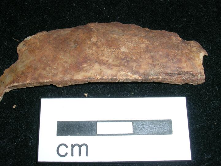
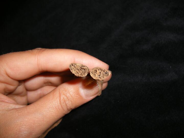
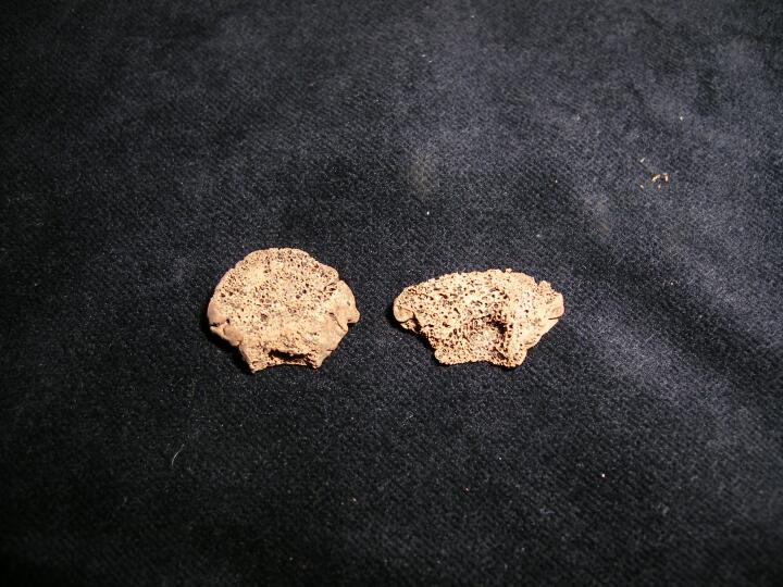
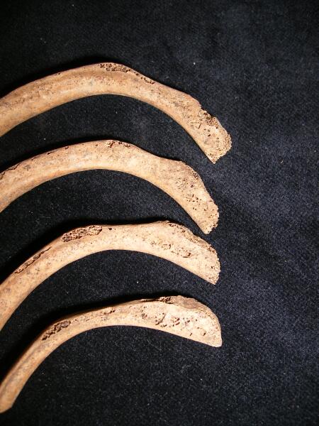
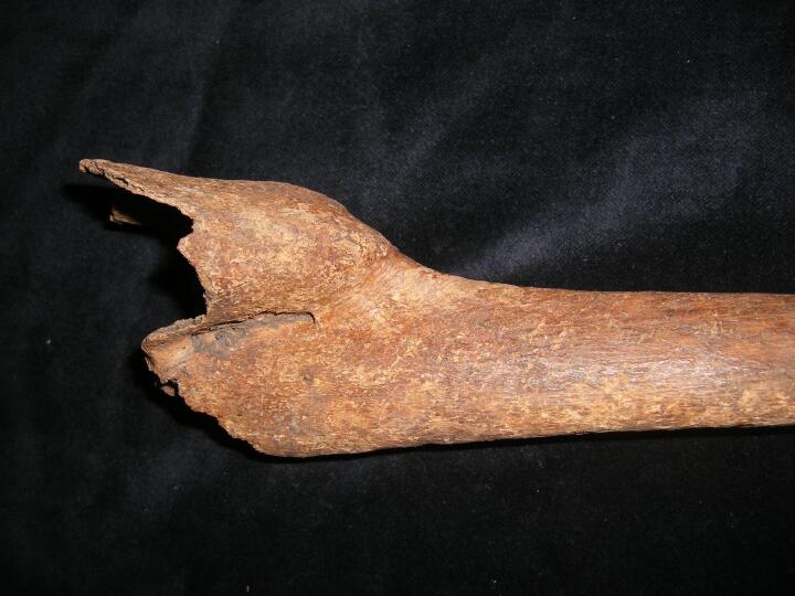
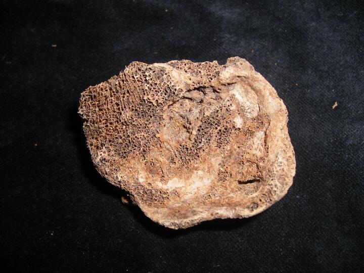
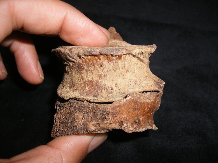
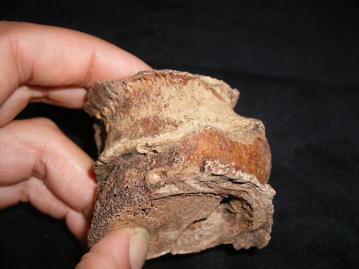
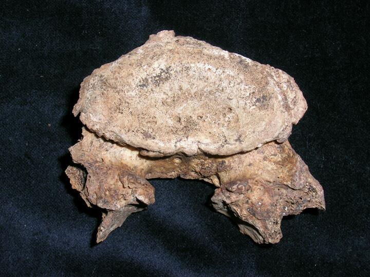
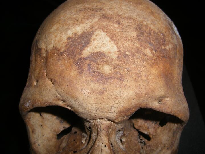
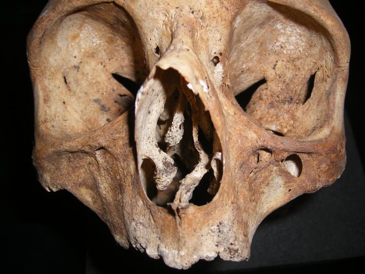
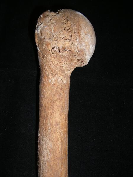
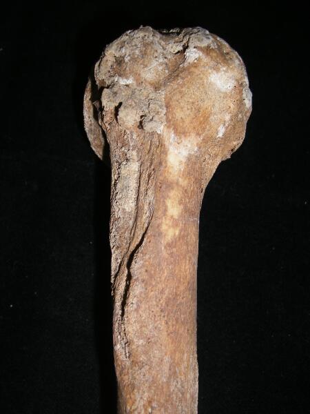
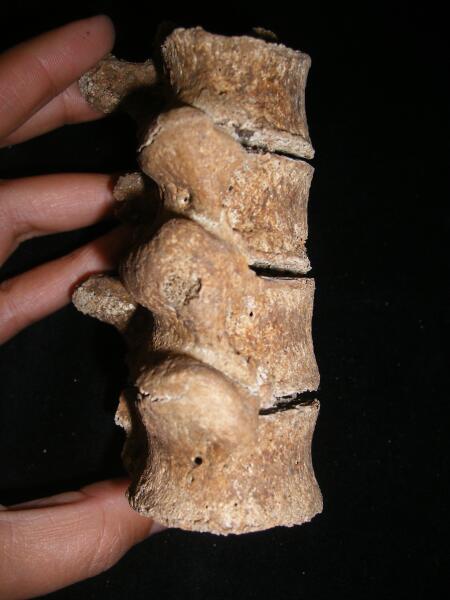
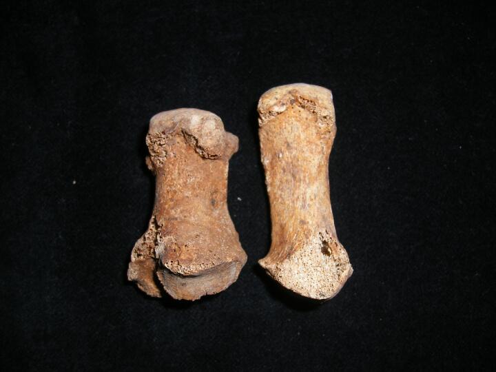
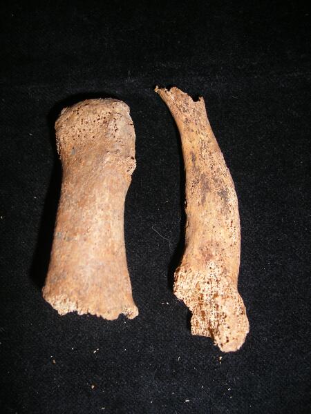
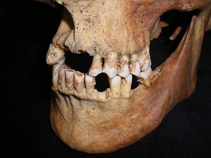
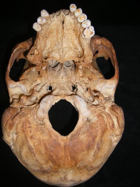
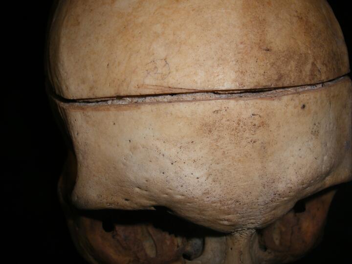
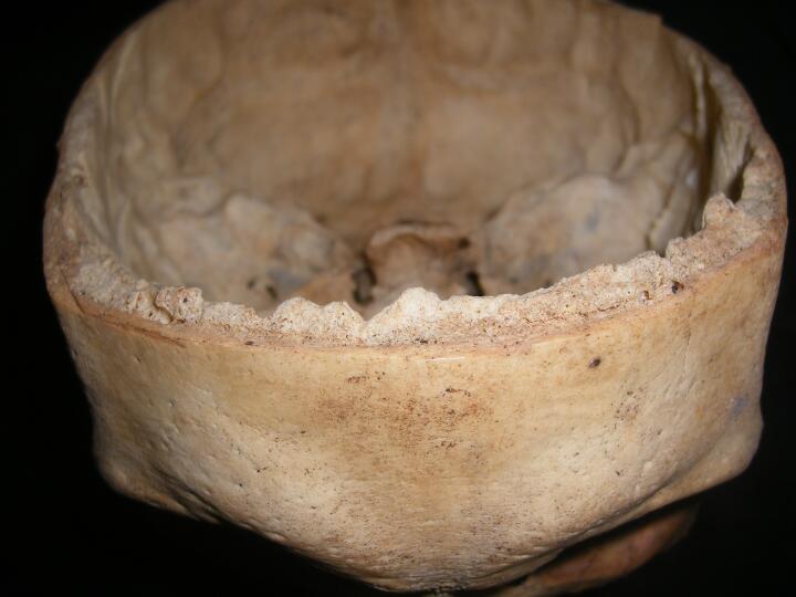
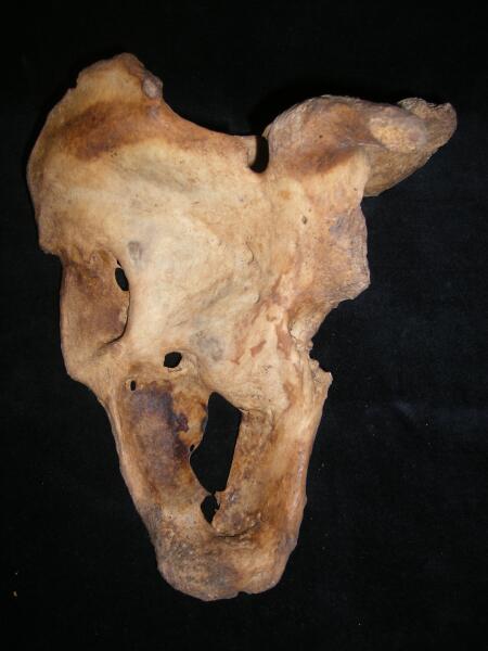
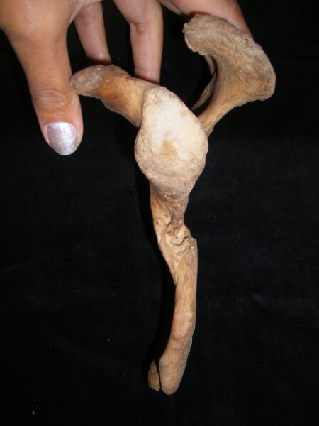
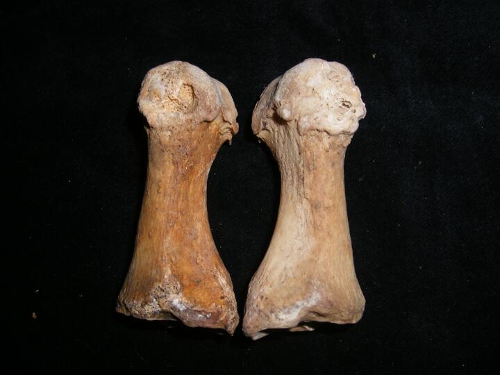
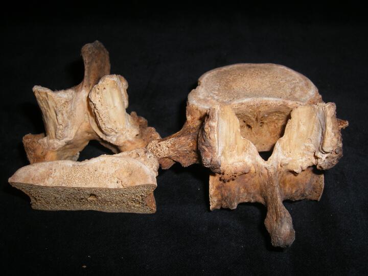
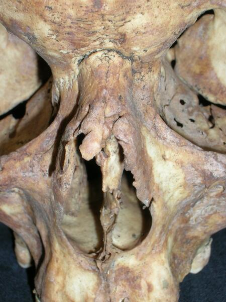
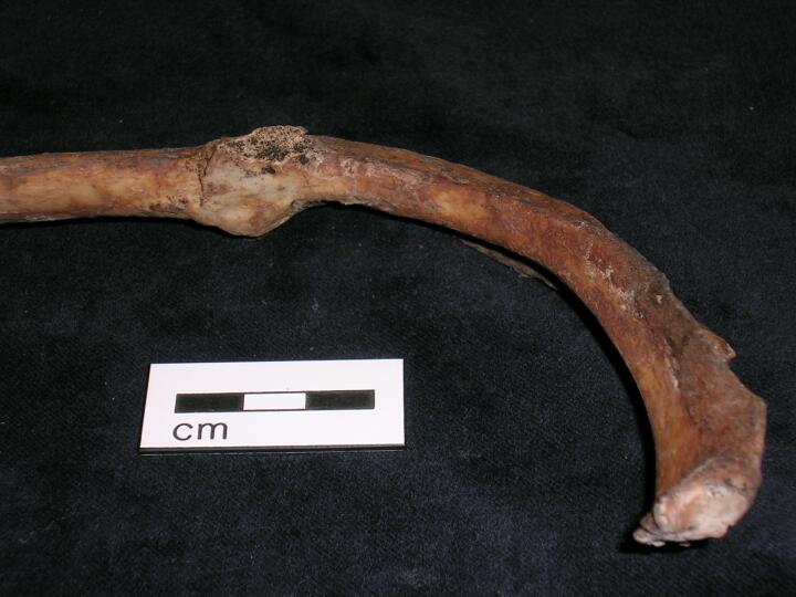
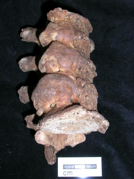
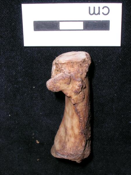
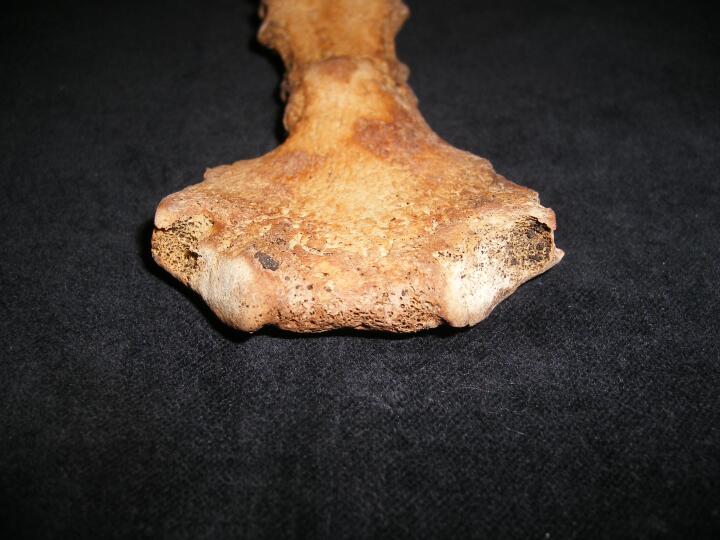
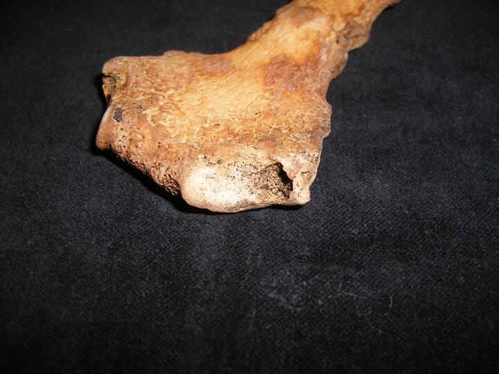
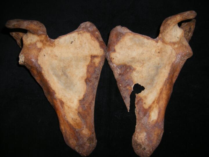
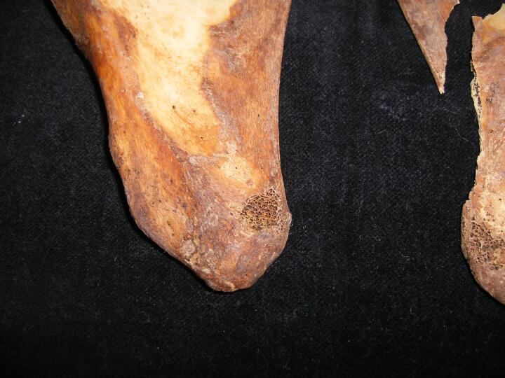
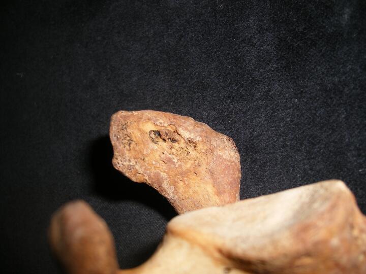
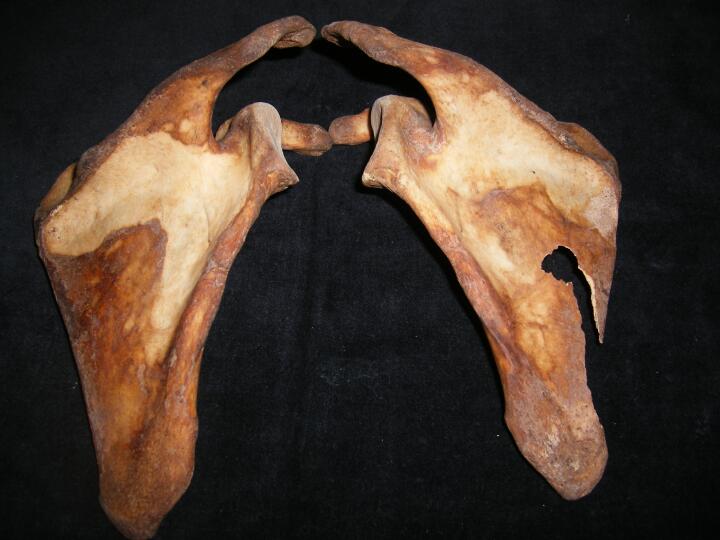
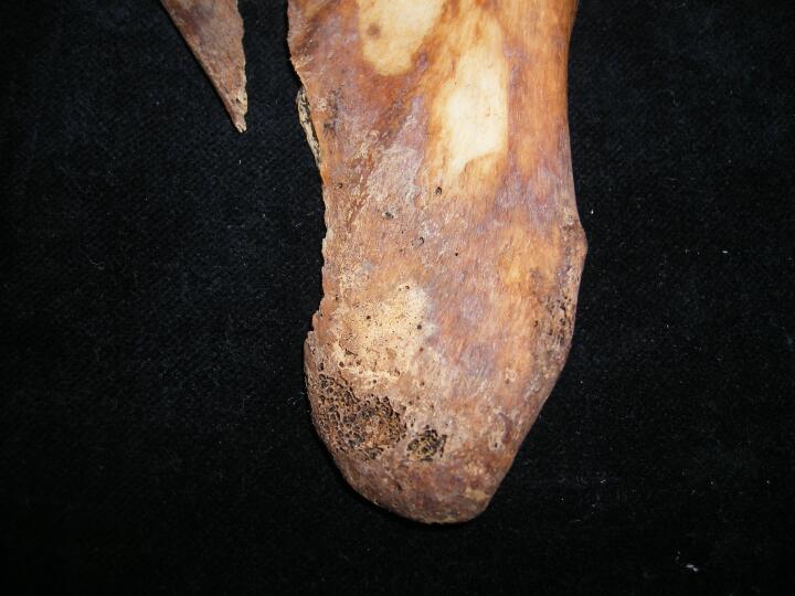
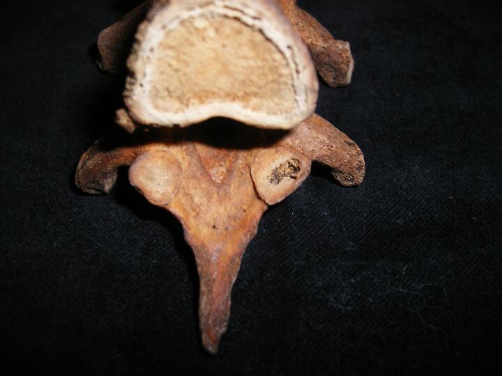
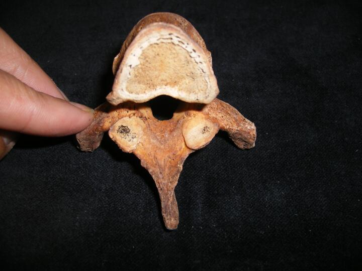
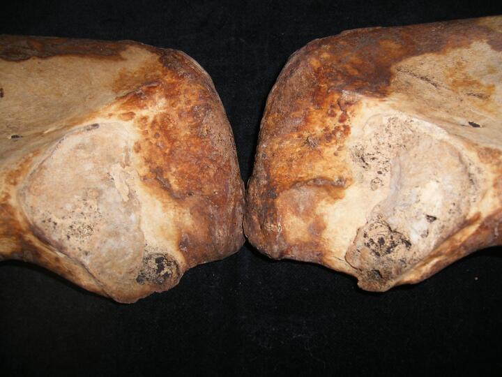
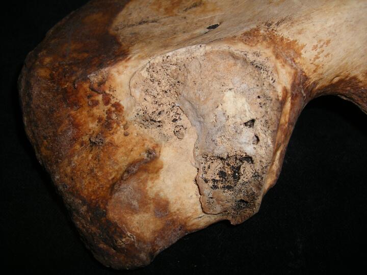
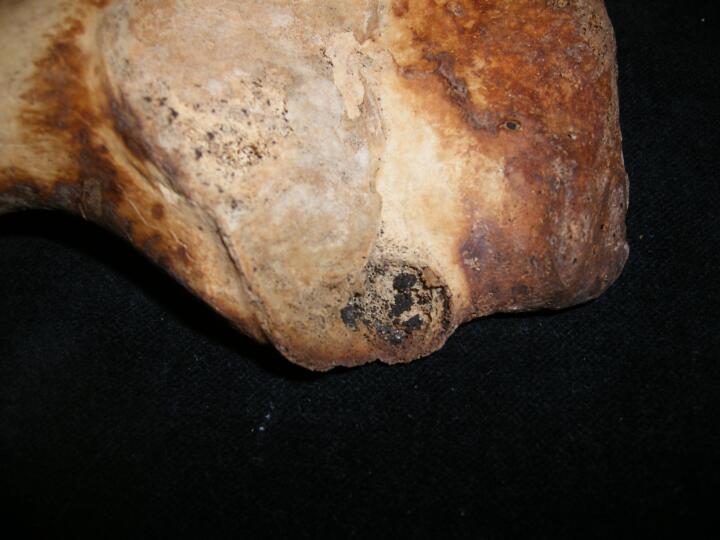
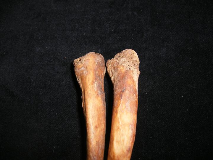
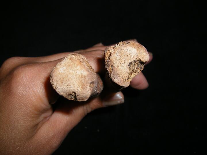
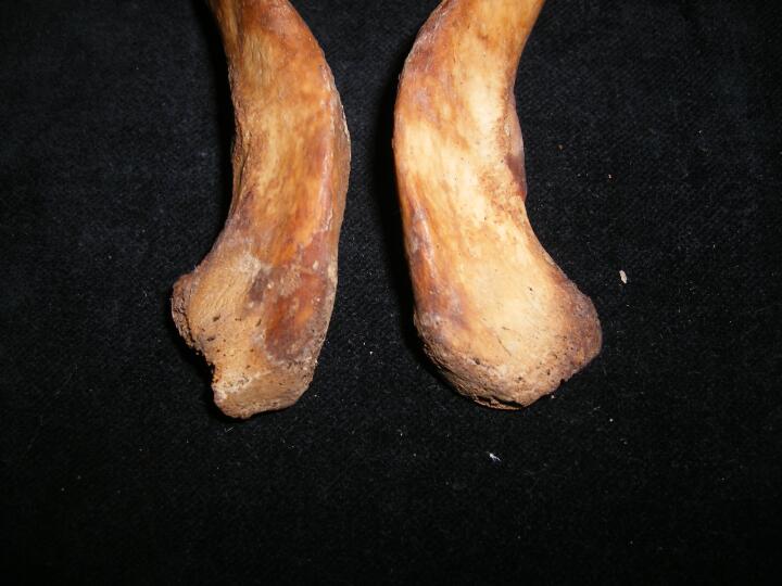
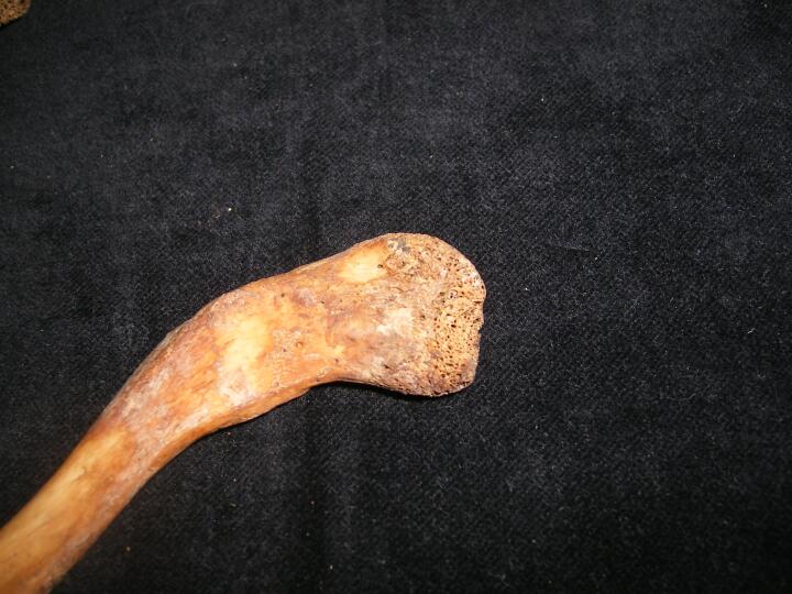
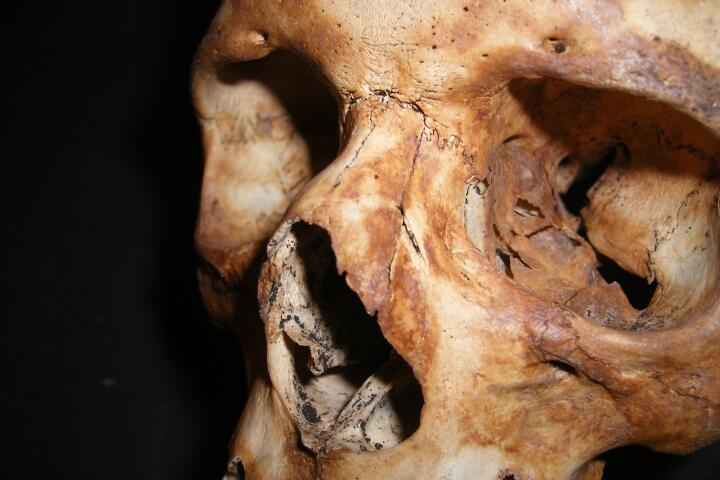
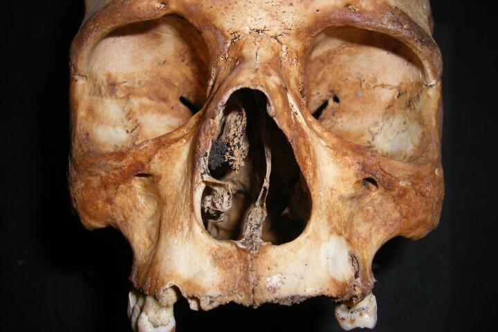
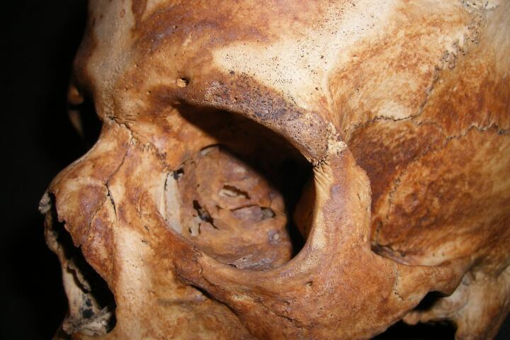
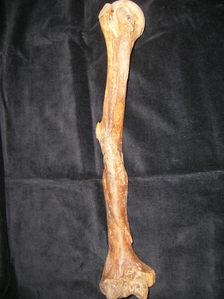
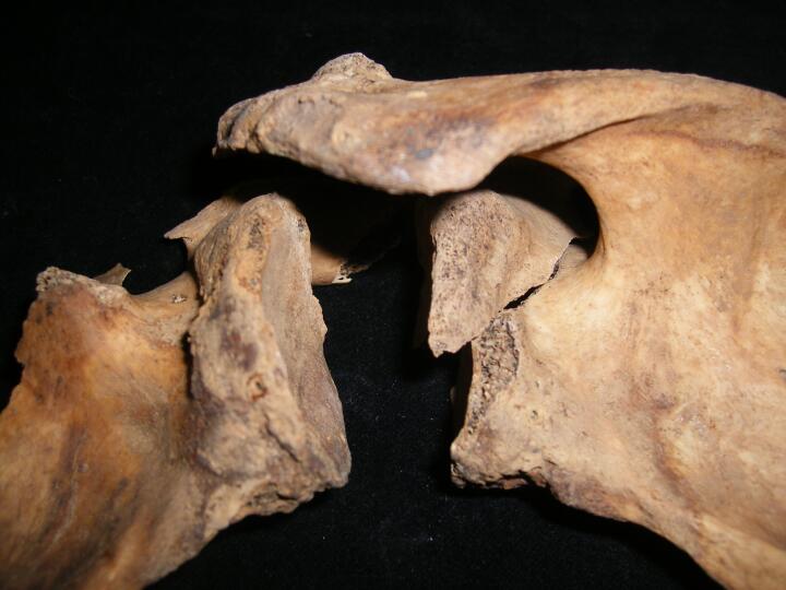
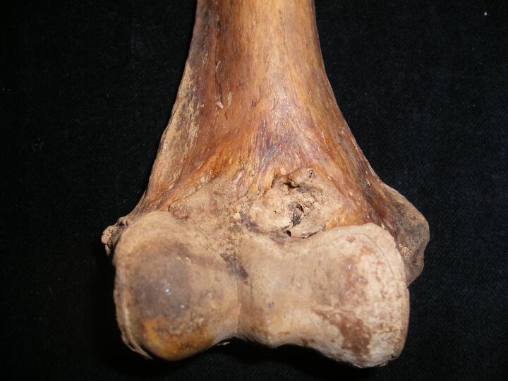
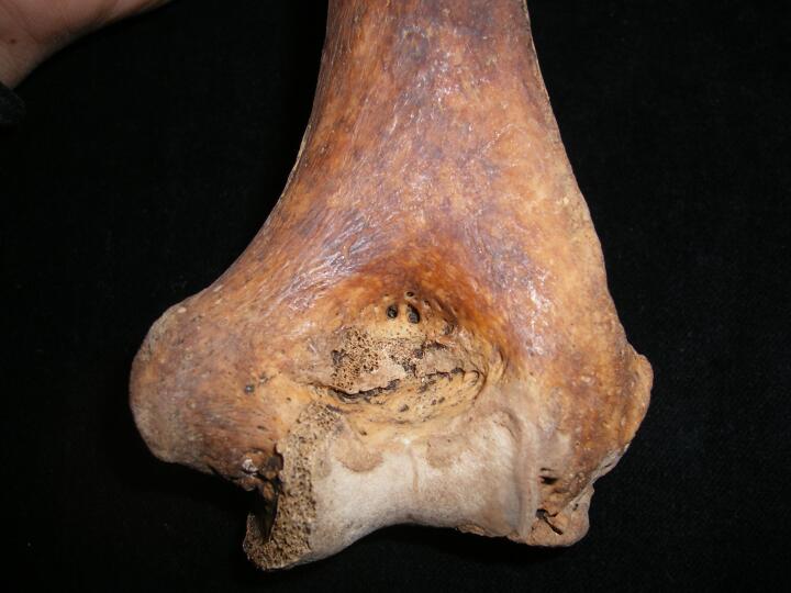
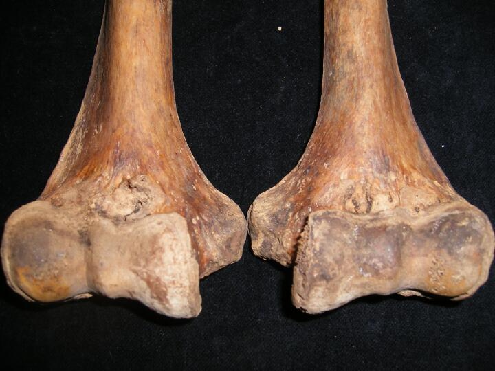
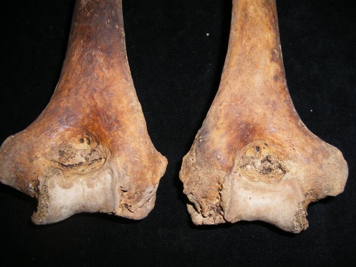
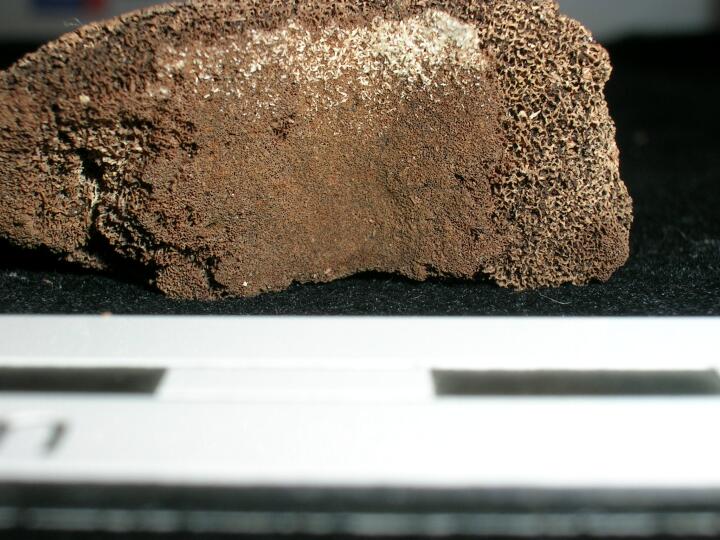
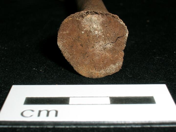
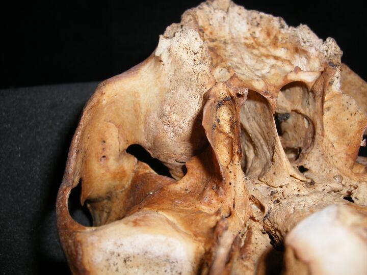
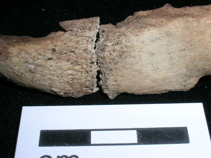
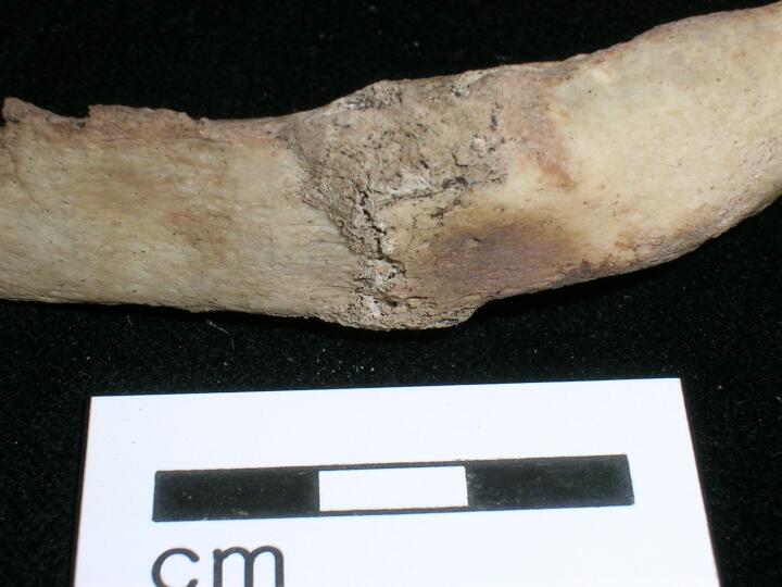
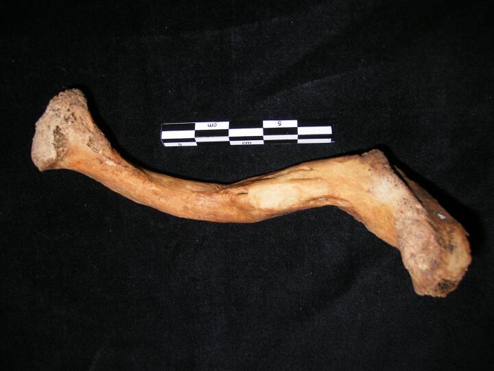
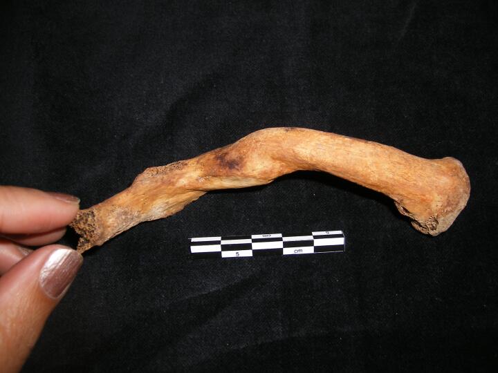
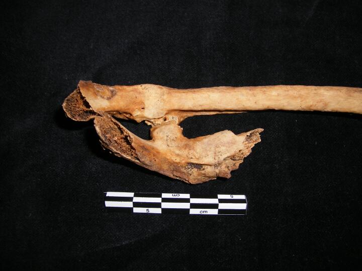
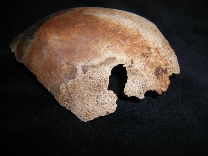
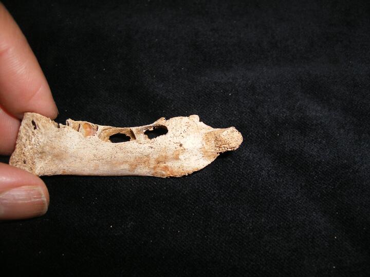
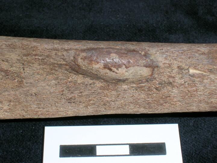
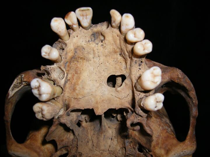
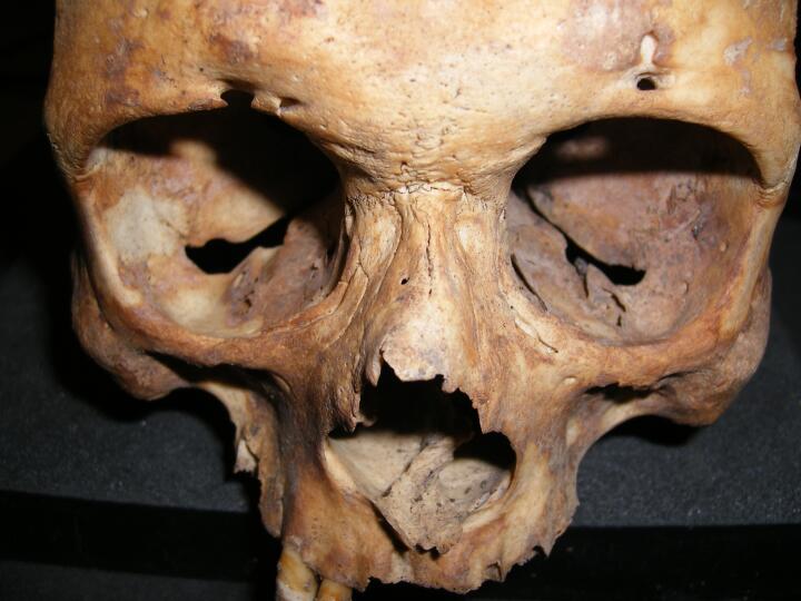
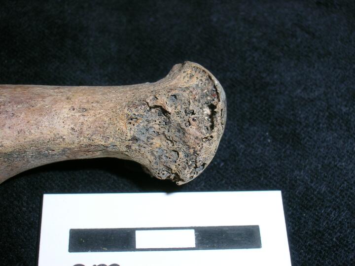
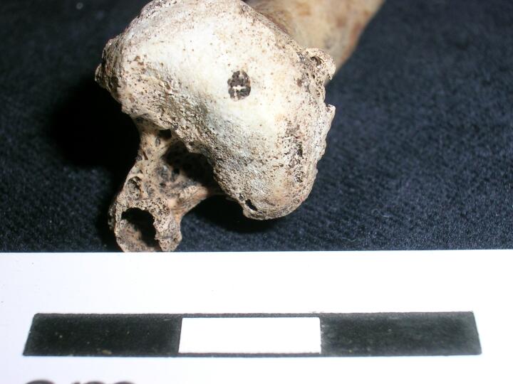
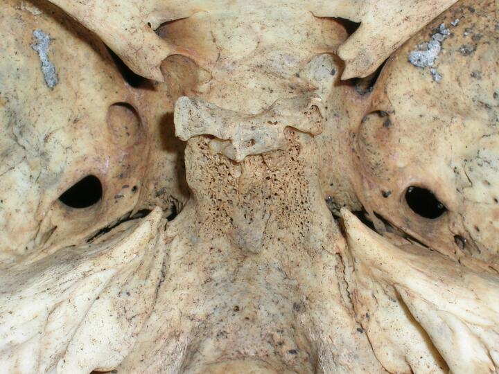
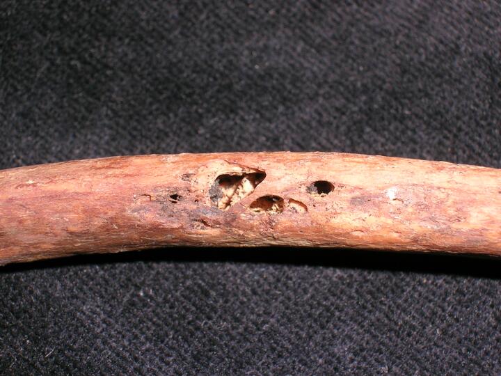
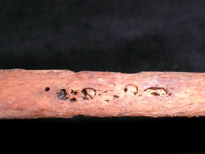
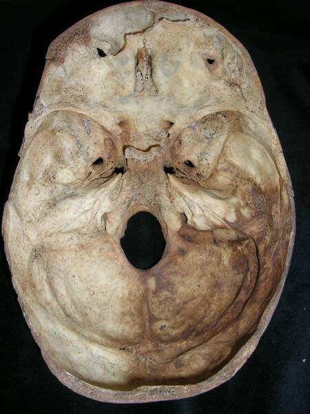
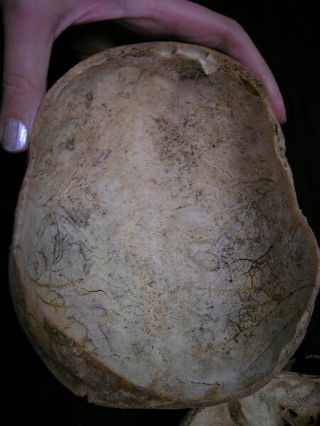
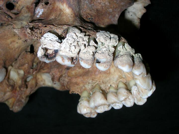
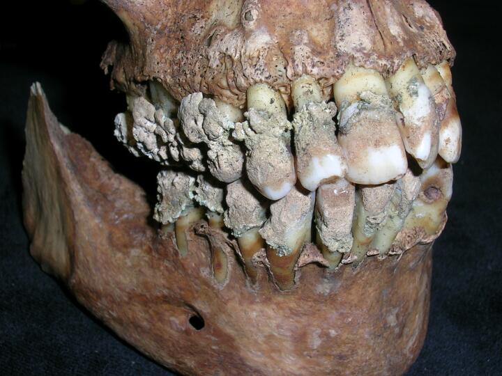
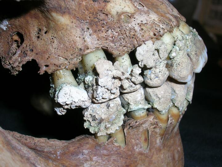
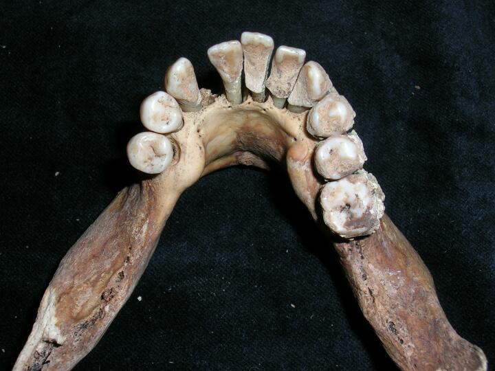
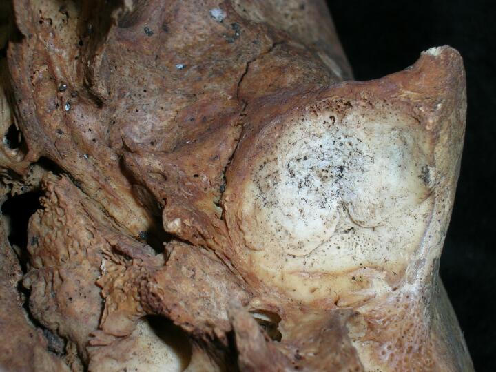
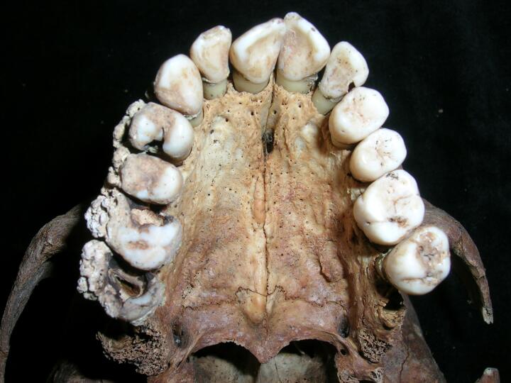
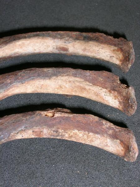
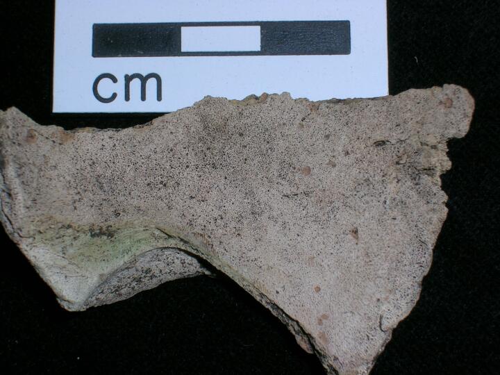
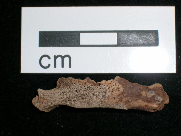
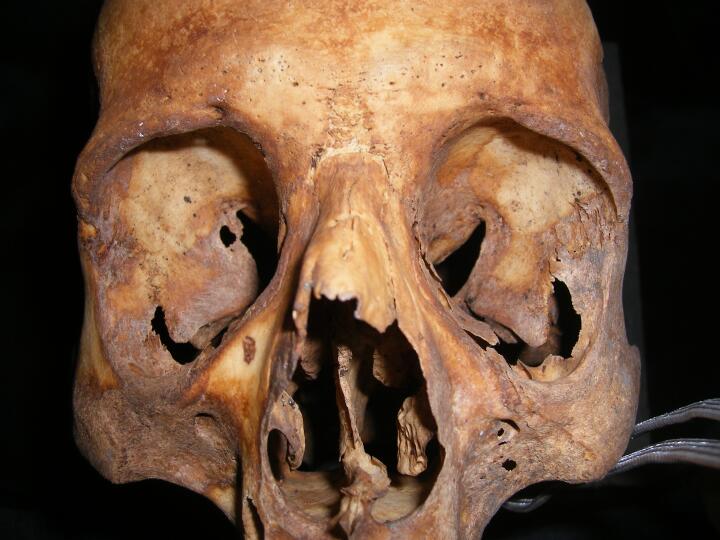
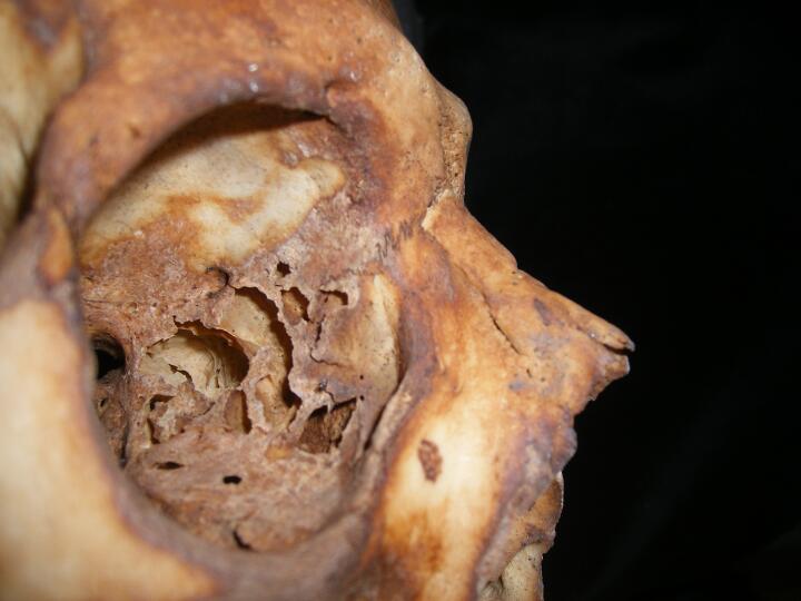
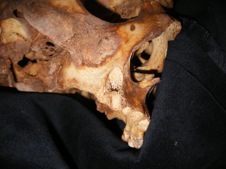
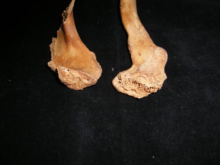
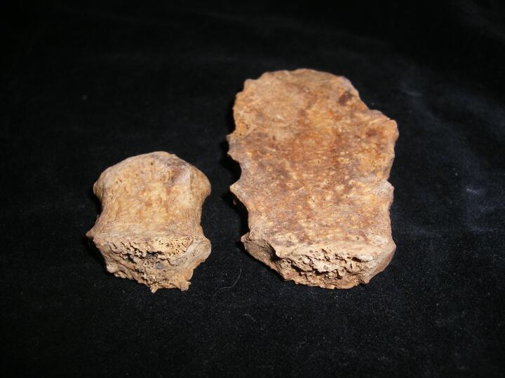
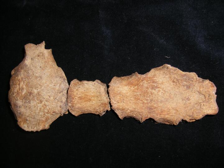
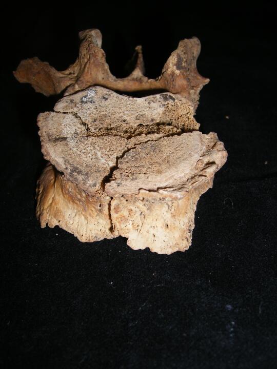
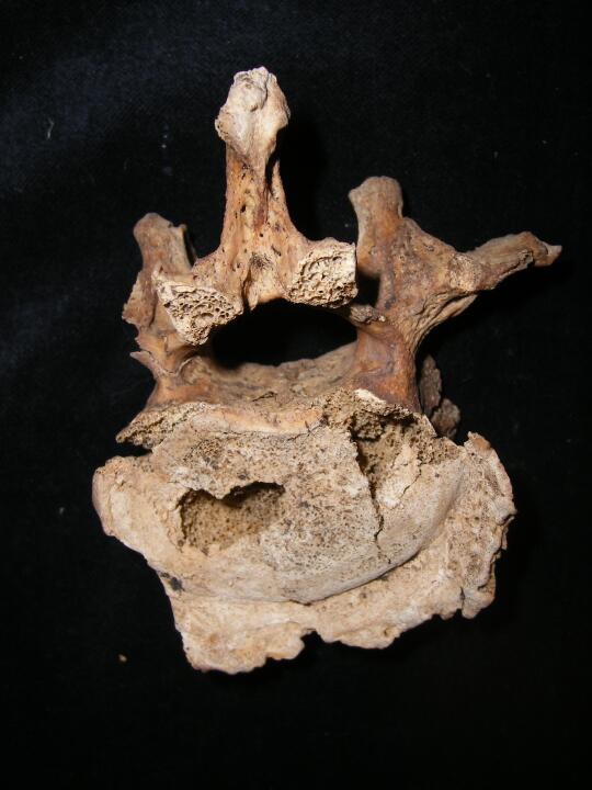
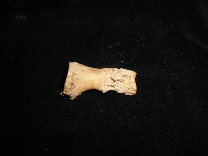
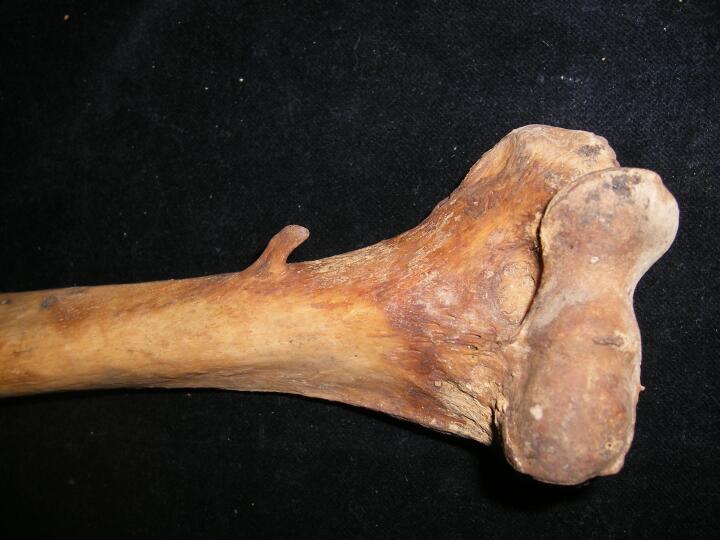
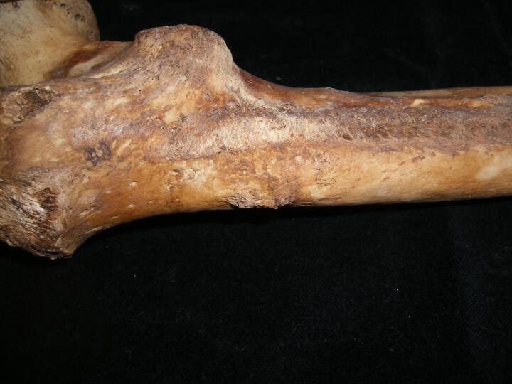
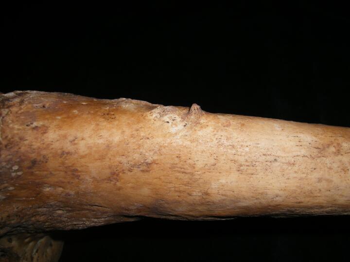
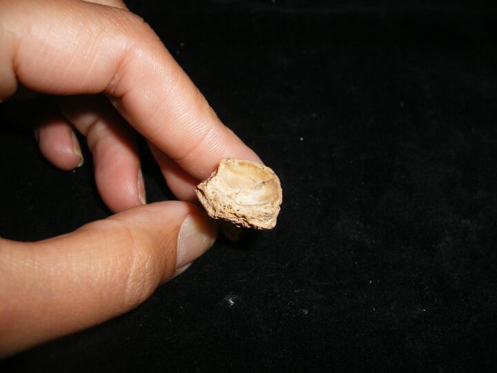
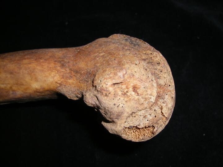
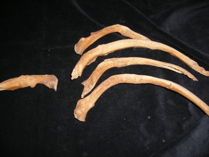
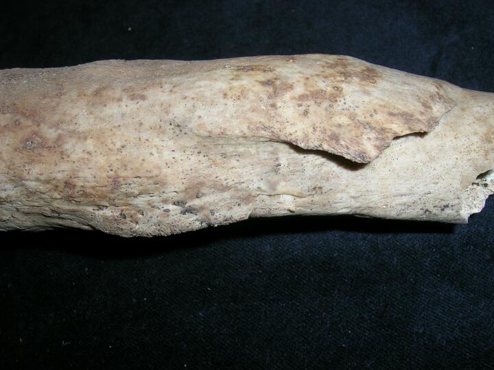
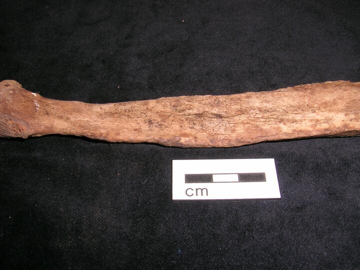
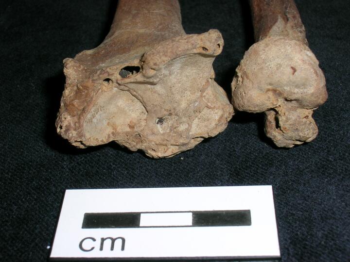
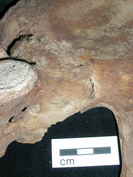
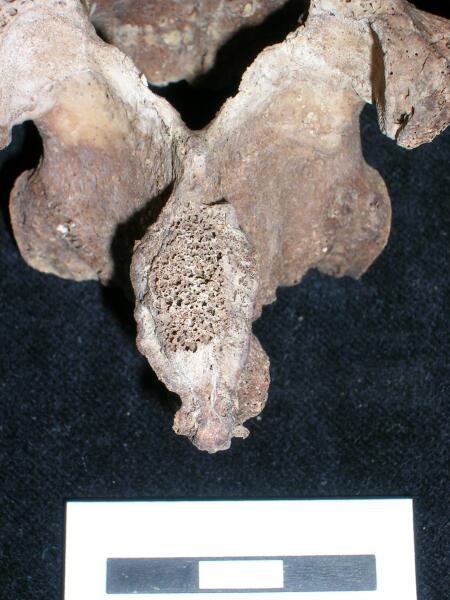
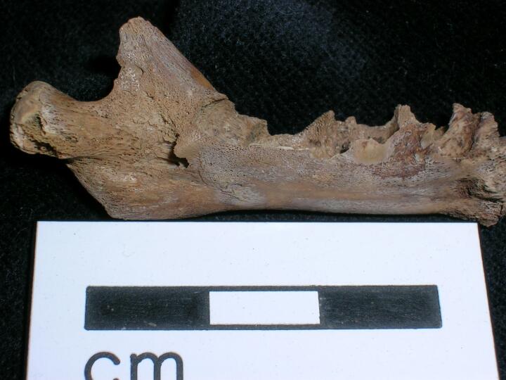
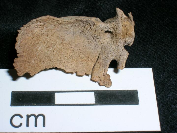
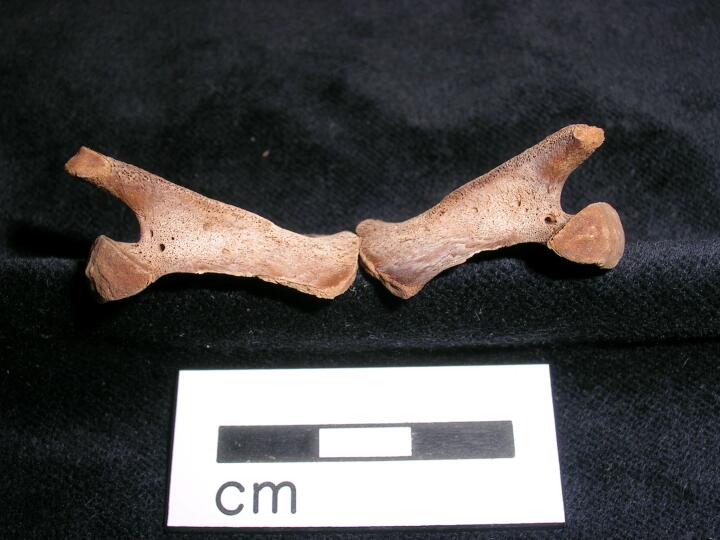
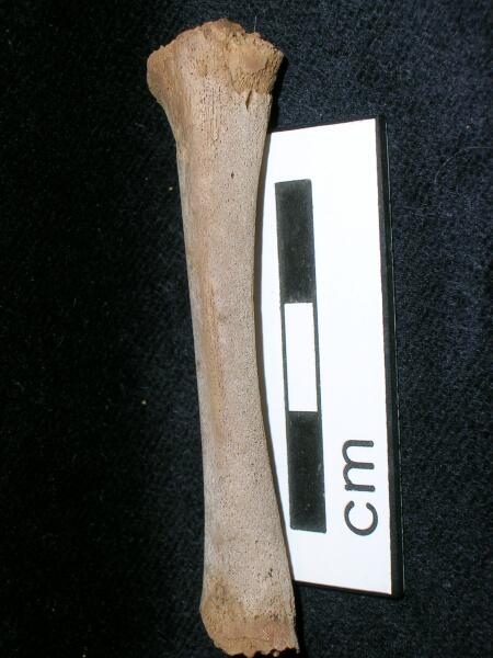
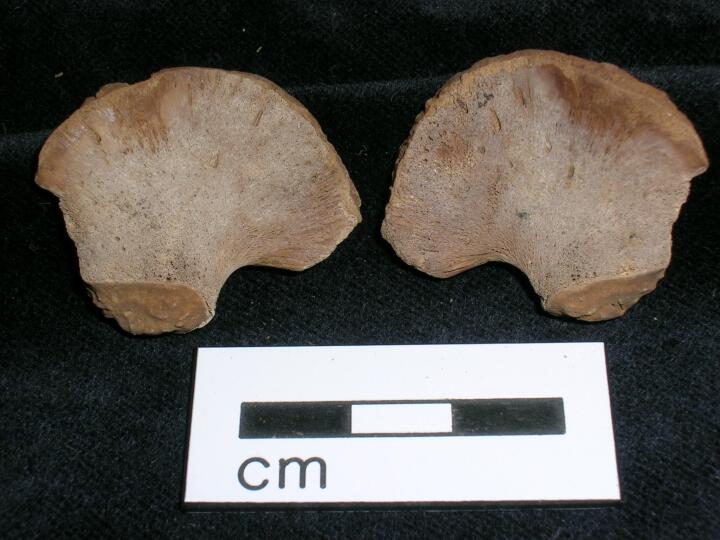
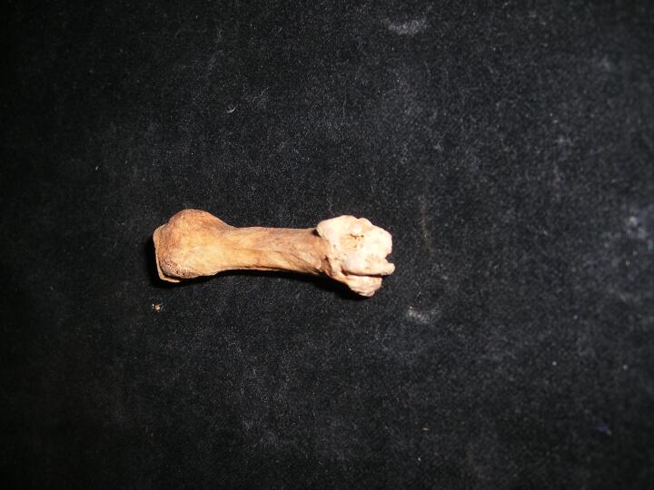
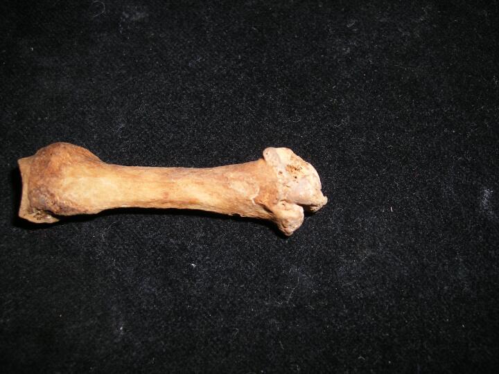
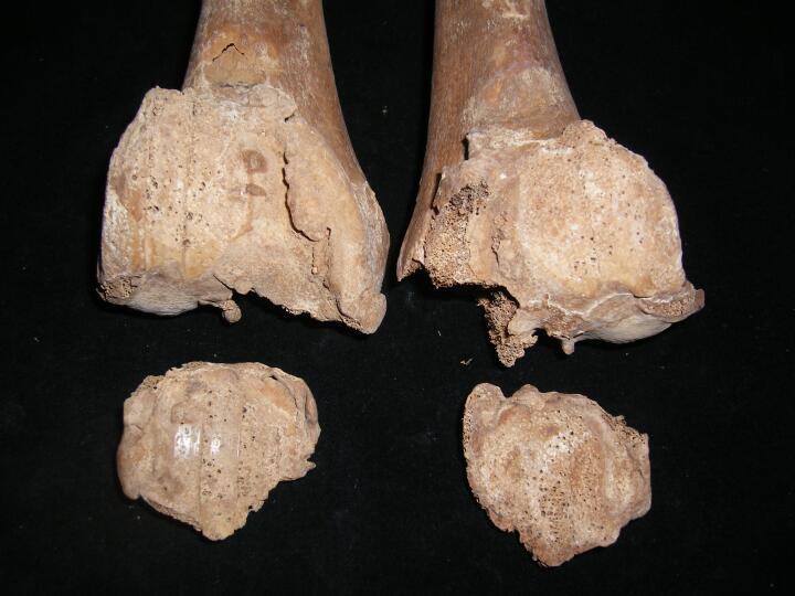
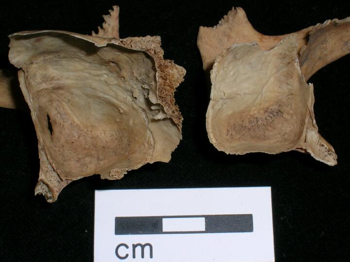
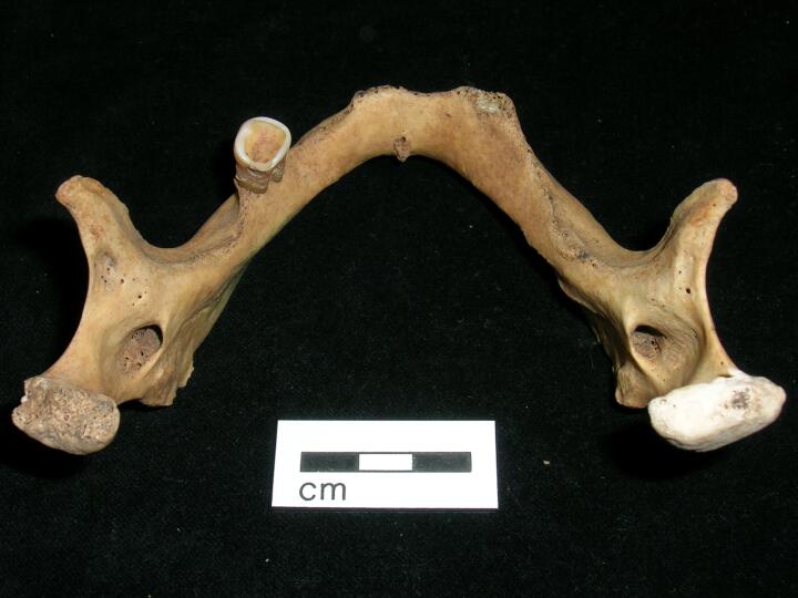
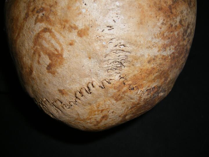
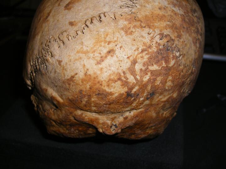
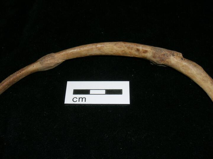
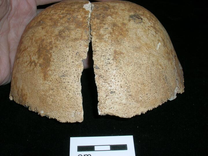
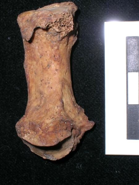
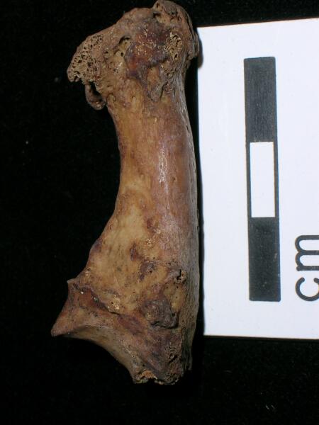
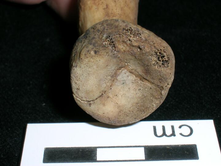
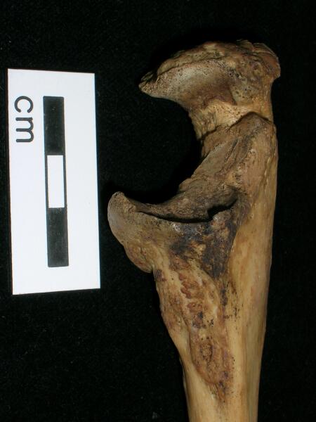
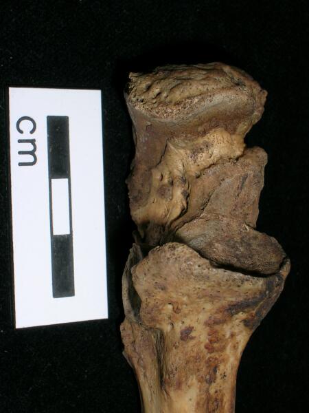
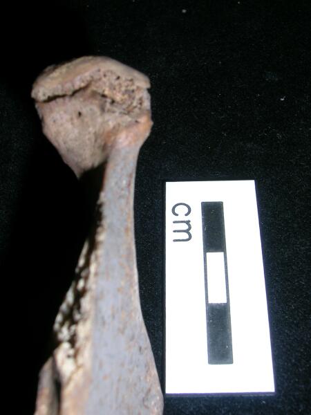
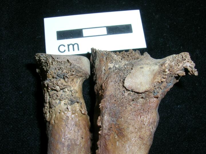
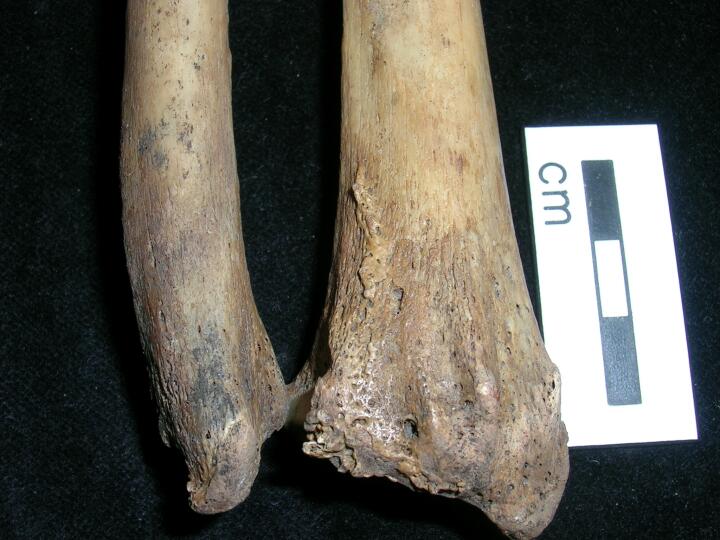
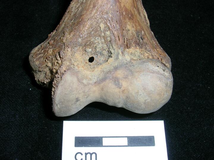
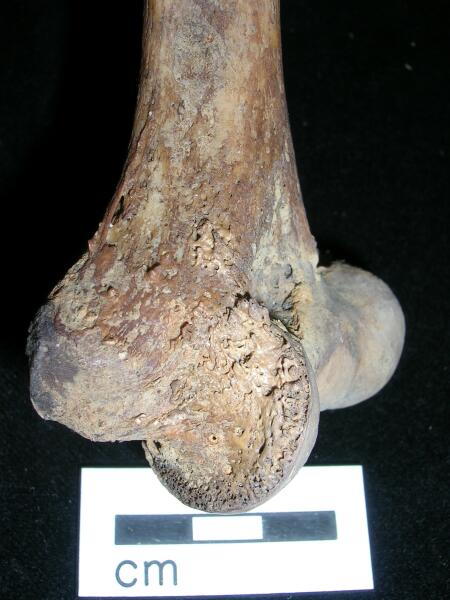
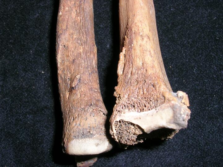
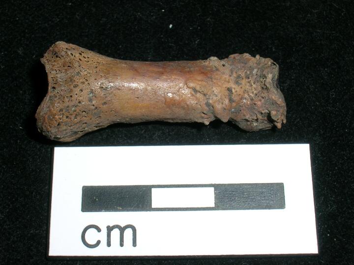
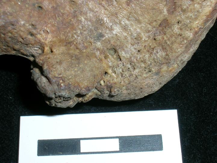
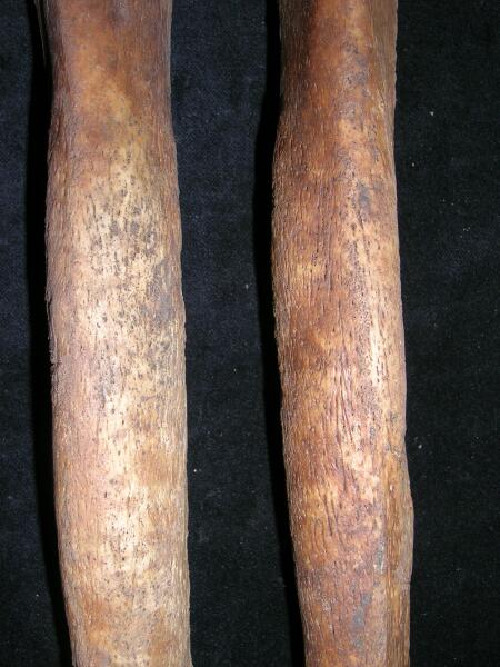
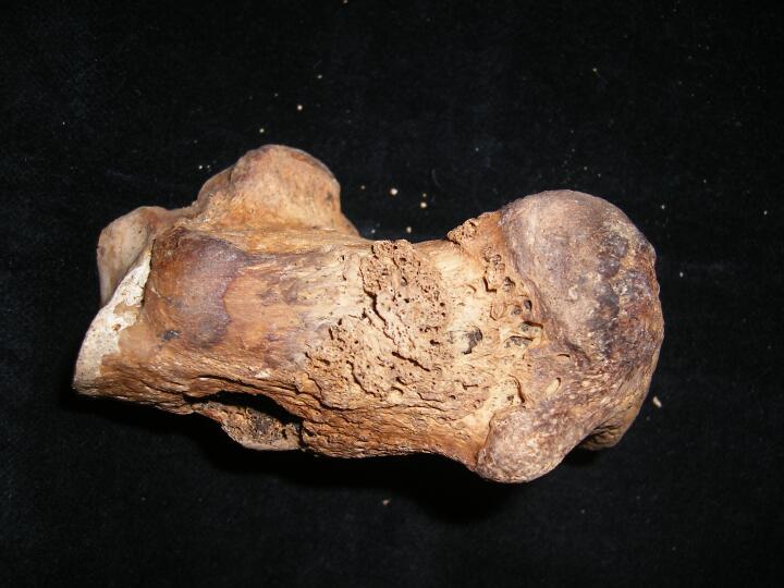
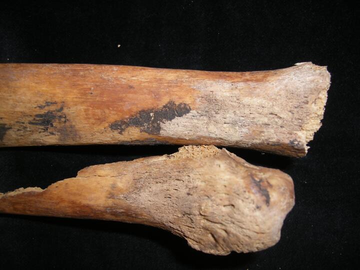
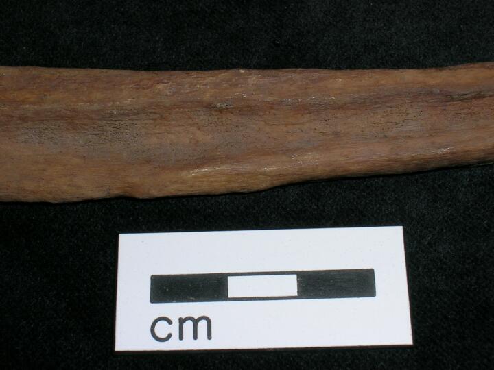
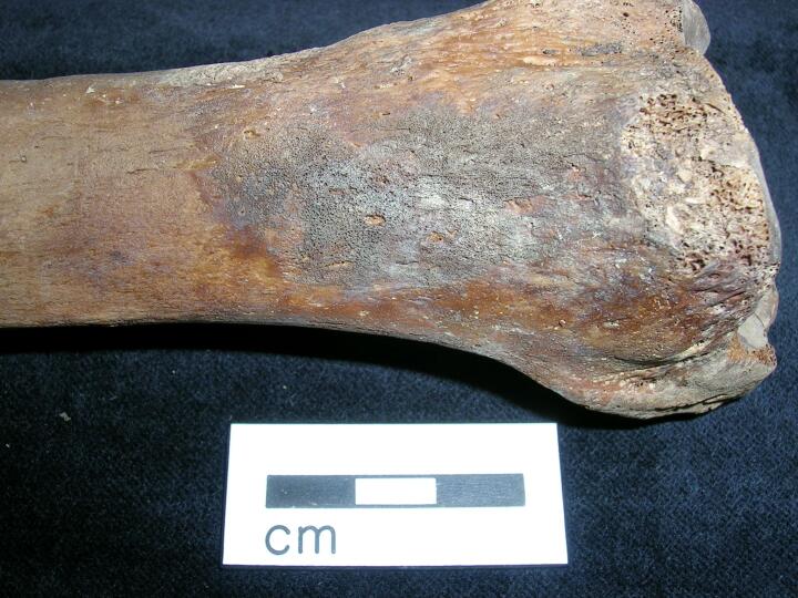
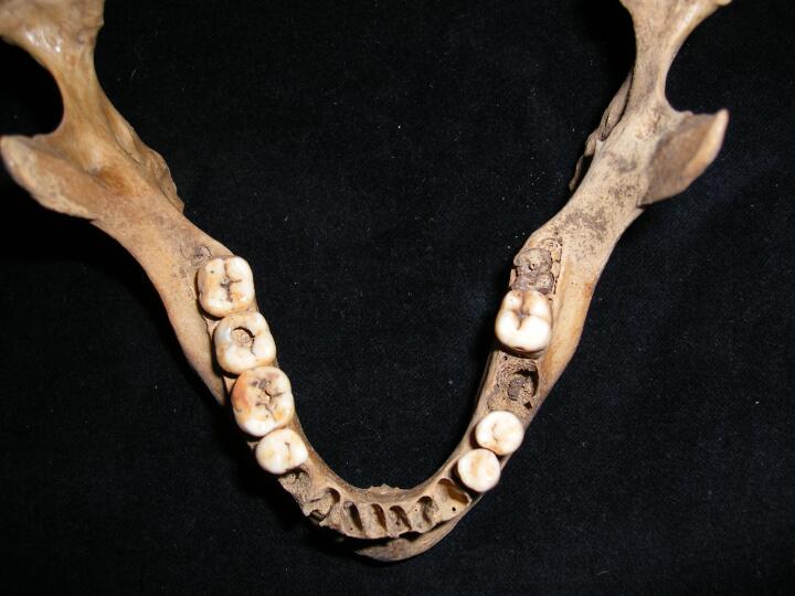
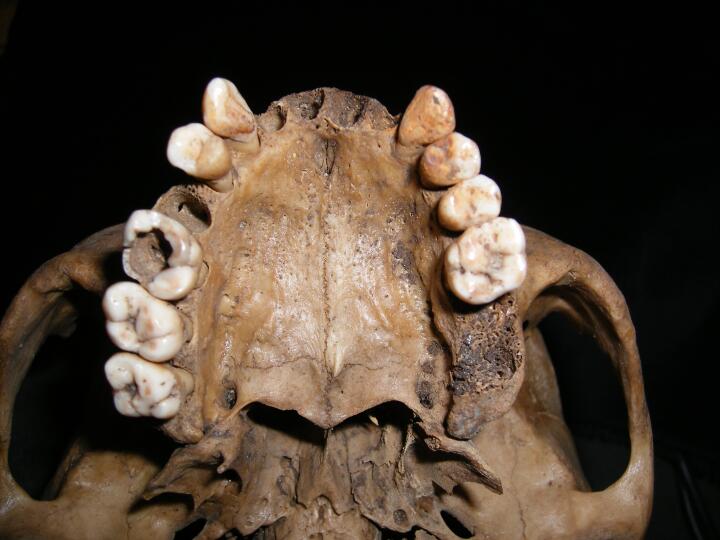
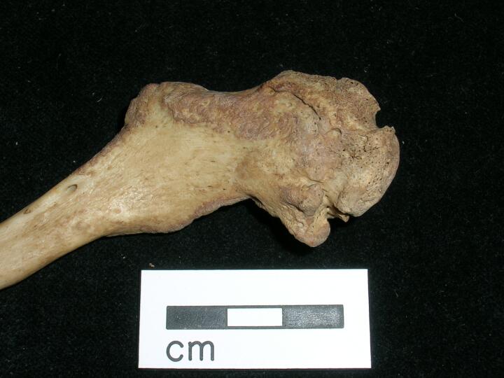
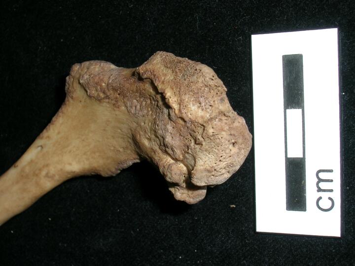
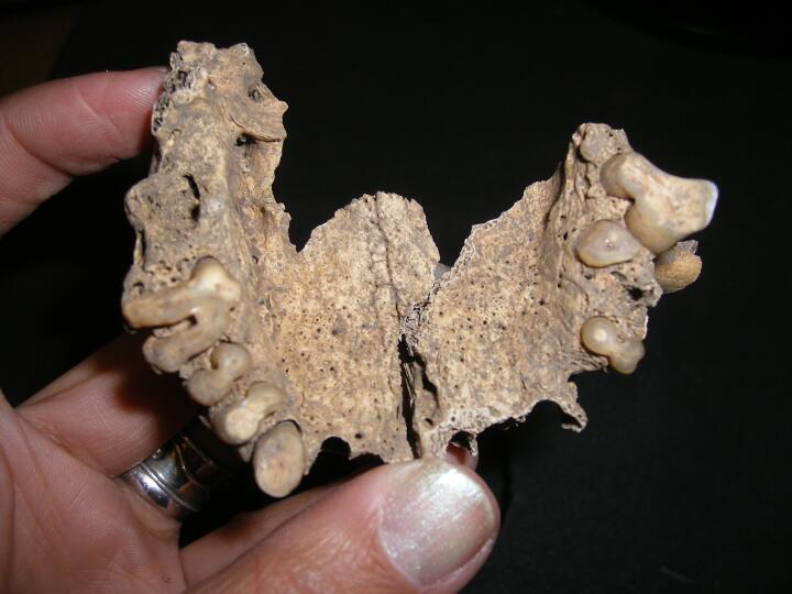
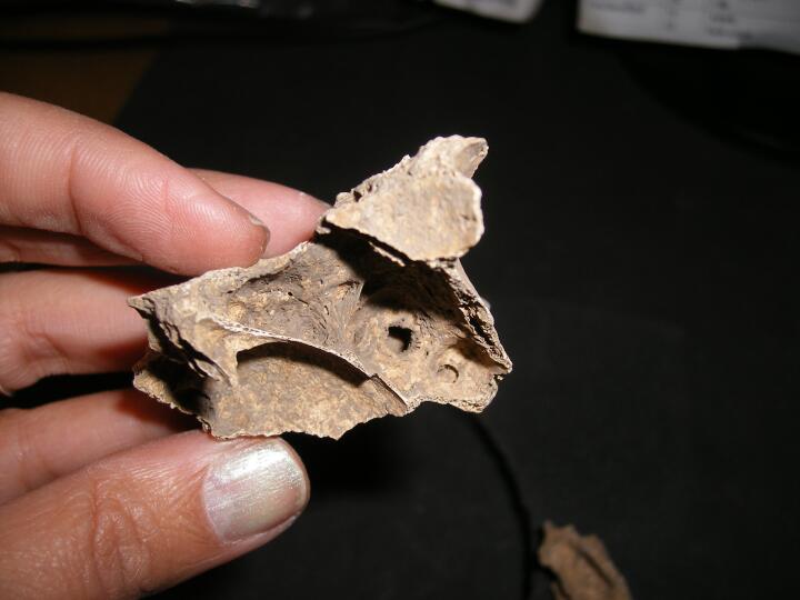