| Site code
|
Context
|
Frame number
|
Photo
|
Description
|
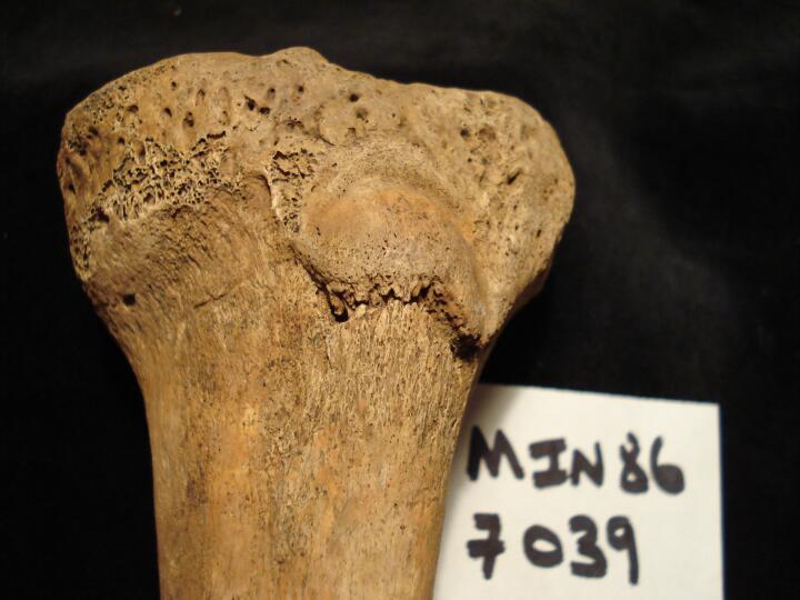
| MIN86
|
7039
|
2
|
MIN86_7039_2.jpg
|
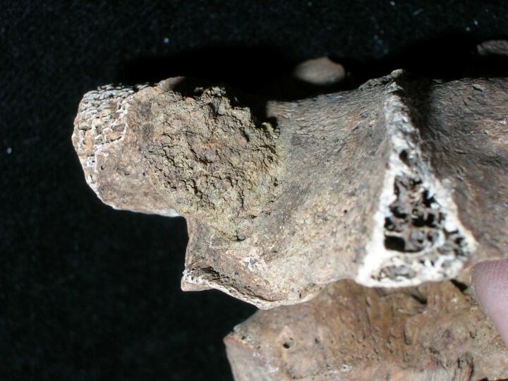
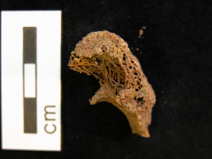
Left tibia Osgood Schlatter disease & groove transversely across tibal tuberosity (anterior surface/close up)
|
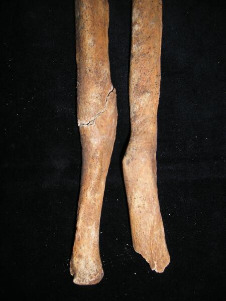
| MIN86
|
7102
|
1
|
MIN86_7102_1.jpg
|
Healed fractures of left radius and ulna
|
| MIN86
|
7102
|
2
|
MIN86_7102_2.jpg
|
Avulsion injury of L femoropatellar joint
|
| MIN86
|
7102
|
3
|
MIN86_7102_3.jpg
|
Non-specific osteoitis on distal shaft of L femur
|
| MIN86
|
7314
|
1
|
MIN86_7314_1.jpg
|
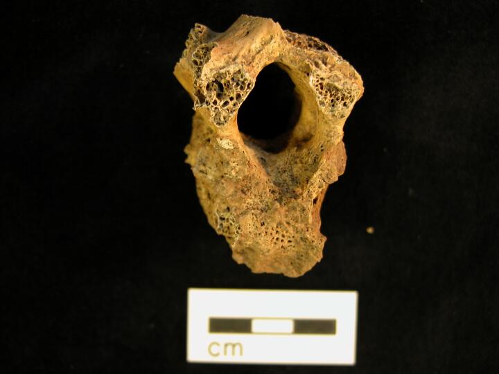
Projectile injury, ferrous object penetrating through T5 the inferior facet
|
| MIN86
|
7314
|
2
|
MIN86_7314_2.jpg
|
Projectile injury, ferrous object penetrating through T5 the inferior facet
|
| MIN86
|
7314
|
3
|
MIN86_7314_3.jpg
|
Projectile injury, ferrous object penetrating through T5 the inferior facet
|
| MIN86
|
7314
|
4
|
MIN86_7314_4.jpg
|
Projectile injury, ferrous object penetrating through T5 the inferior facet
|
| MIN86
|
7314
|
5
|
MIN86_7314_5.jpg
|
Projectile injury, ferrous object penetrating through T5 the inferior facet
|
| MIN86
|
7314
|
6
|
MIN86_7314_6.jpg
|
Blunt force trauma to right parietal
|
| MIN86
|
7314
|
7
|
MIN86_7314_7.jpg
|
Left lateral view of skull
|
| MIN86
|
7314
|
8
|
MIN86_7314_8.jpg
|
Anterior view of skull
|
| MIN86
|
9327
|
1
|
MIN86_9327_1.jpg
|
Osteoarthritic changes to the right temporomandibular joint. Bony outgrowth visible (dorsal view)
|
| MIN86
|
9327
|
2
|
MIN86_9327_2.jpg
|
Close up of osteoarthritic changes to the right temporomandibular joint. Bony outgrowth protruding anteriorly and subchondral cysts are visible (superior view of the joint surface)
|
| MIN86
|
9327
|
3
|
MIN86_9327_3.jpg
|
Osteoarthritis of the left knee. Slight osteophytic lipping around the rim of the articular surface and slight eburnation to the left portion of the joint surface.
|
| MIN86
|
9327
|
4
|
MIN86_9327_4.jpg
|
Osteoarthritis of the left knee. Slight osteophytic lipping around the rim of the patella and slight eburnation to the left portion of the joint surface.
|
| MIN86
|
9373
|
1
|
MIN86_9373_1.jpg
|
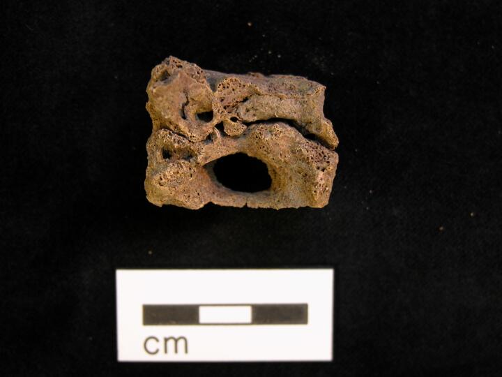
Butterfly vertebra, T11 superior view
|
| MIN86
|
9373
|
2
|
MIN86_9373_2.jpg
|
Butterfly vertebra T11, posterior view
|
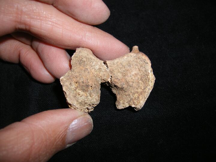
| MIN86
|
9395
|
1
|
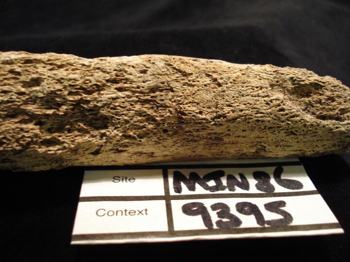
MIN86_9395_1.jpg
|
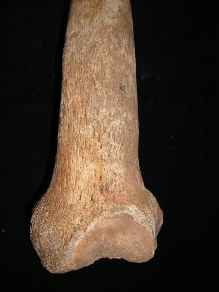
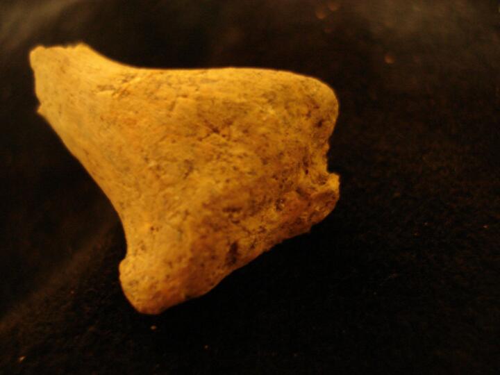
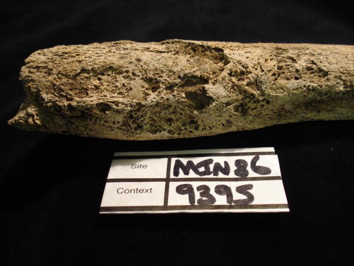
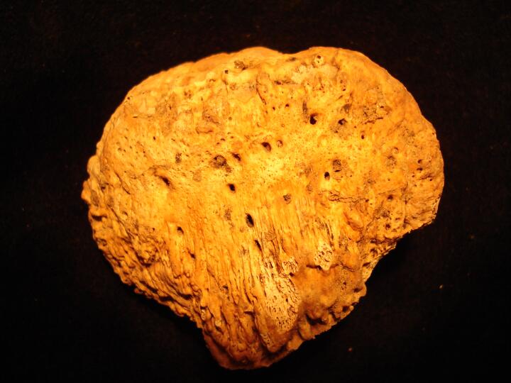
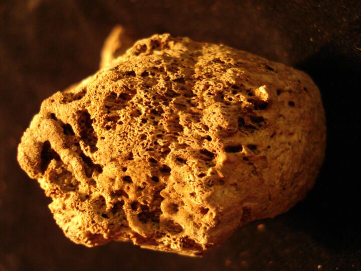
Left femur (anterior view) bones changes possibly venereal syphilis
|
| MIN86
|
9395
|
2
|
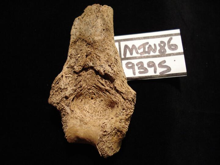
MIN86_9395_2.jpg
|
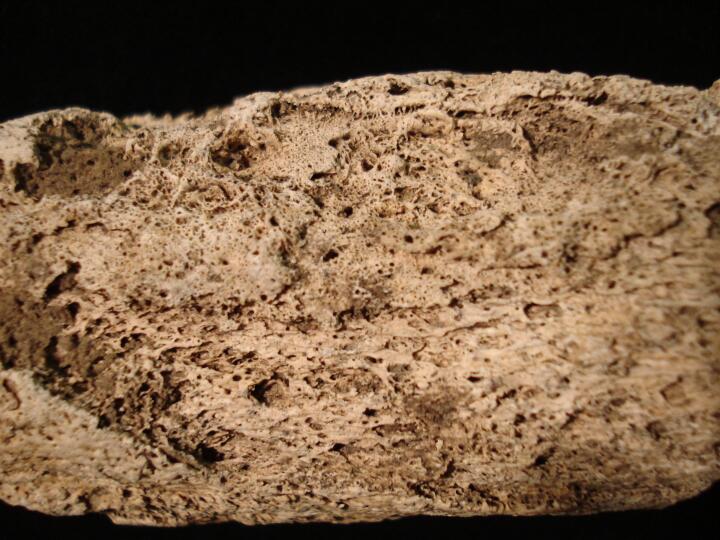
Left femur (anterior view) close up of bone changes possibly venereal syphilis
|
| MIN86
|
9395
|
3
|
MIN86_9395_3.jpg
|
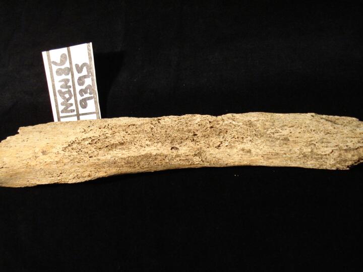
Left femur (posterior view) bones changes possibly venereal syphilis
|
| MIN86
|
9395
|
4
|
MIN86_9395_4.jpg
|
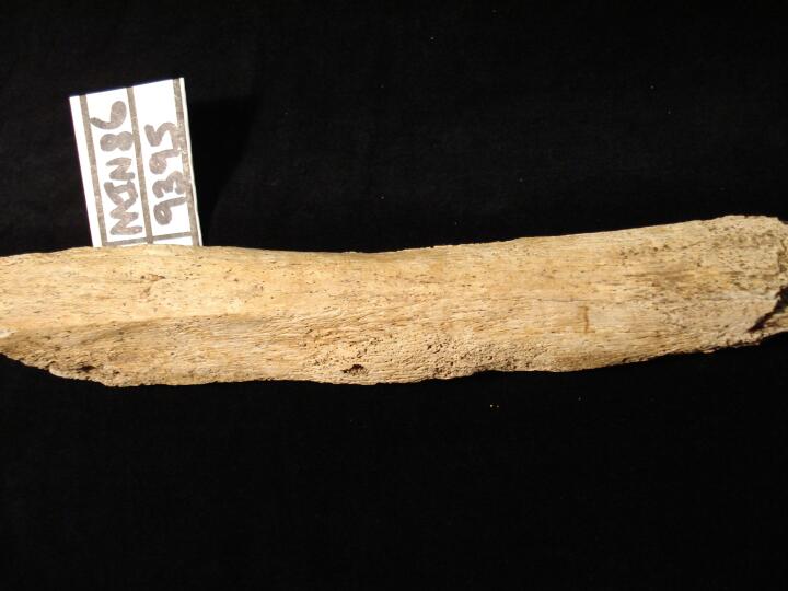
Left femur (medial view) close up of bone changes possibly venereal syphilis
|
| MIN86
|
9395
|
5
|
MIN86_9395_5.jpg
|
Left femur (lateral view) close up of bones changes possibly venereal syphilis
|
| MIN86
|
9395
|
6
|
MIN86_9395_6.jpg
|
Left femur (anterior view)close up of bone changes possibly venereal syphilis
|
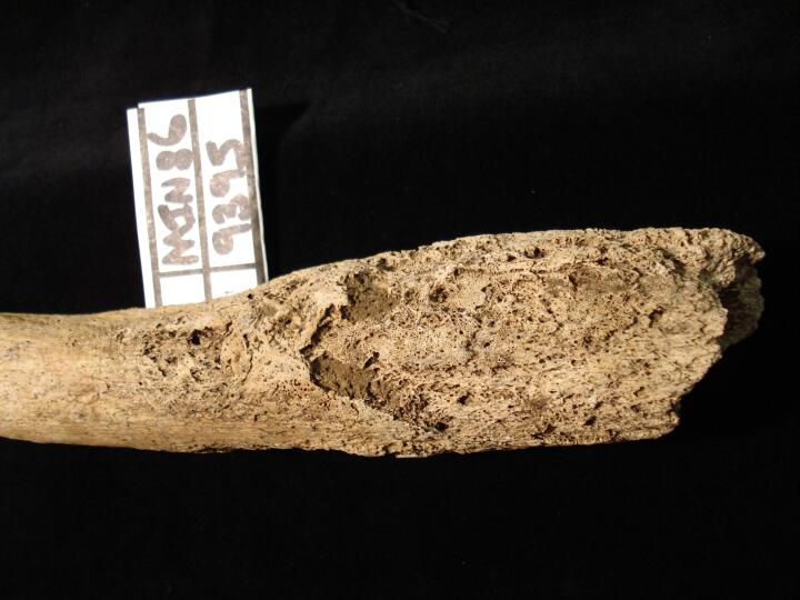
| MIN86
|
9395
|
7
|
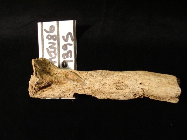
MIN86_9395_7.jpg
|
Left tibia (anterior view) bones changes possibly venereal syphilis
|
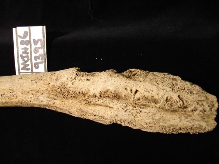
| MIN86
|
9395
|
8
|
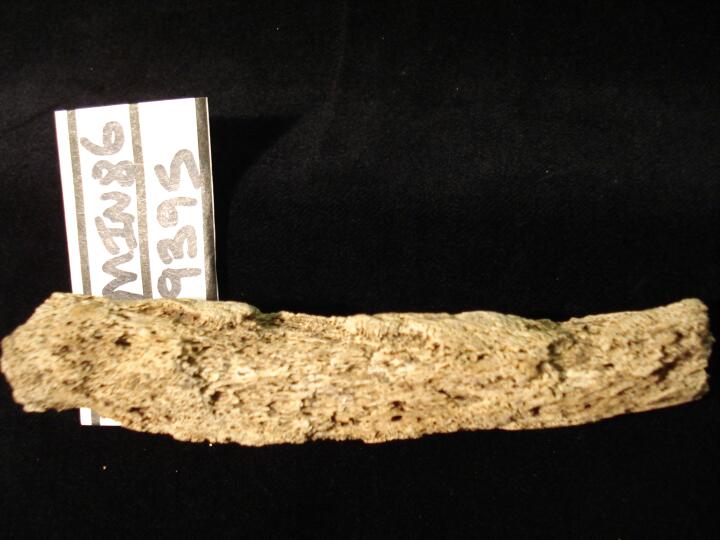
MIN86_9395_8.jpg
|
Left tibia (medial view) bones changes possibly venereal syphilis
|
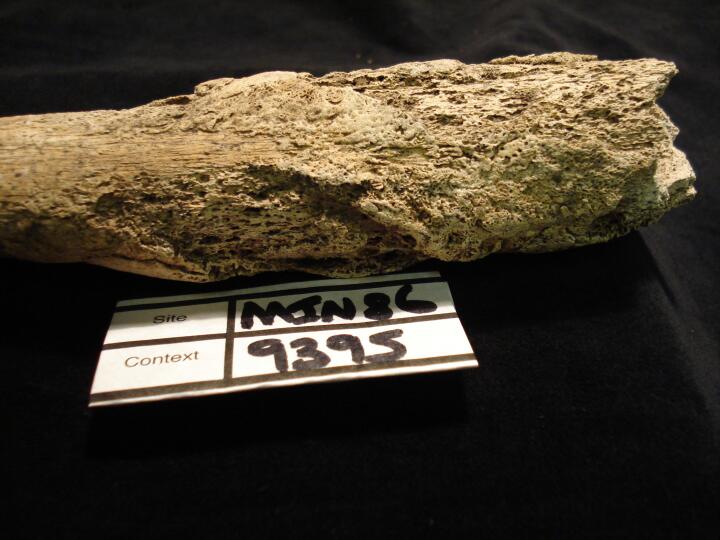
| MIN86
|
9395
|
9
|
MIN86_9395_9.jpg
|
Left tibia (lateral view) bones changes possibly venereal syphilis
|
| MIN86
|
9395
|
10
|
MIN86_9395_10.jpg
|
Left tibia (medial view) close up of bones changes possibly venereal syphilis
|
| MIN86
|
9395
|
11
|
MIN86_9395_11.jpg
|
Left femur & tibia (anterior view) bones changes possibly venereal syphilis
|
| MIN86
|
9395
|
12
|
MIN86_9395_12.jpg
|
Left humerus distal end (anterior view) bones changes possibly venereal syphilis
|
| MIN86
|
9395
|
13
|
MIN86_9395_13.jpg
|
Left humerus distal end (posterior view) bones changes possibly venereal syphilis
|
| MIN86
|
9395
|
14
|
MIN86_9395_14.jpg
|
Left ulna (anterior view) bones changes possibly venereal syphilis
|
| MIN86
|
9395
|
15
|
MIN86_9395_15.jpg
|
Left ulna (posterior view) bones changes possibly venereal syphilis
|
| MIN86
|
9403
|
1
|
MIN86_9403_1.jpg
|
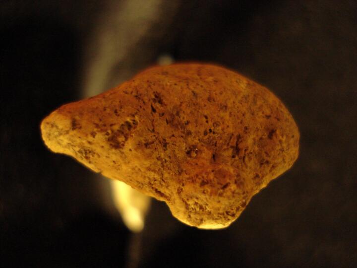
Pronounced demarcated depression on the anterior proximal humerus in the attachment area of the subscapularis muscle.
|
| MIN86
|
9403
|
2
|
MIN86_9403_2.jpg
|
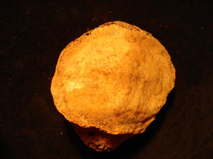
Pronounced demarcated depression on the anterior proximal humerus in the attachment area of the subscapularis muscle (close up).
|
| MIN86
|
9662
|
1
|
MIN86_9662_1.jpg
|
Left ulna distal end, flattened articular surface with osteoarthritis
|
| MIN86
|
9662
|
2
|
MIN86_9662_2.jpg
|
Left ulna distal end, flattened articular surface with osteoarthritis
|
| MIN86
|
10145
|
1
|
MIN86_10145_1.jpg
|
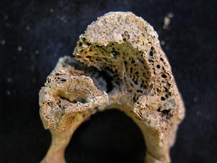
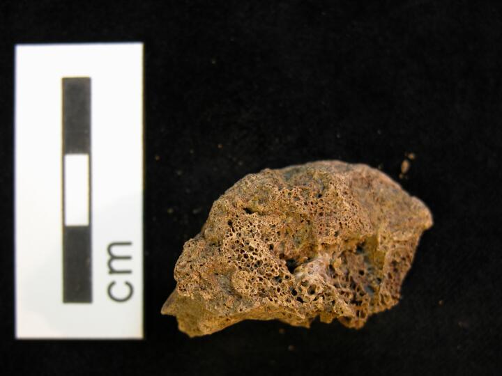
Tuberculosis? Deep penetrating lesion on central aspect of S1
|
| MIN86
|
11436
|
1
|
MIN86_11436_1.jpg
|
Thoracic vertebra, lytic lesion, ? juvenile tuberculosis
|
| MIN86
|
11436
|
2
|
MIN86_11436_2.jpg
|
Thoracic vertebra, lytic lesion, ? juvenile tuberculosis
|
| MIN86
|
11436
|
3
|
MIN86_11436_3.jpg
|
Thoracic vertebrae destruction & collapse, ?juvenile tuberculosis
|
| MIN86
|
11436
|
4
|
MIN86_11436_4.jpg
|
Thoarcic vertebra (superior/anterior view) destruction of centrum, ? Juvenile tuberculosis
|
| MIN86
|
11436
|
5
|
MIN86_11436_5.jpg
|
Thoarcic vertebra (superior/anterior view) destruction of centrum, ? Juvenile tuberculosis
|
| MIN86
|
11436
|
6
|
MIN86_11436_6.jpg
|
Thoarcic vertebra (superior/anterior view) destruction of centrum, ? Juvenile tuberculosis
|
| MIN86
|
12005
|
1
|
MIN86_12005_1.jpg
|
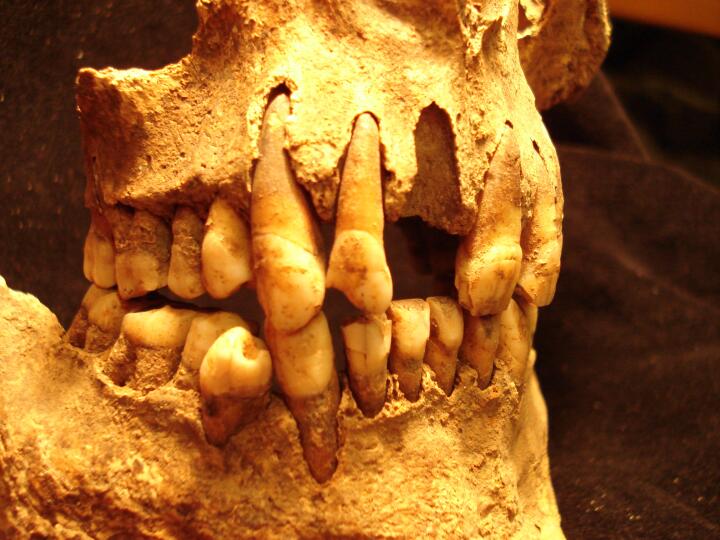
Unusual wear on the maxillary and mandibular right lateral incisors. Over-bite also visible.
|
| MIN86
|
12005
|
2
|
MIN86_12005_2.jpg
|
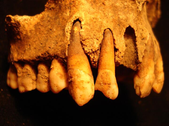
Unusual wear on the maxillary right canine and lateral incisor (T13 and T14)
|
| MIN86
|
12005
|
3
|
MIN86_12005_3.jpg
|
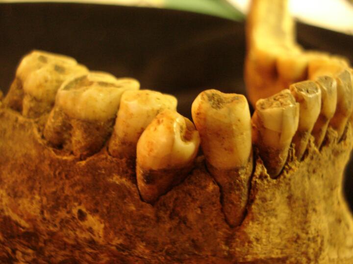
Overcrowding of the 3rd right mandibular premolar (T44)
|
| MIN86
|
12437
|
1
|
MIN86_12437_1.jpg
|
Possible soft tissue trauma to the anterior surface of the right patella. New bone growth across the surface of the patella.
|
| MIN86
|
12437
|
2
|
MIN86_12437_2.jpg
|
Osteophytic lipping around the edges of the inferior articular surfaces of the left talus.
|
| MIN86
|
12437
|
3
|
MIN86_12437_3.jpg
|
Possible soft tissue trauma to the posterior surface of the left calcaneus in the area of the achilles tendon attachment. Increased porosity and bony outgrowths are visible.
|
| MIN86
|
12437
|
4
|
MIN86_12437_4.jpg
|
Osteophytic lipping around the edges of all the articular surfaces of the left calcaneus.
|
| MIN86
|
12455
|
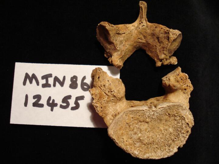
1
|
MIN86_12455_1.jpg
|
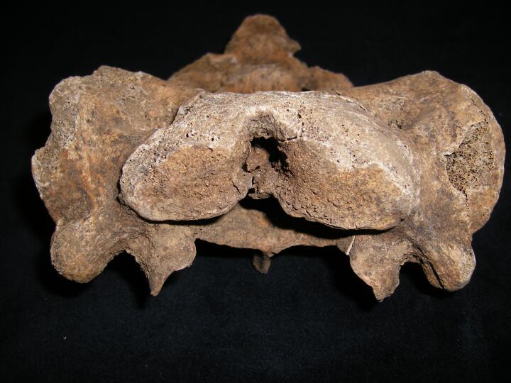
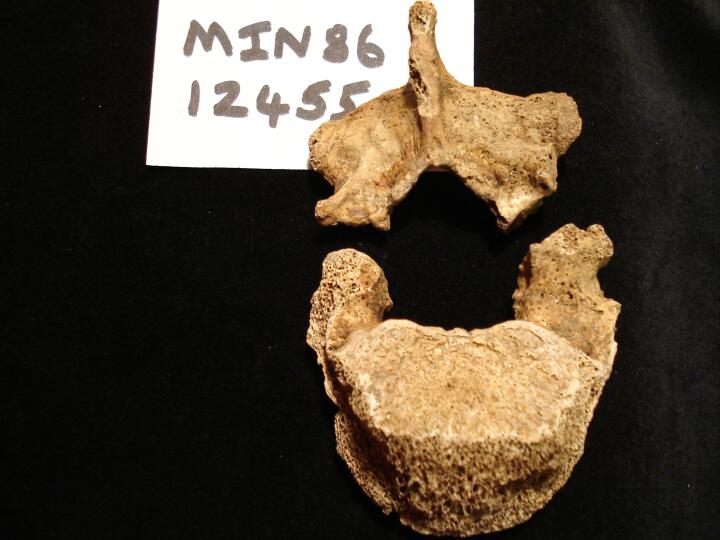
Bilateral spondylolisis of lumbar vertebra L5 (superior view) with pars interarticularis
|
| MIN86
|
12455
|
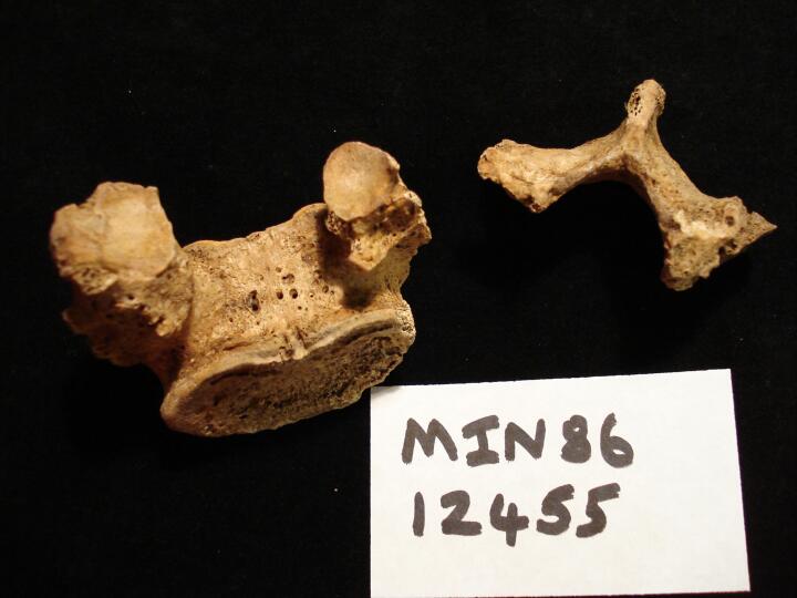
2
|
MIN86_12455_2.jpg
|
Bilateral spondylolisis of lumbar vertebra L5 (inferior view) with pars interarticularis
|
| MIN86
|
12455
|
3
|
MIN86_12455_3.jpg
|
Bilateral spondylolisis of lumbar vertebra L5 (superior view) with pars interarticularis
|
| MIN86
|
12696
|
1
|
MIN86_12696_1.jpg
|
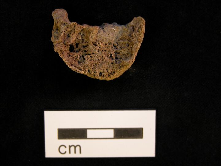
Cervical vertebra (anterior view) ?desructive scalloped lesions/Tuberculosis
|
| MIN86
|
12696
|
2
|
MIN86_12696_2.jpg
|
Thoracic vertebra (left side view) collapse & fusion of bodies
|
| MIN86
|
12696
|
3
|
MIN86_12696_3.jpg
|
Thoracic vertebra, possible destructive lesion, Tuberculosis
|
| MIN86
|
12696
|
4
|
MIN86_12696_4.jpg
|
Thoracic vertebrae fusion of spinous processes (anterior view)
|
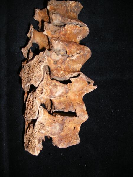
| MIN86
|
12696
|
5
|
MIN86_12696_5.jpg
|
Thoracic vertebrae fusion of spinous & transverse processes (posterior view)
|
| MIN86
|
12696
|
6
|
MIN86_12696_6.jpg
|
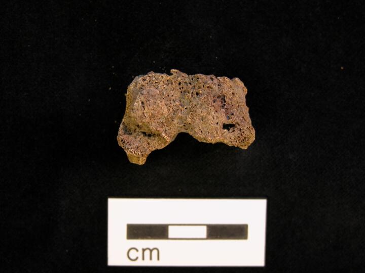
Thoracic vertebra disorganised structure of trabecula, ?lytic focus
|
| MIN86
|
12696
|
7
|
MIN86_12696_7.jpg
|
Thoracic vertebra disorganised structure of trabecula, ?lytic focus
|
| MIN86
|
12696
|
8
|
MIN86_12696_8.jpg
|
Thoracic vertebra disorganised structure of trabecula, ?lytic focus
|
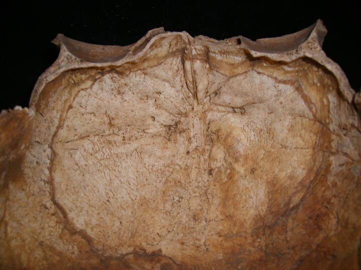
| MIN86
|
13518
|
1
|
MIN86_13518_1.jpg
|
Hyperostosis frontalis interna, view endocranial aspect of frontal bone
|
| MIN86
|
13518
|
2
|
MIN86_13518_2.jpg
|
DISH, lateral view
|
| MIN86
|
13518
|
3
|
MIN86_13518_3.jpg
|
DISH, anterior view
|
| MIN86
|
16052
|
1
|
MIN86_16052_1.jpg
|
Cervical vertebra cut mark from decapition
|
| MIN86
|
16052
|
2
|
MIN86_16052_2.jpg
|
Cervical vertebra cut mark from decapition
|
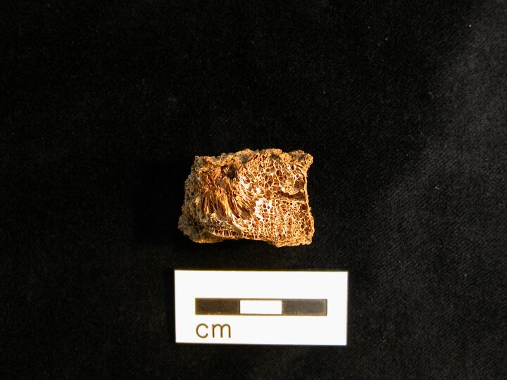
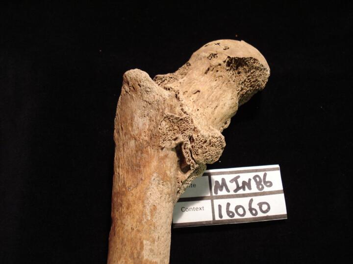
| MIN86
|
16060
|
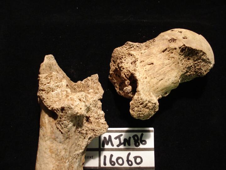
1
|
MIN86_16060_1.jpg
|
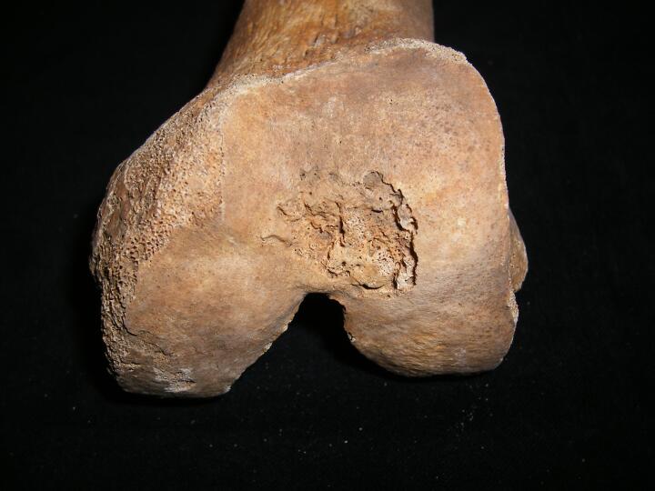
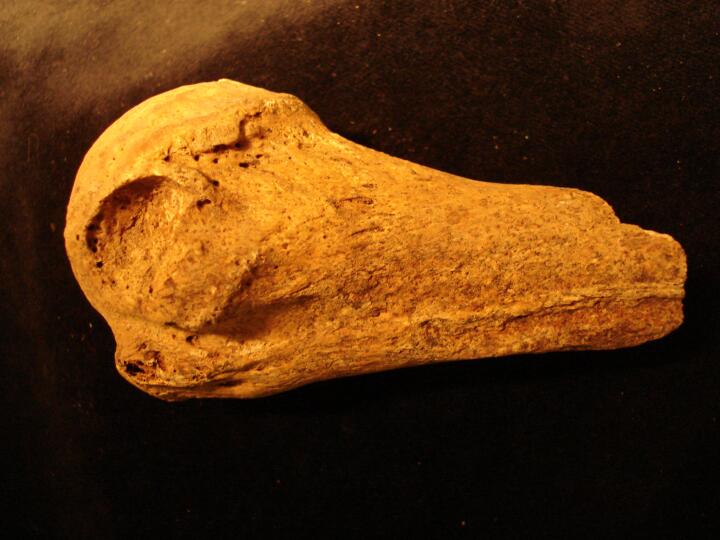
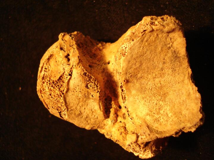
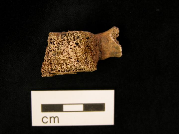
Right femur fracture of femoral neck with non-union of fracture (anterior view) of femoral head & neck
|
| MIN86
|
16060
|
2
|
MIN86_16060_2.jpg
|
Right femur fracture of femoral neck with non-union of fracture (anterior view) two separate bone fragments from fracture ite
|
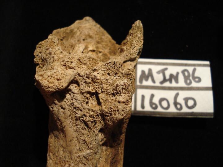
| MIN86
|
16060
|
3
|
MIN86_16060_3.jpg
|
Right femur (anterior view) shaft surface
|
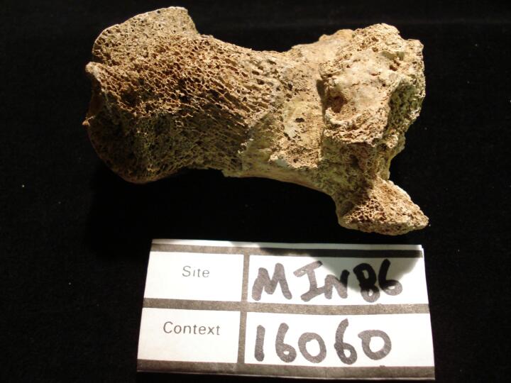
| MIN86
|
16060
|
4
|
MIN86_16060_4.jpg
|
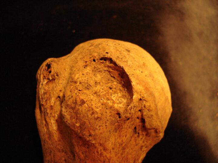
Right femur, femoral head (posterior view) showing non-united fracture site
|
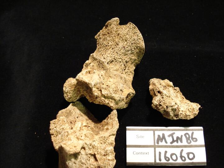
| MIN86
|
16060
|
5
|
MIN86_16060_5.jpg
|
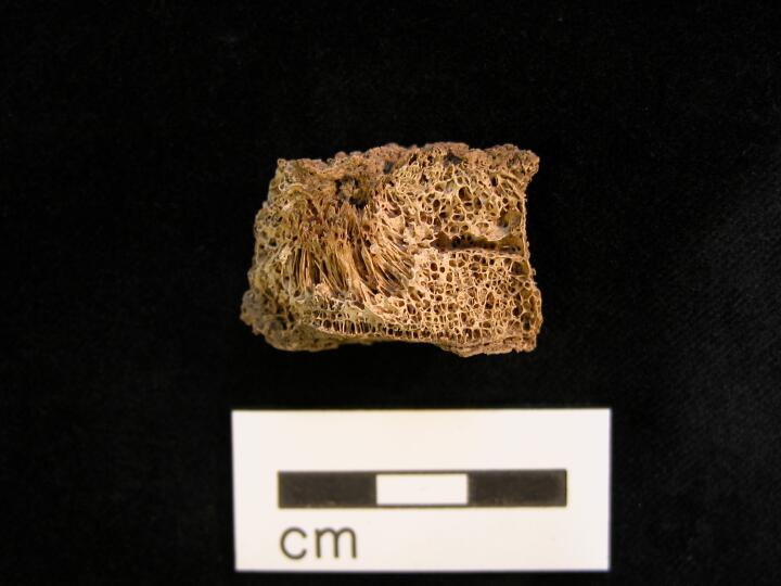
Right femur non-united fracture of femoral neck (medial view) three separate bone fragments from fracture site
|
| MIN86
|
16060
|
6
|
MIN86_16060_6.jpg
|
Right femur (anterior/medial view) three separate bone fragments of frature
|
| MIN86
|
16098
|
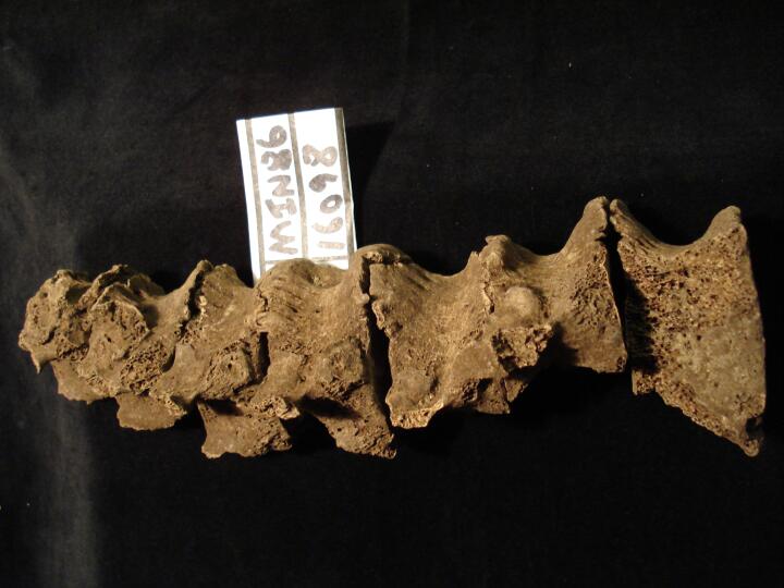
1
|
MIN86_16098_1.jpg
|
Thoracic vertebrae, Th7 to Th12 (right side) some post mortem damage, indication of DISH
|
| MIN86
|
16098
|
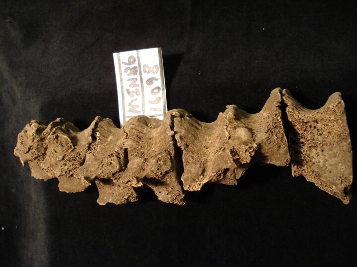
2
|
MIN86_16098_2.jpg
|
Thoracic vertebrae, Th7 to Th12 (right side) some post mortem damage, indication of DISH
|
| MIN86
|
16098
|
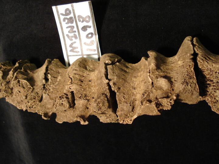
3
|
MIN86_16098_3.jpg
|
Thoracic vertebrae, Th7 to Th12 (right side/close up) some post mortem damage, indication of DISH
|
| MIN86
|
16332
|
1
|
MIN86_16332_1.jpg
|
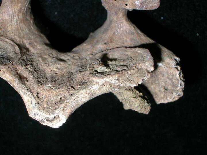
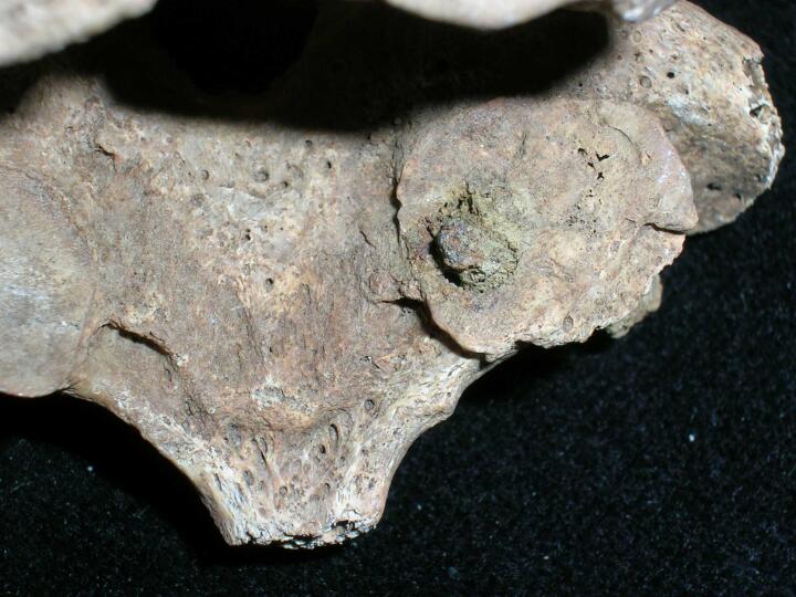
Left acetabulum pitting & porosity, DJD
|
| MIN86
|
16332
|
2
|
MIN86_16332_2.jpg
|
Left acetabulum pitting & porosity, DJD
|
| MIN86
|
16344
|
1
|
MIN86_16344_1.jpg
|
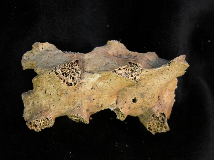
DISH. 'Dripping candle wax' like osteophytes resulting in fusion down the right anterior surface of 5 upper thoracic vertebrae. Disc spaces are retained. Large amount of post-mortem damage to a number of the vertebral bodies.
|


