Our London Wall site is now closed to visitors
but is still available for venue hire and private events.
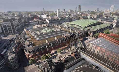
Our new museum coming in 2026 will be situated at the heart of the Smithfield area. Until then, the fun continues at our Docklands museum!
Explore more Delve into fascinating stories of Londoners through videos, articles and more
VIEW ALL
Be the first to hear about new exhibitions and events

Donate to the museum to help us tell the extraordinary story of London and its people – past, present and future
Donate now