| Site code
|
Context
|
Frame number
|
Photo
|
Description
|
| MIN86
|
5206
|
1
|
MIN86_5206_1.jpg
|
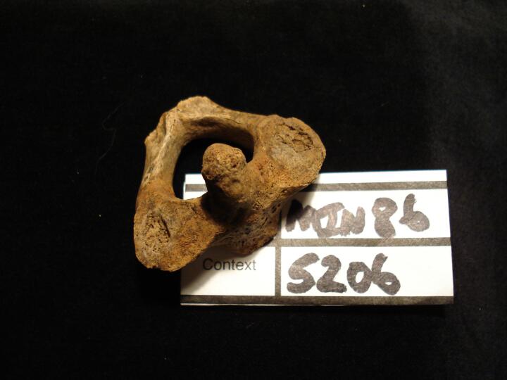
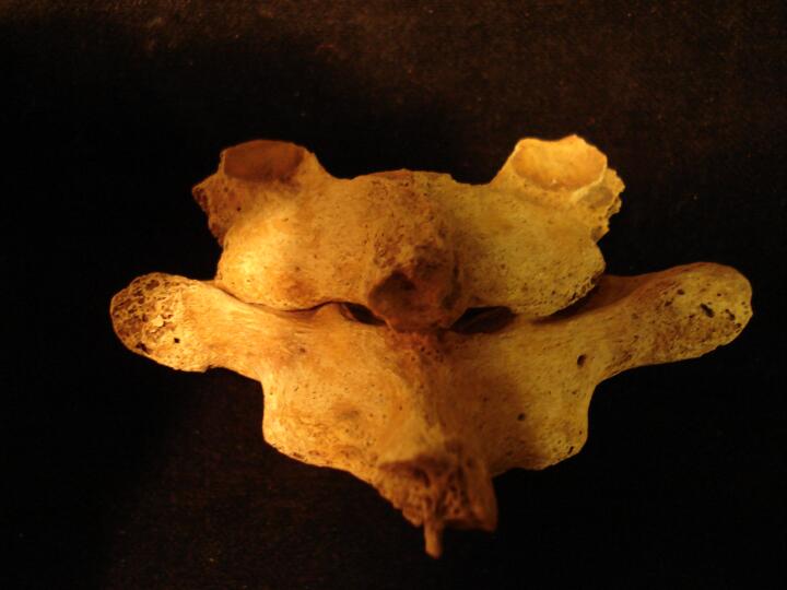
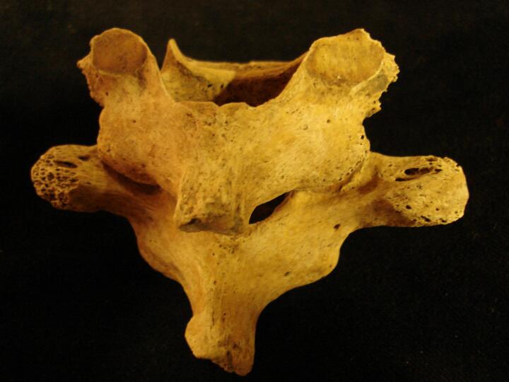
Axis vertebra (superior view) smooth circular defects, possibly developmental
|
| MIN86
|
5265
|
1
|
MIN86_5265_1.jpg
|
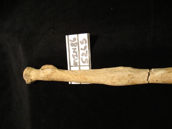
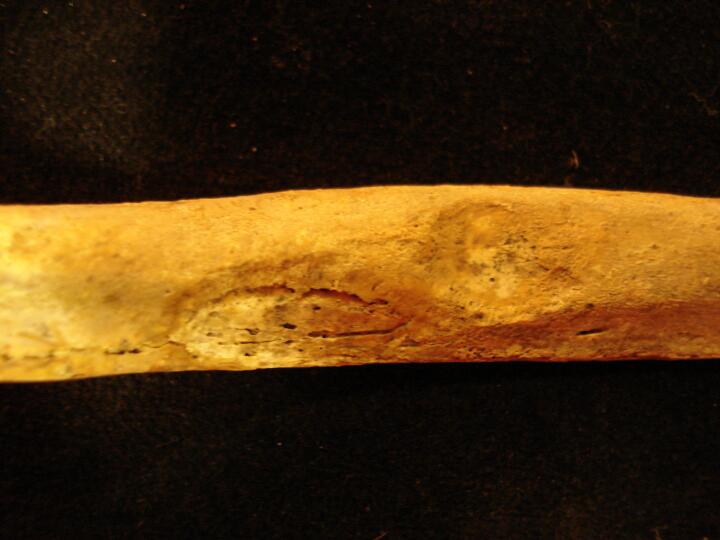
Right radius healed closed fracture mid shaft (anterior view)
|
| MIN86
|
5265
|
2
|
MIN86_5265_2.jpg
|
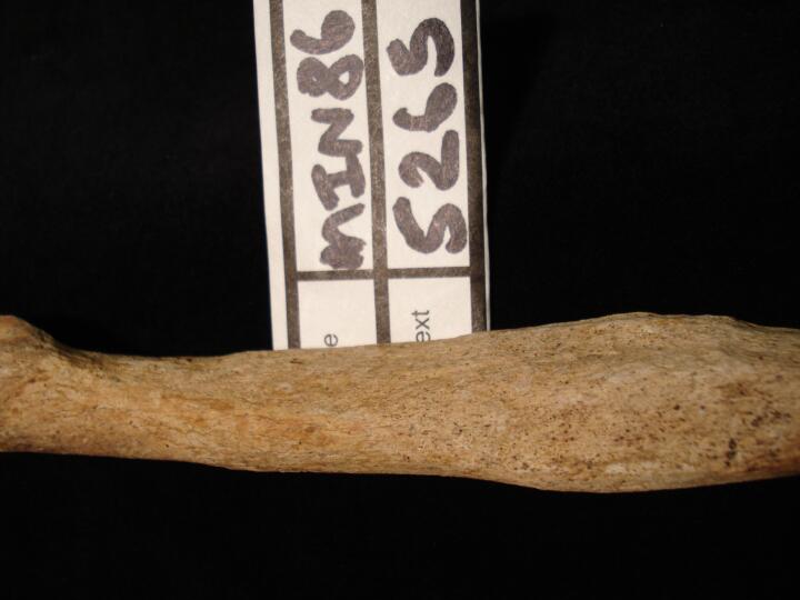
Right radius healed closed fracture mid shaft (anterior view) close up of fracture site
|
| MIN86
|
5279
|
1
|
MIN86_5279_1.jpg
|
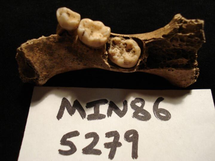
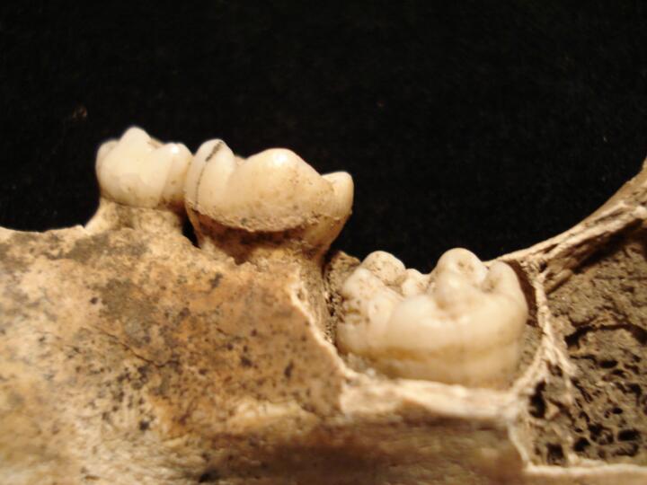
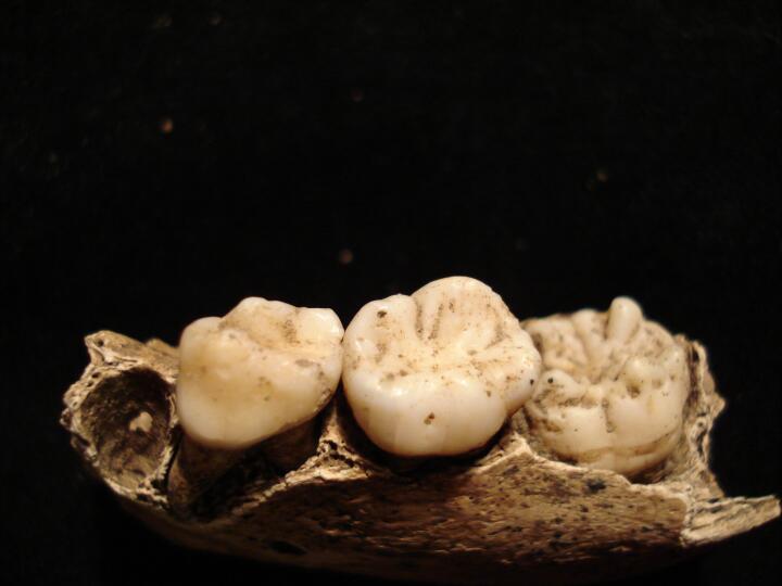
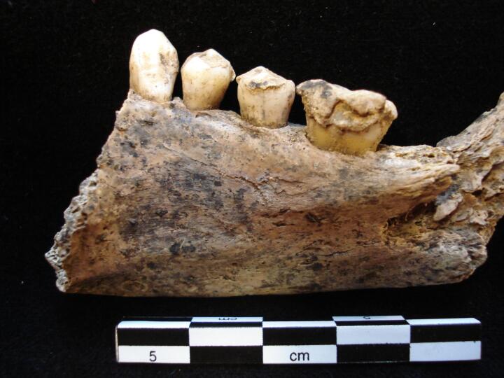
Right side of mandible (lingual view) 1st permanent molar enamel defects possibly associated with congenital syphilis
|
| MIN86
|
5279
|
2
|
MIN86_5279_2.jpg
|
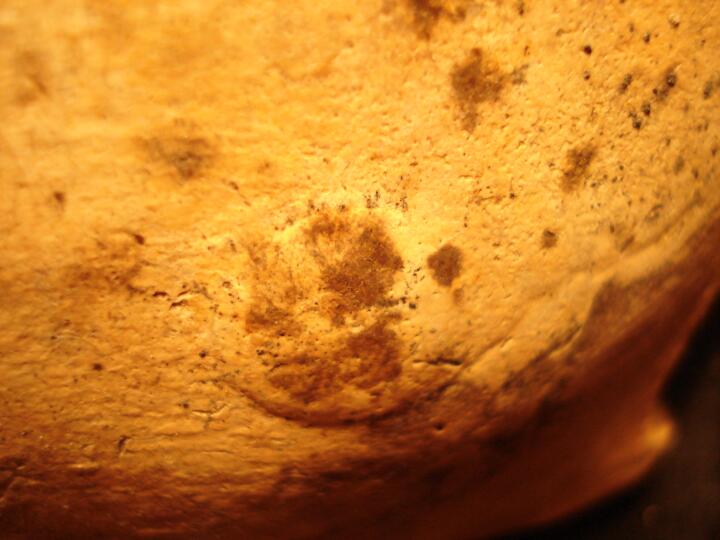
Right side of mandible (lingual view/close up) 1st permanent molar enamel defects possibly associated with congenital syphilis
|
| MIN86
|
5279
|
3
|
MIN86_5279_3.jpg
|
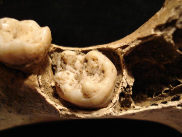
Right side of mandible (occlusal view) 1st permanent molar enamel defects possibly associated with congenital syphilis
|
| MIN86
|
5279
|
4
|
MIN86_5279_4.jpg
|
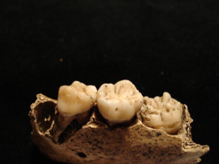
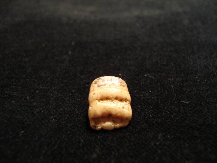
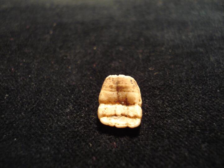
Left side of mandible (buccal view) 1st permanent molar enamel hypoplastic defects (?mulberry molars) possibly associated with specific treponemal infection, congenital syphilis
|
| MIN86
|
5279
|
5
|
MIN86_5279_5.jpg
|
Left side of mandible (buccal view/close up) 1st permanent molar enamel hypoplastic defects (?mulberyy molars) possibly associated with congenital syphilis
|
| MIN86
|
5279
|
6
|
MIN86_5279_6.jpg
|
Left side of mandible (occlusal view) 1st permanent molar enamel hypoplastic defects possibly associated with specific treponemal infection, congenital syphilis
|
| MIN86
|
5279
|
7
|
MIN86_5279_7.jpg
|
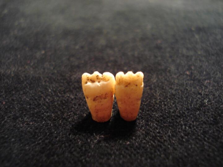
Developing (loose) permanent maxillary lateral incisor & left central incisor hypoplastic defects (buccal surface) possibly associated with specific treponemal infection, congenital syphilis
|
| MIN86
|
5279
|
8
|
MIN86_5279_8.jpg
|
Left maxillary permanent central incisor (buccal surface) linear hypoplastic defects (?Hutchinson incisor) possibly associated with congenital syphilis
|
| MIN86
|
5279
|
9
|
MIN86_5279_9.jpg
|
Left maxillary permanent central incisor (lingual surface) linear hypoplastic defects (?Hutchinson incisor) possibly associated with congenital syphilis
|
| MIN86
|
5343
|
1
|
MIN86_5343_1.jpg
|
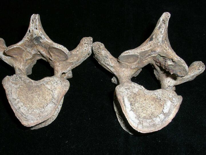
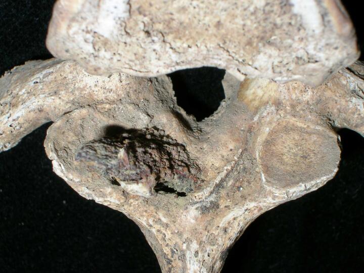
Projectile injury, ferrous object embedded in spinous process
|
| MIN86
|
5343
|
2
|
MIN86_5343_2.jpg
|
Projectile injury, ferrous object embedded, inferior view
|
| MIN86
|
5343
|
3
|
MIN86_5343_3.jpg
|
Projectile injury, ferrous object embedded, superior view
|
| MIN86
|
5343
|
4
|
MIN86_5343_4.jpg
|
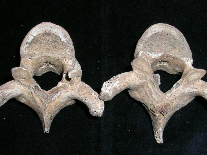
Projectile injury, ferrous object penetrated to vertebra below showing healing of bone
|
| MIN86
|
5343
|
5
|
MIN86_5343_5.jpg
|
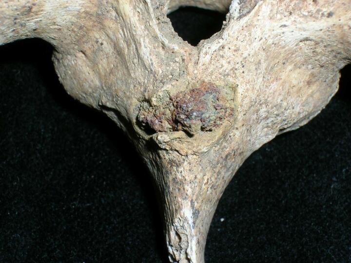
Projectile injury penetrating through the spinous process
|
| MIN86
|
5343
|
6
|
MIN86_5343_6.jpg
|
Projectile injury, ferrous object embedded in spinous process
|
| MIN86
|
5728
|
1
|
MIN86_5728_1.jpg
|
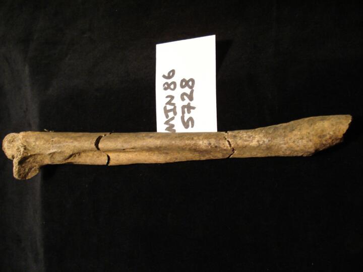
Right ulna healed 'Parry' fracture (anterior view from medial aspect)
|
| MIN86
|
5728
|
2
|
MIN86_5728_2.jpg
|
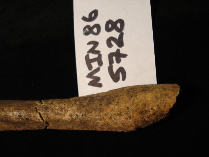
Right ulna healed 'Parry' fracture (anterior view from medial aspect) close up of fracture site
|
| MIN86
|
5728
|
3
|
MIN86_5728_3.jpg
|
Right ulna healed 'Parry' fracture (anterior view from lateral aspect)
|
| MIN86
|
5902
|
1
|
MIN86_5902_1.jpg
|
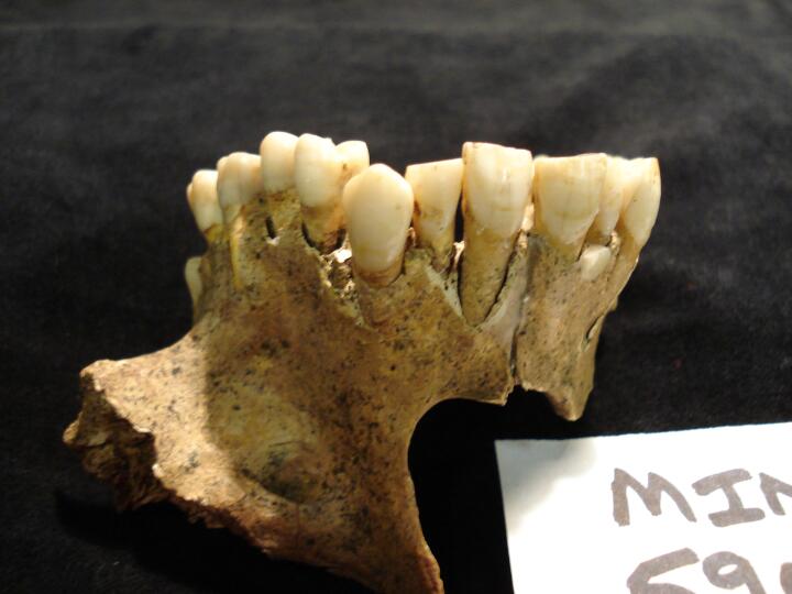
Maxilla (buccal view) crowding of maxillary teeth, some post mortem damage but indication of either super nummary tooth or deciduous retention (between left lateral incisor & canine)
|
| MIN86
|
5902
|
2
|
MIN86_5902_2.jpg
|
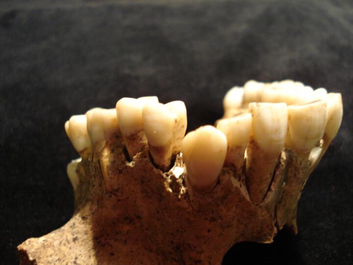
Maxilla (buccal view) close up of crowding of maxillary teeth
|
| MIN86
|
5940
|
1
|
MIN86_5940_1.jpg
|
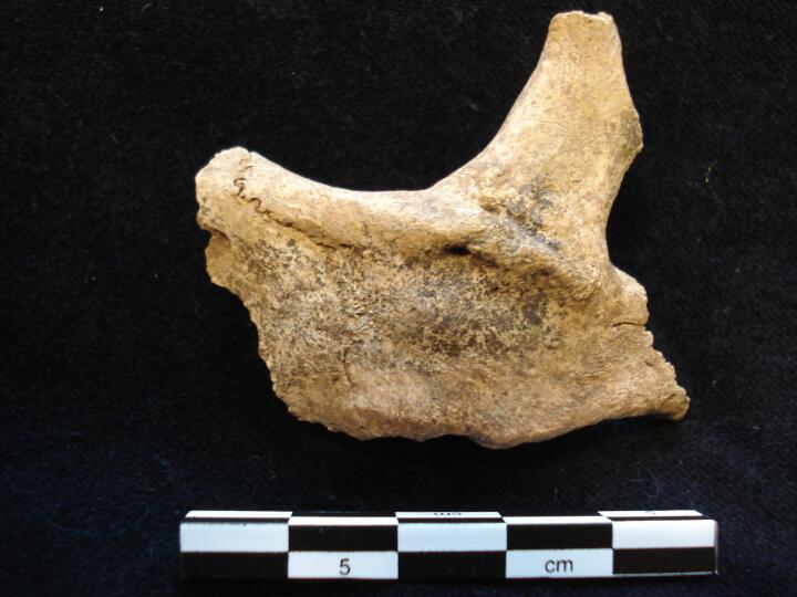
Left zygomatic process (anterior view) healed fracture
|
| MIN86
|
5940
|
2
|
MIN86_5940_2.jpg
|
Left zygomatic process (anterior view/close up) healed fracture
|
| MIN86
|
6097
|
1
|
MIN86_6097_1.jpg
|
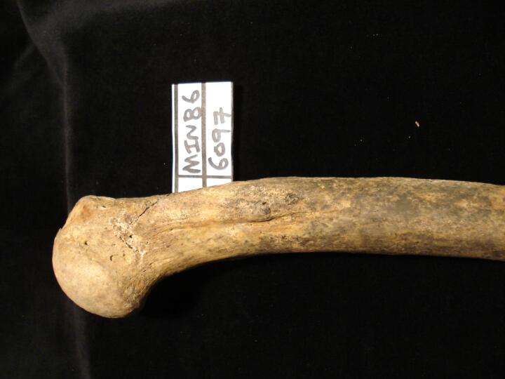
Left humerus proximal end healed malaligned fracture (anterior view)
|
| MIN86
|
6097
|
2
|
MIN86_6097_2.jpg
|
Left humerus proximal end healed malaligned fracture (posterior view)
|
| MIN86
|
6097
|
3
|
MIN86_6097_3.jpg
|
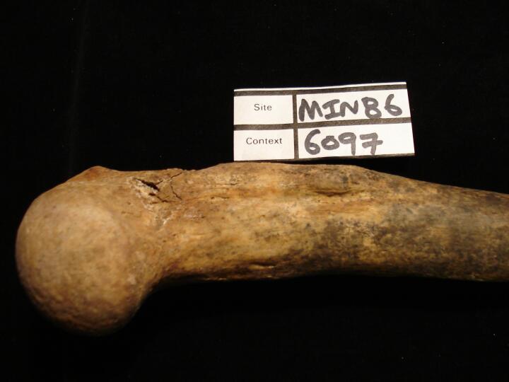
Left humerus proximal end healed malaligned fracture (medial view)
|
| MIN86
|
6097
|
4
|
MIN86_6097_4.jpg
|
Left humerus proximal end healed malaligned fracture (close up from medial aspect)
|
| MIN86
|
6412
|
1
|
MIN86_6412_1.jpg
|
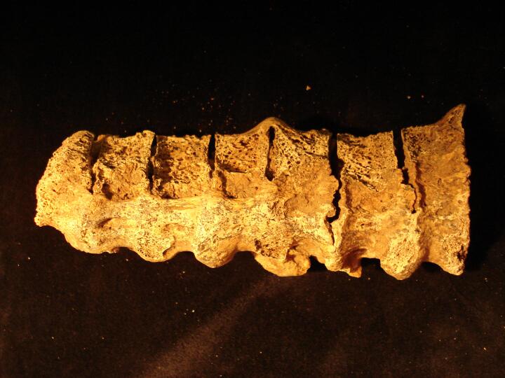
DISH. 'Dripping candle wax' like osteophytes causing fusion of T5-11 down the right anterior surface of the vertebral bodies. Disc spaces retained. (Dorsal view)
|
| MIN86
|
6412
|
2
|
MIN86_6412_2.jpg
|
DISH. 'Dripping candle wax' like osteophytes causing fusion T5-11 down the right anterior surface of the vertebral bodies. Disc spaces retained. (right view)
|
| MIN86
|
6412
|
3
|
MIN86_6412_3.jpg
|
Well healed new bone formation to the posterior distal left tibia. Bony outgrowths and porosity visible.
|
| MIN86
|
6412
|
4
|
MIN86_6412_4.jpg
|
Close up of bony projections and porosity on the medial and anterior distal left tibia (view from the medial malleolus)
|
| MIN86
|
6412
|
5
|
MIN86_6412_5.jpg
|
Close up of bony projections and porosity on the medial malleolus of the distal left tibia.
|
| MIN86
|
6412
|
6
|
MIN86_6412_6.jpg
|
Bony projections and porosity on the lateral distal left tibia tracking down the area of attachment for the interosseous crest.
|
| MIN86
|
6412
|
7
|
MIN86_6412_7.jpg
|
Traumatic arthritis. Osteoarthritic changes to the distal articular surface of the left tibia. Osteophytic lipping, subchondral cysts and an irregular surface area are observable.
|
| MIN86
|
6452
|
1
|
MIN86_6452_1.jpg
|
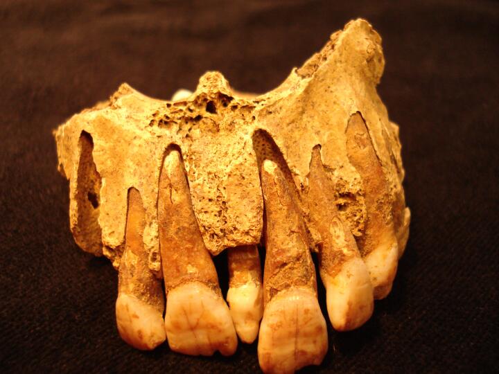
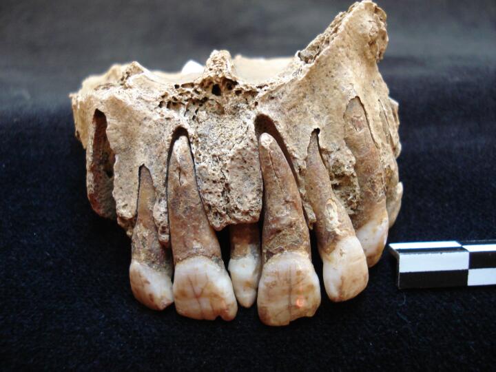
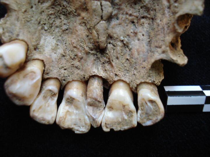
Maxilla with visible supernummery peg tooth between the two front incisors. (anterior view)
|
| MIN86
|
6452
|
2
|
MIN86_6452_2.jpg
|
Maxilla with visible supernummery peg tooth between the two front incisors and torus of the palate.
|
| MIN86
|
6452
|
3
|
MIN86_6452_3.jpg
|
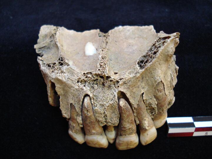
Maxilla with visible supernummery peg tooth between the two front incisors and another protruding into the right portion of the nasal cavity.
|
| MIN86
|
6452
|
4
|
MIN86_6452_4.jpg
|
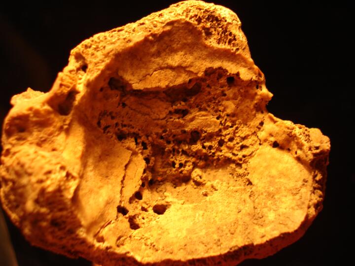
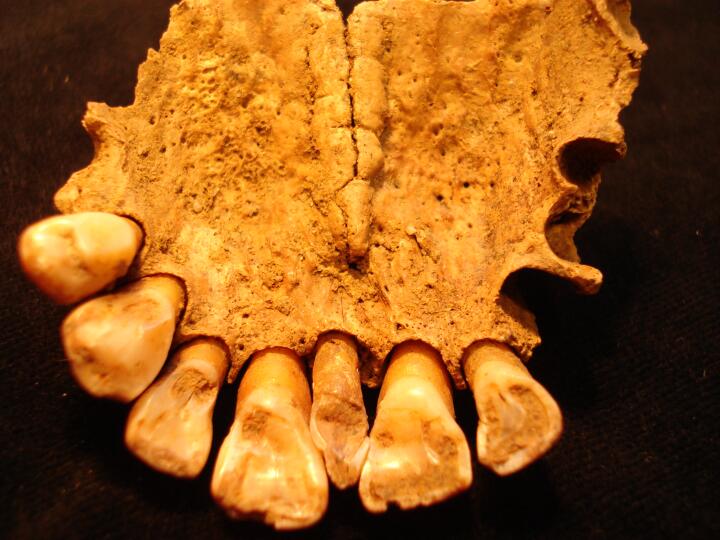
Maxilla (buccal view) supernumerary tooth between central incisors
|
| MIN86
|
6452
|
5
|
MIN86_6452_5.jpg
|
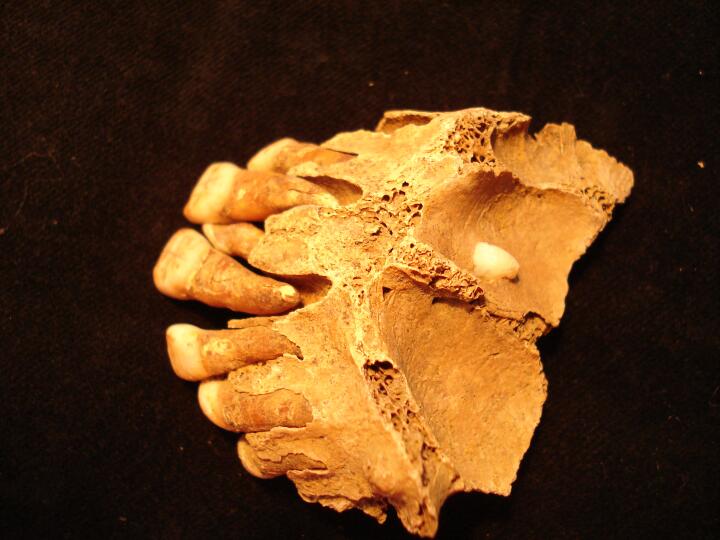
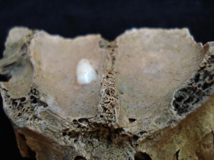
Maxilla (superior view) supernumerary tooth between central incisors & impacted right canine
|
| MIN86
|
6452
|
6
|
MIN86_6452_6.jpg
|
Maxilla (superior view) close up of impacted right canine
|
| MIN86
|
6452
|
7
|
MIN86_6452_7.jpg
|
Maxilla (palatal/lingual view) supernumerary tooth between central incisors & pronounced palatine torus
|
| MIN86
|
6545
|
1
|
MIN86_6545_1.jpg
|
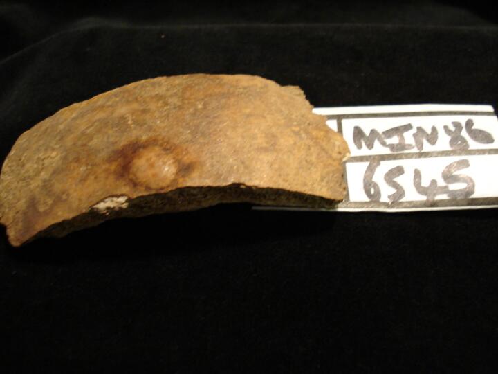
Skull left parietal fragment (ectocranial surface) button osteoma
|
| MIN86
|
6674
|
1
|
MIN86_6674_1.jpg
|
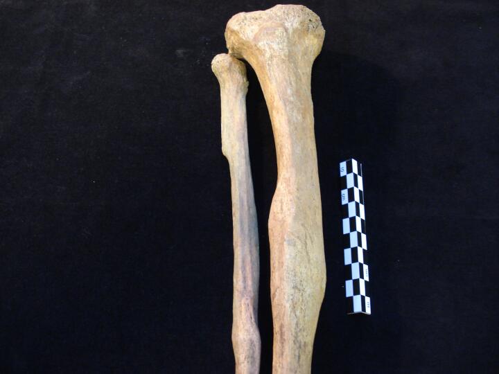
Right tibia & fibula (anterior view) healed fractures of the mid shaft of tibia & distal end fibula
|
| MIN86
|
6674
|
2
|
MIN86_6674_2.jpg
|
Right tibia & fibula (anterior view)close up of healed fractures of the mid shaft of tibia & distal end fibula
|
| MIN86
|
6674
|
3
|
MIN86_6674_3.jpg
|
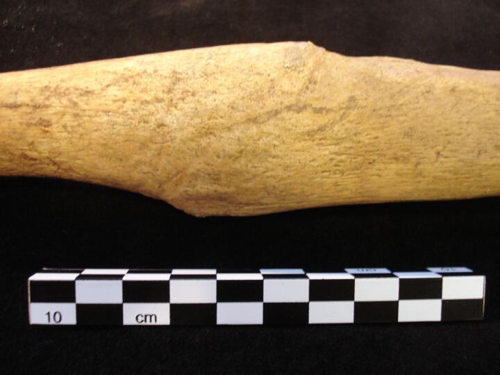
Right tibia close up of calus of healed fracture (anterior view)
|
| MIN86
|
6674
|
4
|
MIN86_6674_4.jpg
|
Right tibia close up of calus of healed fracture (medial view)
|
| MIN86
|
6674
|
5
|
MIN86_6674_5.jpg
|
Right tibia close up of calus of healed fracture (posterior view)
|
| MIN86
|
6674
|
6
|
MIN86_6674_6.jpg
|
Right fibula close up of calus of healed fracture (anterior view)
|
| MIN86
|
6674
|
7
|
MIN86_6674_7.jpg
|
Right fibula close up of calus of healed fracture (posterior view)
|
| MIN86
|
6674
|
8
|
MIN86_6674_8.jpg
|
Right tibia & fibula close up of fracture sites with healed remodelled calus (medial view)
|
| MIN86
|
6674
|
9
|
MIN86_6674_9.jpg
|
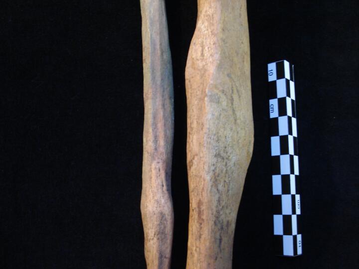
Comparison between left tibia and right tibia with fracture (anteior view)
|
| MIN86
|
6674
|
10
|
MIN86_6674_10.jpg
|
Right and left fibula to show comparison with bony change of right at proximal end & healed fracture compared to left fibula (medial view)
|
| MIN86
|
6674
|
11
|
MIN86_6674_11.jpg
|
Right and left fibula (distal ends) to show comparison of healed fracture of right compared to left fibula (medial view)
|
| MIN86
|
7248
|
1
|
MIN86_7248_1.jpg
|
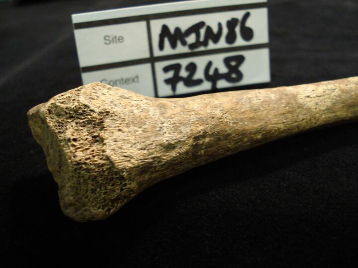
Right radius (anterior view) very well healed & remodelled 'Colles' fracture
|
| MIN86
|
7248
|
2
|
MIN86_7248_2.jpg
|
Right radius (medial view) very well healed & remodelled 'Colles' fracture
|
| MIN86
|
8057
|
1
|
MIN86_8057_1.jpg
|
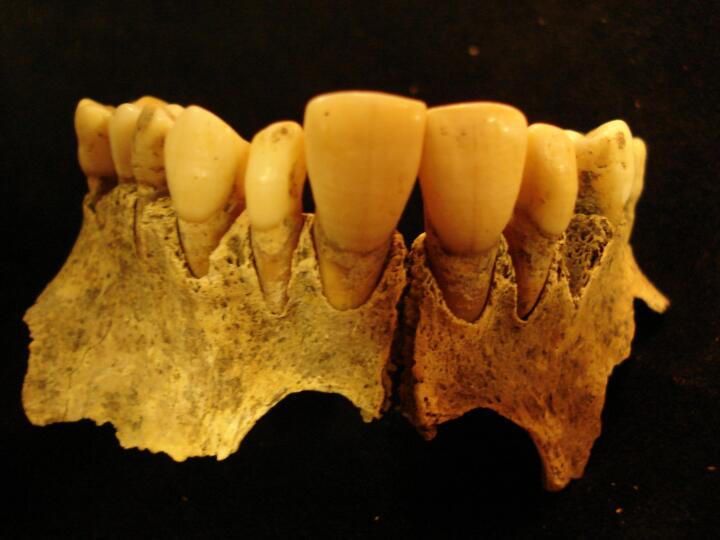
Maxilla with peg shaped lateral incisors in situ
|
| MIN86
|
8057
|
2
|
MIN86_8057_2.jpg
|
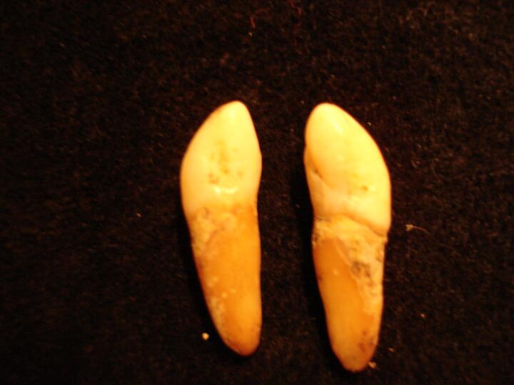
Peg shaped maxillary lateral incisors.
|
| MIN86
|
8075
|
1
|
MIN86_8075_1.jpg
|
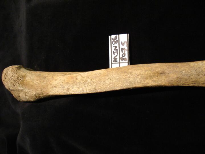
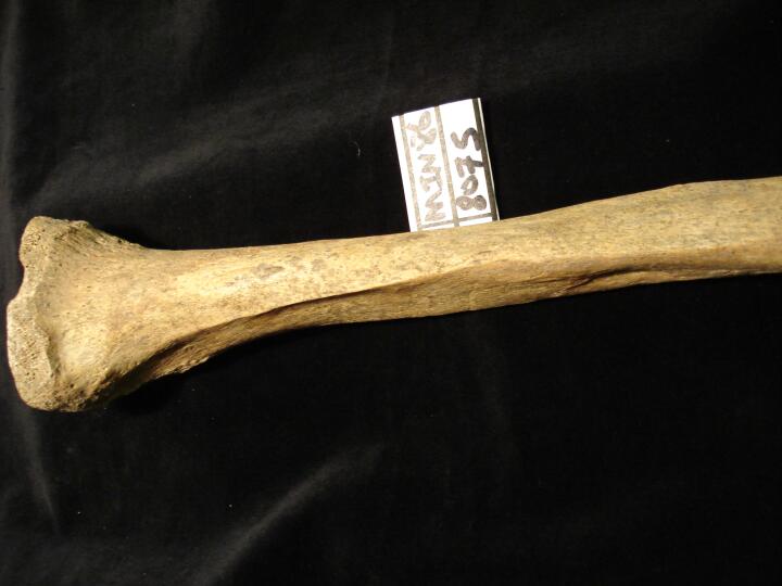
Left tibia healed simple fracture (?oblique) with area of depressed bone (lateral view)
|
| MIN86
|
8075
|
2
|
MIN86_8075_2.jpg
|
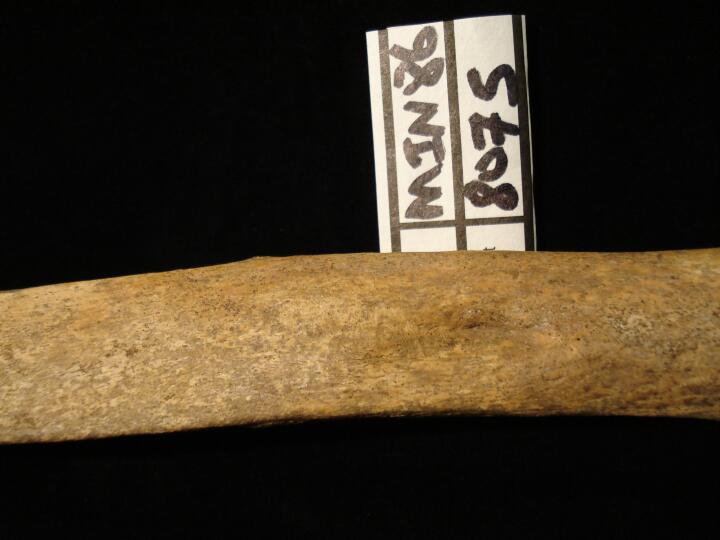
Left tibia healed simple fracture (?oblique) close up of depression (lateral view)
|
| MIN86
|
8075
|
3
|
MIN86_8075_3.jpg
|
Left tibia healed simple fracture close up (lateral view) of distal end
|
| MIN86
|
8075
|
4
|
MIN86_8075_4.jpg
|
Left tibia healed simple fracture ?oblique (posterior view)
|
| MIN86
|
8099
|
1
|
MIN86_8099_1.jpg
|
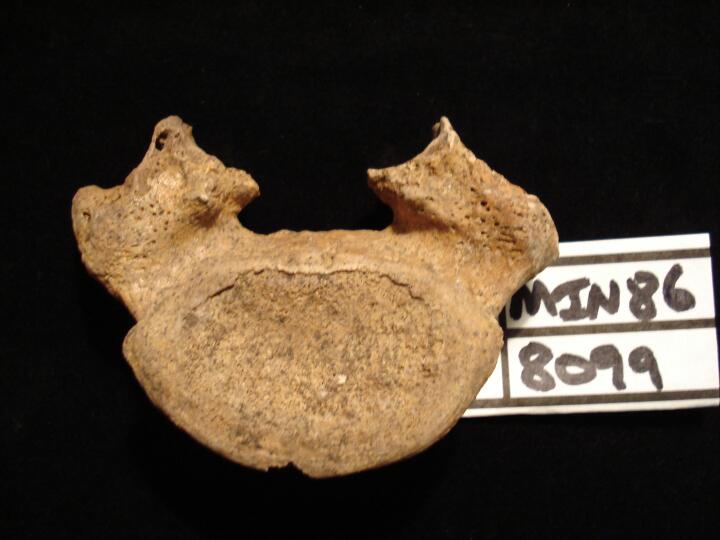
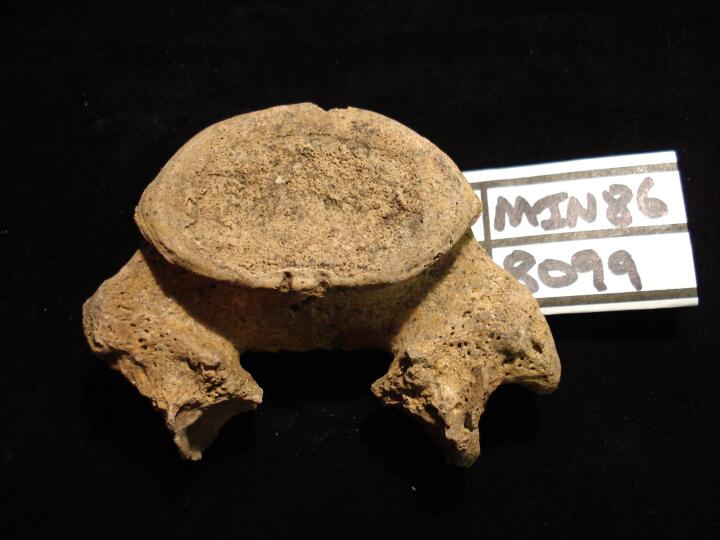
Spondylolisis of lumbar vertebra L5 (Inferior surface/anterior view)
|
| MIN86
|
8099
|
2
|
MIN86_8099_2.jpg
|
Spondylolisis of lumbar vertebra L5 (Inferior surface/posterior view)
|
| MIN86
|
8281
|
1
|
MIN86_8281_1.jpg
|
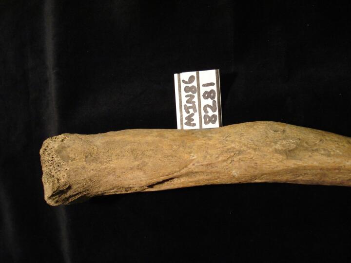
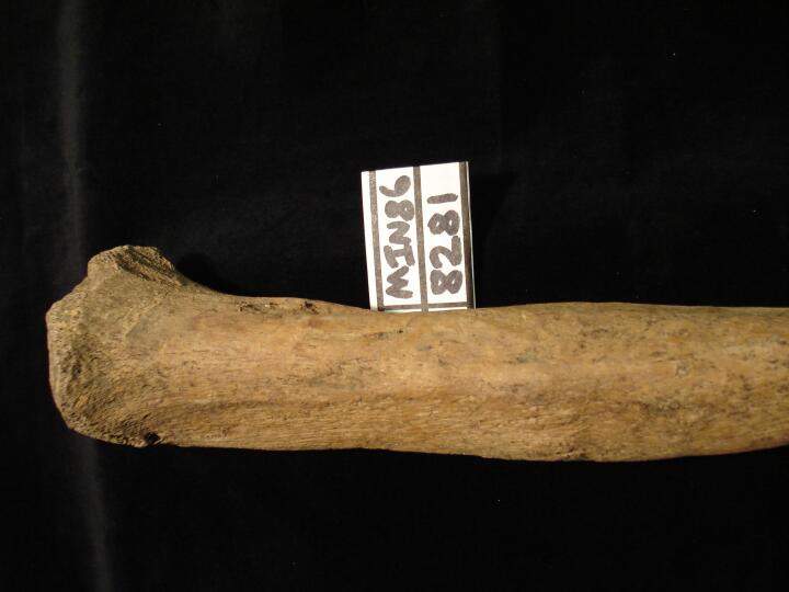
Left tibia healed fracture (anterior view)
|
| MIN86
|
8281
|
2
|
MIN86_8281_2.jpg
|
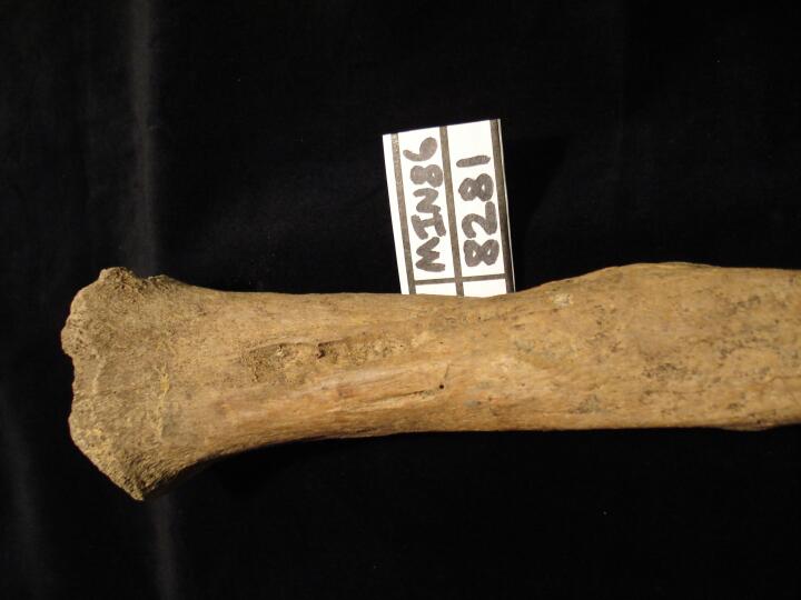
Left tibia healed fracture (medial view)
|
| MIN86
|
8281
|
3
|
MIN86_8281_3.jpg
|
Left tibia healed fracture (lateral view)
|
| MIN86
|
8281
|
4
|
MIN86_8281_4.jpg
|
Left tibia healed fracture (posterior view)
|
| MIN86
|
8281
|
5
|
MIN86_8281_5.jpg
|
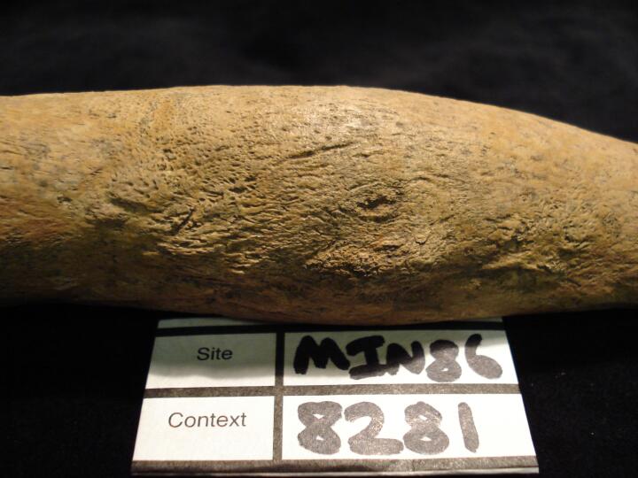
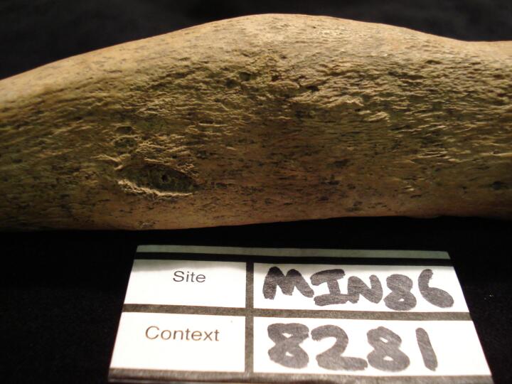
Left tibia healed fracture (medial view) close up of fracture site
|
| MIN86
|
8281
|
6
|
MIN86_8281_6.jpg
|
Left tibia healed fractue (lateral view) close up of fracture site
|
| MIN86
|
8281
|
7
|
MIN86_8281_7.jpg
|
Left tibia healed fracture (anterior view) close up of fracture
|
| MIN86
|
8317
|
1
|
MIN86_8317_1.jpg
|
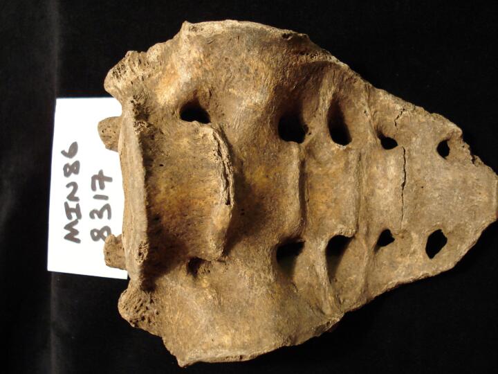
Sacrum (anterior view) sacralisation of lumbar vertebra L6
|
| MIN86
|
8317
|
2
|
MIN86_8317_2.jpg
|
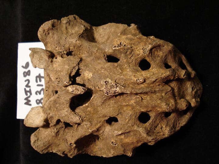
Sacrum (posterior view) sacralisation of lumbar vertebra L6 & clefting
|
| MIN86
|
8317
|
3
|
MIN86_8317_3.jpg
|
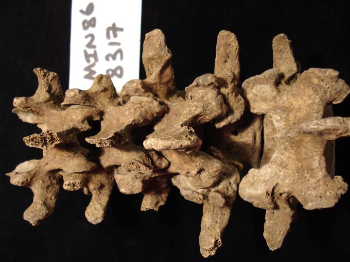
Lumbar vertebrae, articulated L1 to L5, with L4 exhibiting bilateral spondylolisis (posterior view)
|
| MIN86
|
8317
|
4
|
MIN86_8317_4.jpg
|
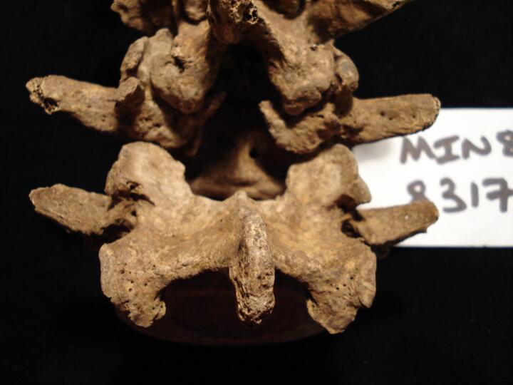
Lumbar vertebrae, articulated L1 to L5, with L4 exhibiting bilateral spondylolisis (posterior view/close up)
|
| MIN86
|
8380
|
1
|
MIN86_8380_1.jpg
|
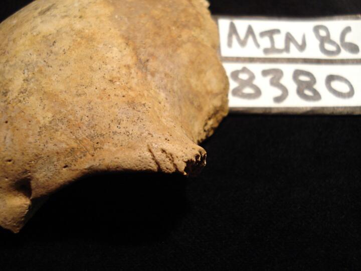
Left orbit sharp force trauma two cut marks at the distal aspect of the orbit (anterior view)
|
| MIN86
|
8380
|
2
|
MIN86_8380_2.jpg
|
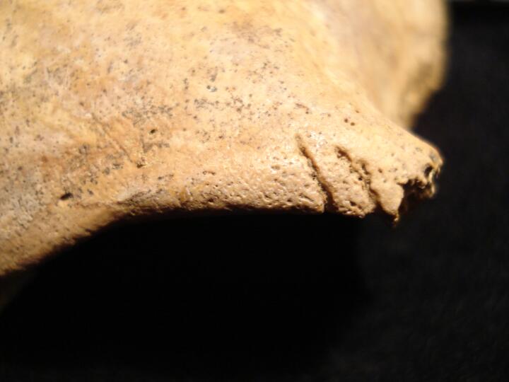
Left orbit sharp force trauma two cut marks at the distal aspect of the orbit (anterior view) close up of cut marks
|
| MIN86
|
8380
|
3
|
MIN86_8380_3.jpg
|
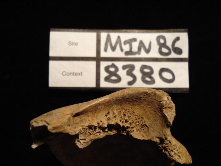
Left orbit sharp force trauma two cut marks & cribra orbitalia (Type 4) (superior view)
|
| MIN86
|
8380
|
4
|
MIN86_8380_4.jpg
|
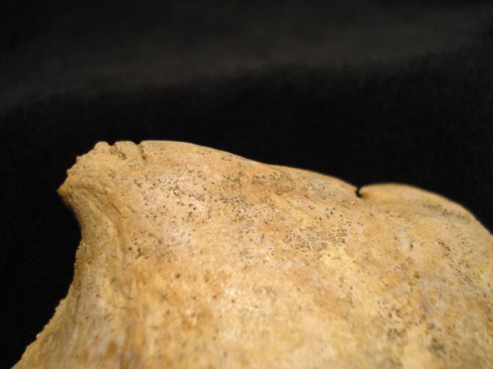
Frontal bone (superior/posterior view) showing cut marks of left orbit
|
| MIN86
|
8393
|
1
|
MIN86_8393_1.jpg
|
Active infection on the anterior sacrum of S1 and S2.
|
| MIN86
|
8450
|
1
|
MIN86_8450_1.jpg
|
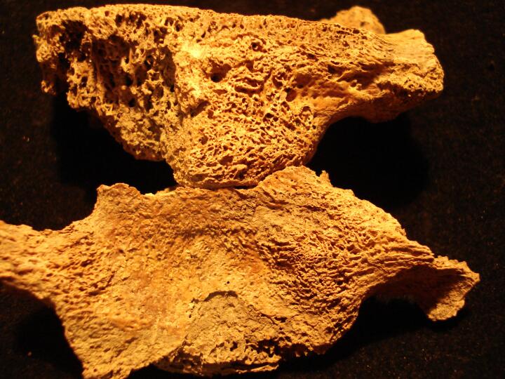
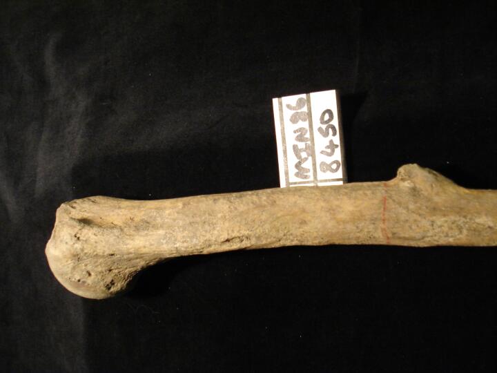
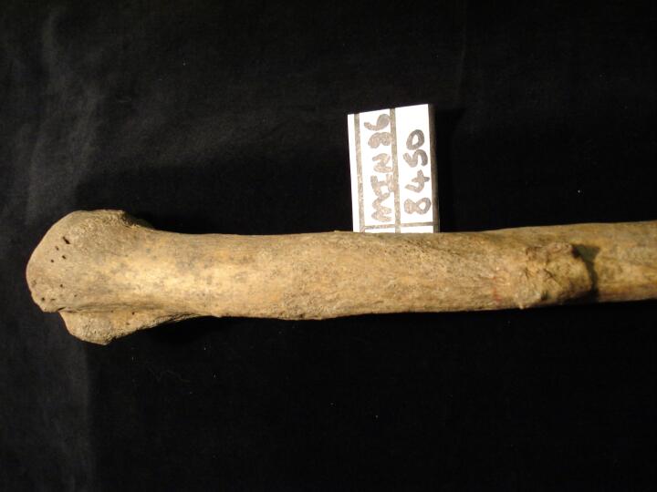
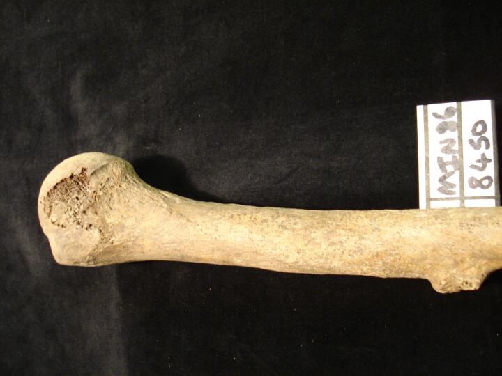
Left humerus metaphyseal surface (anterior view) osteochondroma
|
| MIN86
|
8450
|
2
|
MIN86_8450_2.jpg
|
Left humerus metaphyseal surface (lateral view) osteochondroma
|
| MIN86
|
8450
|
3
|
MIN86_8450_3.jpg
|
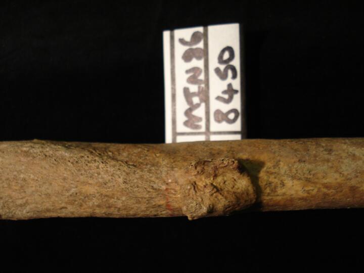
Left humerus metaphyseal surface (lateral view/close up) osteochondroma
|
| MIN86
|
8450
|
4
|
MIN86_8450_4.jpg
|
Left humerus metaphyseal surface (side on from lateral aspect) osteochondroma
|
| MIN86
|
8450
|
5
|
MIN86_8450_5.jpg
|
Left humerus metaphyseal surface (posterior view) osteochondroma
|
| MIN86
|
8456
|
1
|
MIN86_8456_1.jpg
|
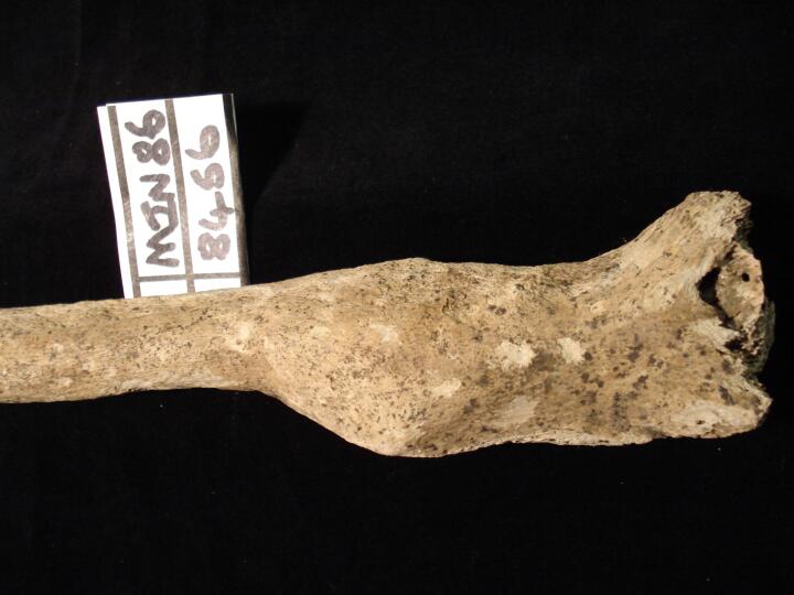
Right humerus (anterior view) healed transverse fracture, malaligned
|
| MIN86
|
8456
|
2
|
MIN86_8456_2.jpg
|
Right humerus (anterior view/close up) healed transverse fracture, malaligned
|
| MIN86
|
8456
|
3
|
MIN86_8456_3.jpg
|
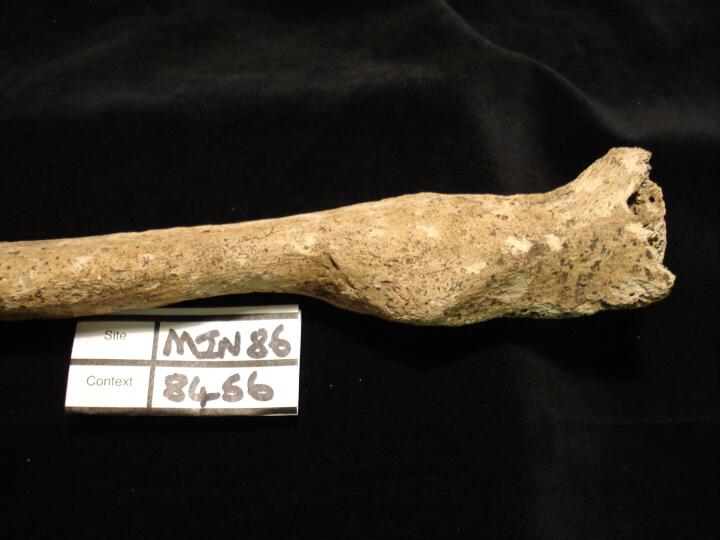
Right humerus distal 1/3 (lateral view) healed transverse fracture
|
| MIN86
|
9035
|
1
|
MIN86_9035_1.jpg
|
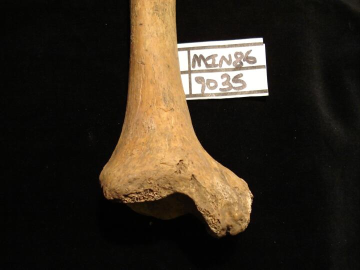
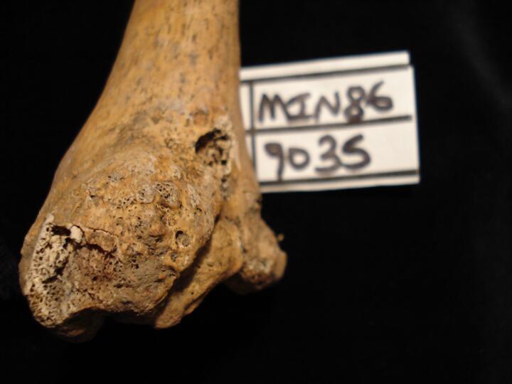
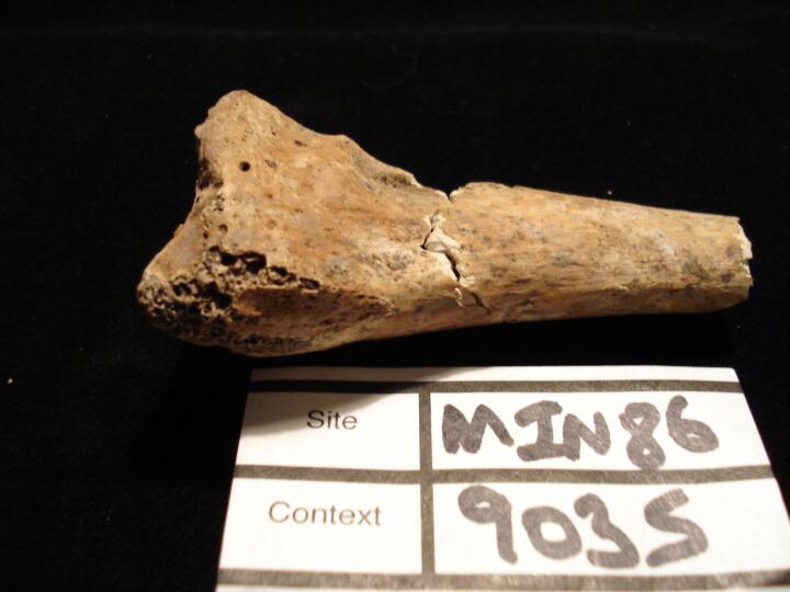
Right tibia distal end (anterior view) healed fracture of medial malleolus
|
| MIN86
|
9035
|
2
|
MIN86_9035_2.jpg
|
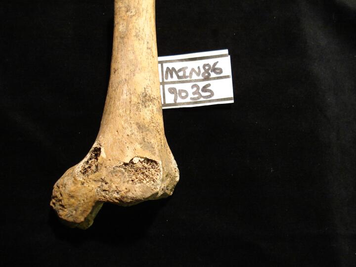
Right tibia distal end (medial view) healed fracture of medial malleolus
|
| MIN86
|
9035
|
3
|
MIN86_9035_3.jpg
|
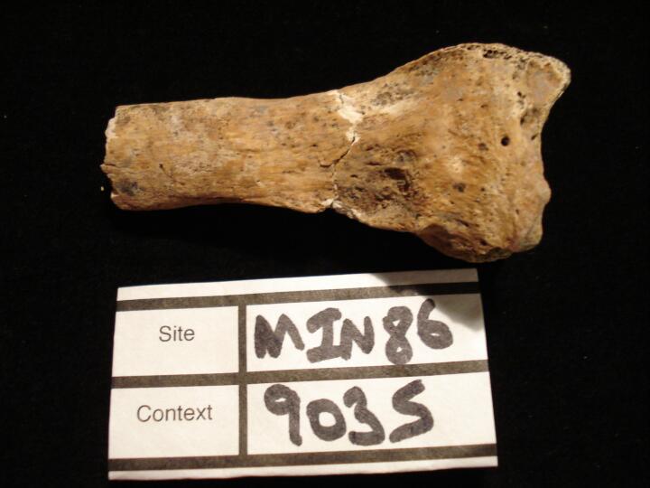
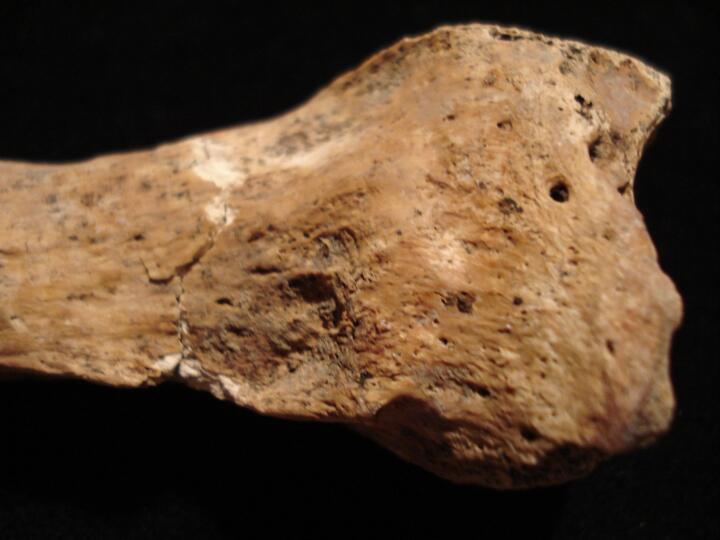
Right tibia distal end (posterior view) healed fracture of medial malleolus
|
| MIN86
|
9035
|
4
|
MIN86_9035_4.jpg
|
Left radius (anterior view) from the medial aspect healed 'Colles' fracture
|
| MIN86
|
9035
|
5
|
MIN86_9035_5.jpg
|
Left radius (anterior view) close up from the medial aspect helaed 'Colles' fracture
|
| MIN86
|
9035
|
6
|
MIN86_9035_6.jpg
|
Left radius (anterior view) looking superiorly from the distal articular surface to show healed 'Colles' fracture
|
| MIN86
|
9041
|
1
|
MIN86_9041_1.jpg
|
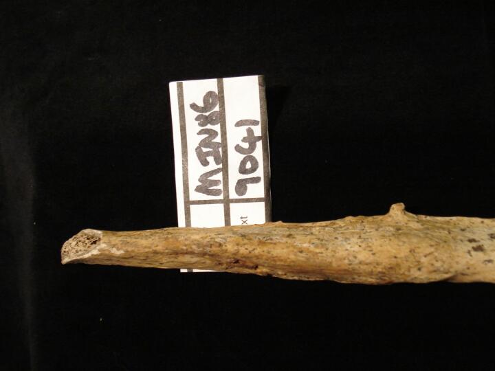
Right fibula proximal end (lateral view) healed oblique fracture with myostisis ossifcans
|
| MIN86
|
9041
|
2
|
MIN86_9041_2.jpg
|
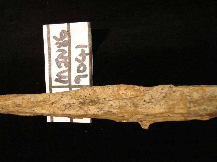
Right fibula proximal end (medial view) healed oblique fracture with myostisis ossificans
|
| MIN86
|
9066
|
1
|
MIN86_9066_1.jpg
|
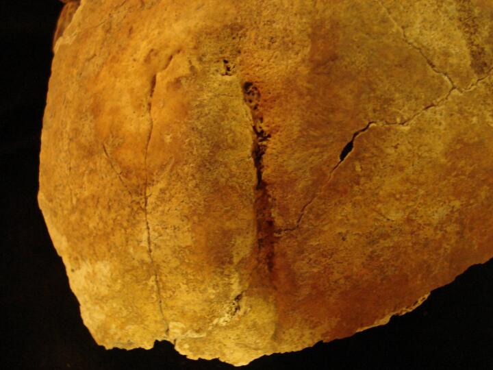
Well healed blade wound to the left parietal bone just proximal to the location of the mastoid process.
|
| MIN86
|
9066
|
2
|
MIN86_9066_2.jpg
|
Close up of a well healed blade wound to the left parietal bone just proximal to the location of the mastoid process.
|
| MIN86
|
9517
|
1
|
MIN86_9517_1.jpg
|
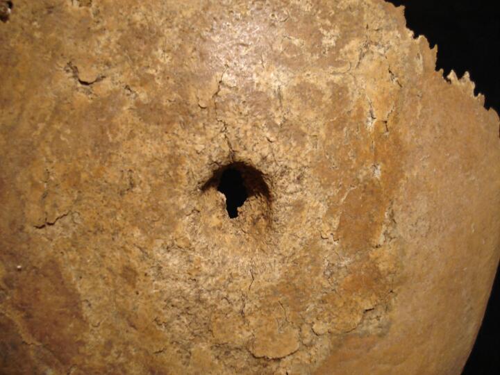
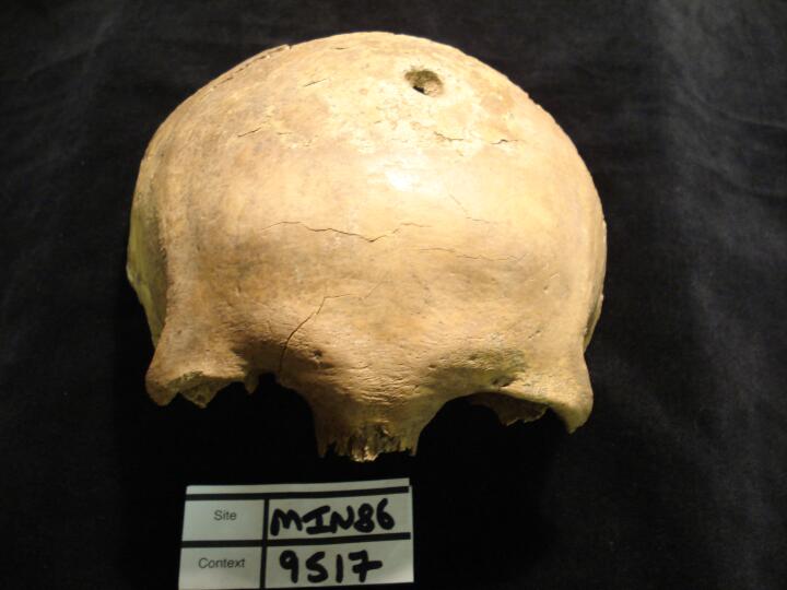
Skull frontal bone healed blunt force trauma (superior view)
|
| MIN86
|
9517
|
2
|
MIN86_9517_2.jpg
|
Skull frontal bone healed blunt force trauma (superior view/close up)
|
| MIN86
|
9517
|
3
|
MIN86_9517_3.jpg
|
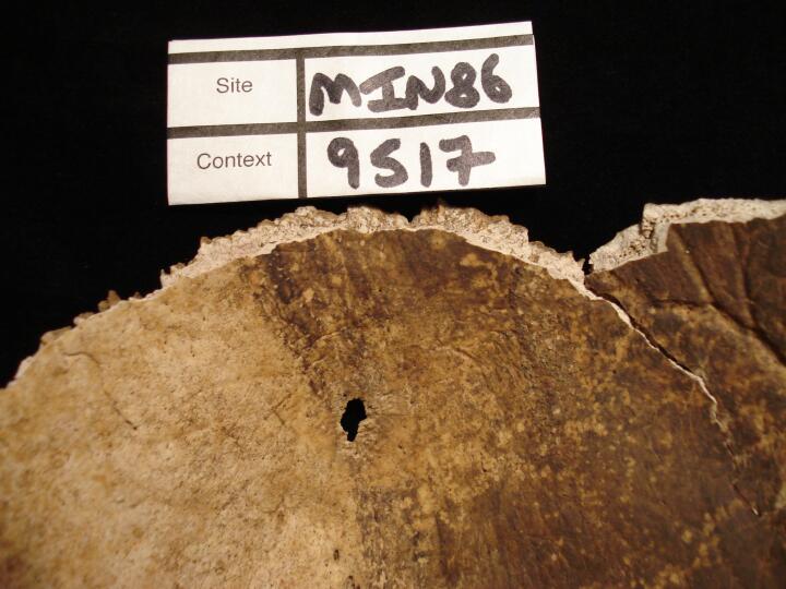
Skull frontal bone (endocranial surface) some post mortem damage
|
| MIN86
|
9517
|
4
|
MIN86_9517_4.jpg
|
Skull frontal bone helaed blunt force trauma (anterior/superior
|
| MIN86
|
9540
|
1
|
MIN86_9540_1.jpg
|
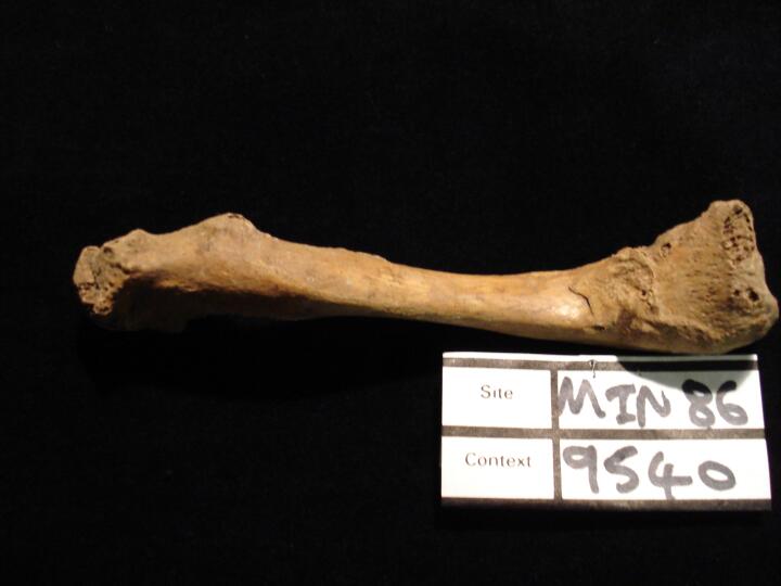
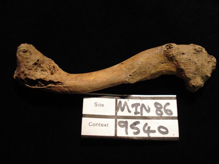
Right clavicle (superior view) healed fracture
|
| MIN86
|
9540
|
2
|
MIN86_9540_2.jpg
|
Right clavicle (posterior view) healed fracture
|
| MIN86
|
9540
|
3
|
MIN86_9540_3.jpg
|
Right radius healed 'Colles' fracture (anterior view)
|
| MIN86
|
9540
|
4
|
MIN86_9540_4.jpg
|
Right radius healed 'Colles' fracture (anterior view) medial aspect
|
| MIN86
|
9540
|
5
|
MIN86_9540_5.jpg
|
Right radius (anterior view) looking superiorly from the distal articular surface to show healed 'Colles' fracture
|
| MIN86
|
9540
|
6
|
MIN86_9540_6.jpg
|
Rib shaft fragment healed fracture (visceral surface)
|
| MIN86
|
9540
|
7
|
MIN86_9540_7.jpg
|
Rib shaft fragment healed fracture (pulmonary surface)
|
| MIN86
|
9540
|
8
|
MIN86_9540_8.jpg
|
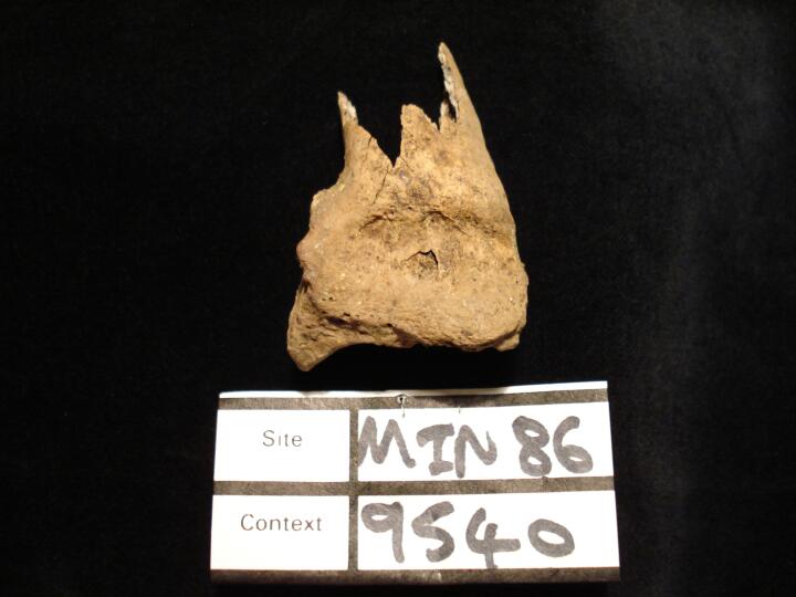
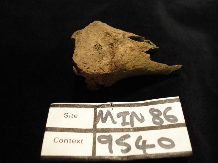
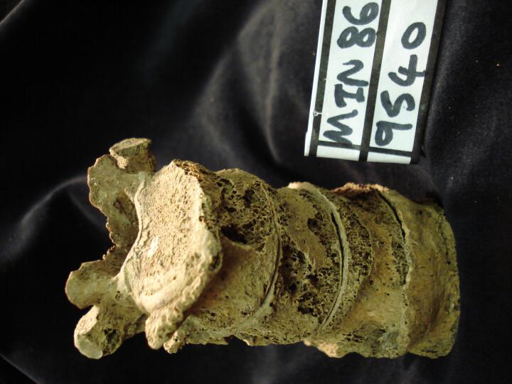
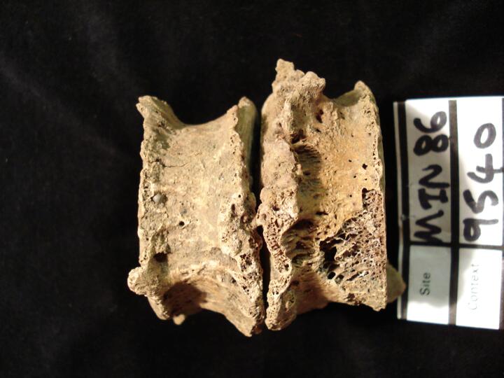
Vertebral compression fracture of thoracic vertenrae Th11 & Th12 (anteior view)
|
| MIN86
|
9540
|
9
|
MIN86_9540_9.jpg
|
Vertebral compression fracture of thoracic vertenrae Th11 & Th12 (right side view)
|
| MIN86
|
9540
|
10
|
MIN86_9540_10.jpg
|
Vertebral compression fracture of thoracic vertenrae Th11 & Th12 (left side view)
|
| MIN86
|
9540
|
11
|
MIN86_9540_11.jpg
|
Vertebral compression fracture articulating vertebrae from anterior aspect
|
| MIN86
|
9540
|
12
|
MIN86_9540_12.jpg
|
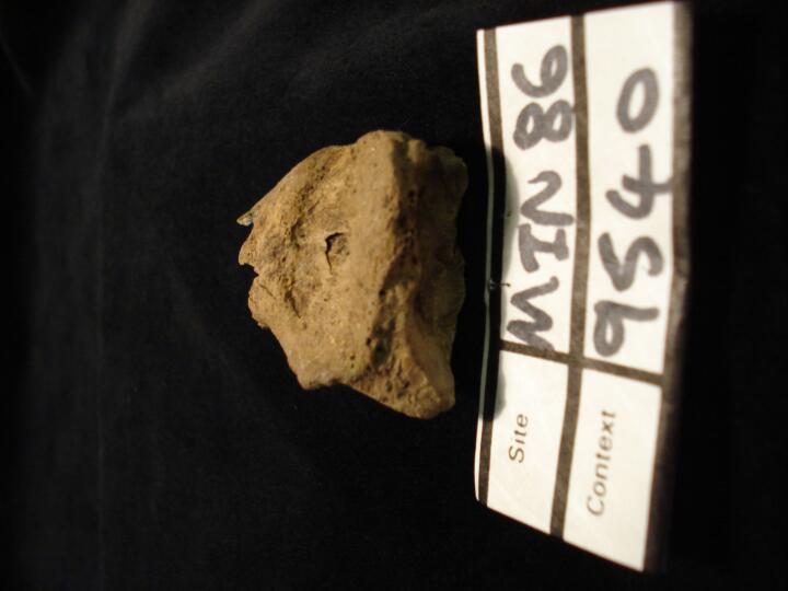
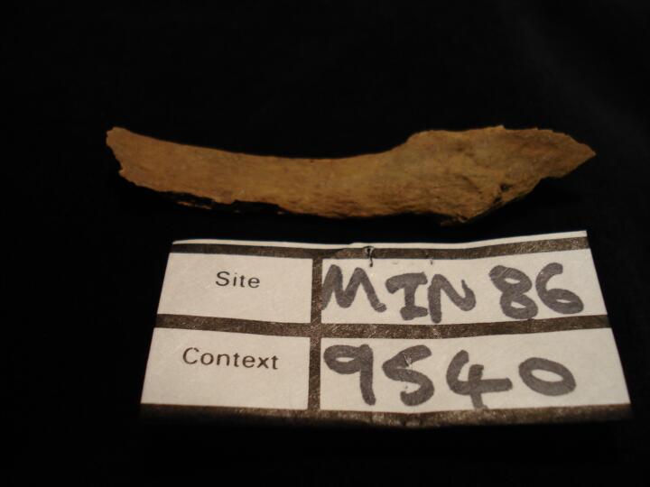
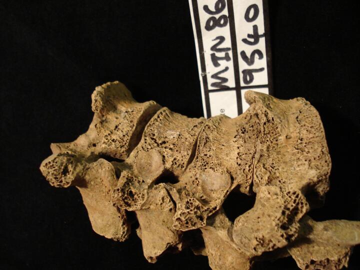
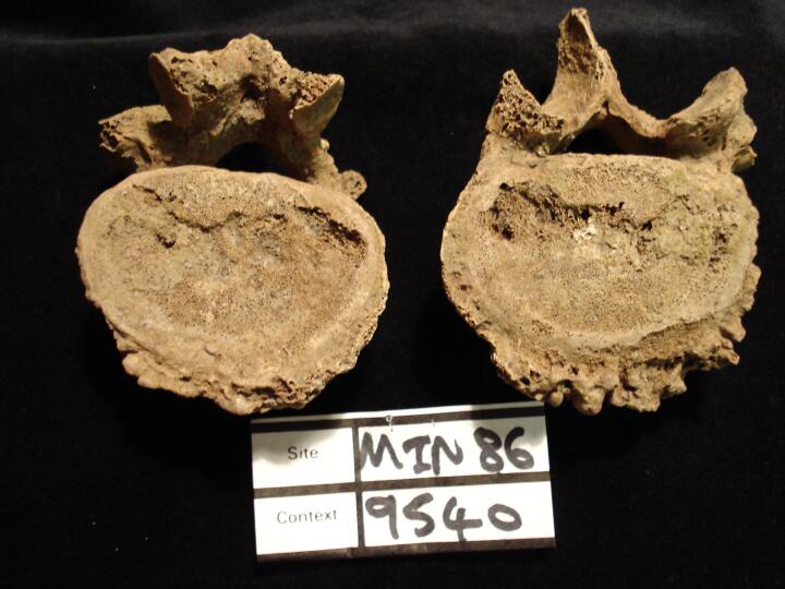
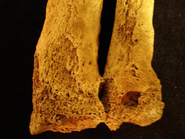
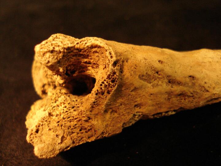
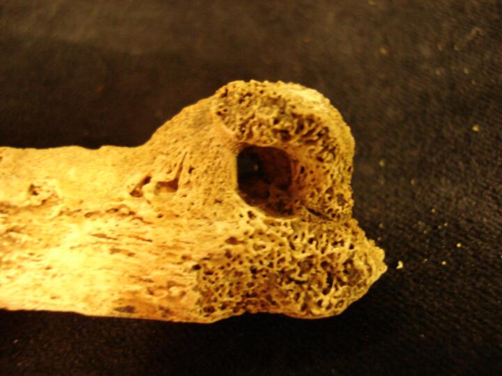
Lumbar vertebrae with destructive lesion possibly associated with specific infection Brucellosis (L3 inferior view, L4 superior view)
|
| MIN86
|
9540
|
13
|
MIN86_9540_13.jpg
|
Articulating lumbar vertebrae L3 & L4 (anterior view) showing bony changes possibly associated with specific infection, Brucellosis
|
| MIN86
|
9540
|
14
|
MIN86_9540_14.jpg
|
Lumbar vertebrae L3 & L4 from the left side showing bony changes possibly associated with specific infection, Brucellosis
|
| MIN86
|
9695
|
1
|
MIN86_9695_1.jpg
|
Sacrum (anterior view) anterior cleft between S1 & S2
|
| MIN86
|
9832
|
1
|
MIN86_9832_1.jpg
|
Right ulna & radius articulates distal ends. Gross change ?osteomyelitis or septic arthropathy
|
| MIN86
|
9832
|
2
|
MIN86_9832_2.jpg
|
Right ulna & radius articulates distal ends (scaphoid view) Gross change ?osteomyelitis or septic arthropathy
|
| MIN86
|
9832
|
3
|
MIN86_9832_3.jpg
|
Right radius (radioulna joint) possible osteomyelitis with cloaca (close up)
|
| MIN86
|
9832
|
4
|
MIN86_9832_4.jpg
|
Right ulna (radioulna joint) possible osteomyelitis with cloaca (close up)
|
| MIN86
|
9832
|
5
|
MIN86_9832_5.jpg
|
Scaphoid & lunate, ankylosed from infection (from radius view)
|
| MIN86
|
9832
|
6
|
MIN86_9832_6.jpg
|
Scaphoid & lunate, ankylosed from infection (from capitate view)
|
| MIN86
|
9832
|
7
|
MIN86_9832_7.jpg
|
Right scaphoid
|
| MIN86
|
9832
|
8
|
MIN86_9832_8.jpg
|
Right capitate
|
| MIN86
|
9832
|
9
|
MIN86_9832_9.jpg
|
Right capitate
|
| MIN86
|
9832
|
10
|
MIN86_9832_10.jpg
|
Right capitate
|
| MIN86
|
11118
|
1
|
MIN86_11118_1.jpg
|
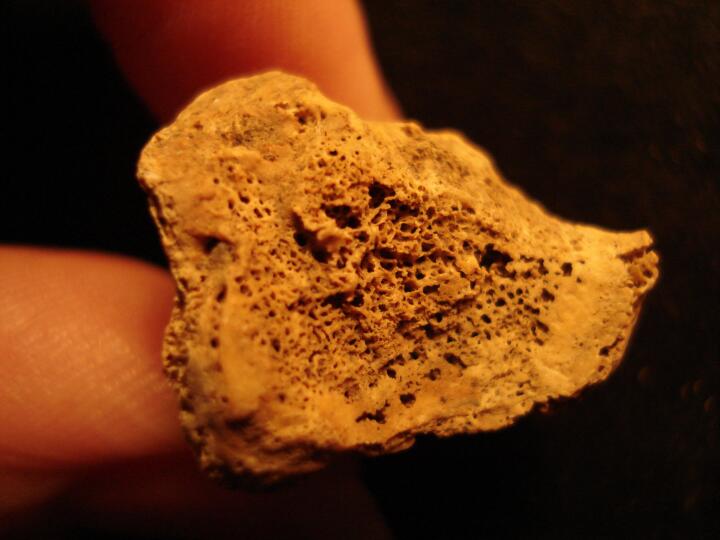
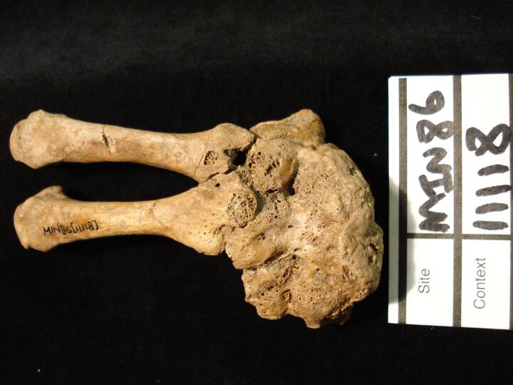
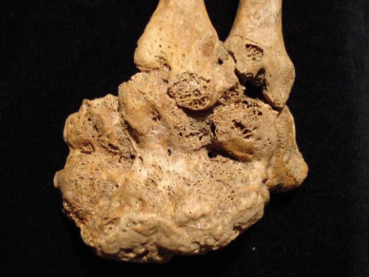
Left hand/wrist complete ankylosis of the carpal bones & two present metacarpals, indicating specific infection, Tuberculosis (dorsal view)
|
| MIN86
|
11118
|
2
|
MIN86_11118_2.jpg
|
Left hand/wrist close up of complete ankylosis of the carpal bones & two present metacarpals, indicating specific infection, Tuberculosis (dorsal view)
|
| MIN86
|
11118
|
3
|
MIN86_11118_3.jpg
|
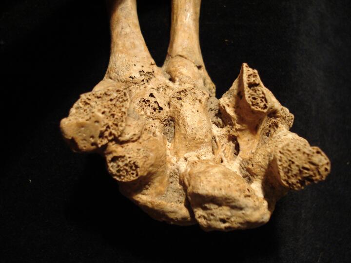
Left hand/wrist close up of complete ankylosis of the carpal bones & two present metacarpals, indicating specific infection, Tuberculosis (palmar view)
|
| MIN86
|
11118
|
4
|
MIN86_11118_4.jpg
|
Ankylosed left carpal bones (palmar view/close up) indicating specific infection, Tuberculosis
|
| MIN86
|
11118
|
5
|
MIN86_11118_5.jpg
|
Left wrist showing ankylosis of carpal bones from the aspect of articulation of the carpal bones & lower arm bones
|
| MIN86
|
11124
|
1
|
MIN86_11124_1.jpg
|
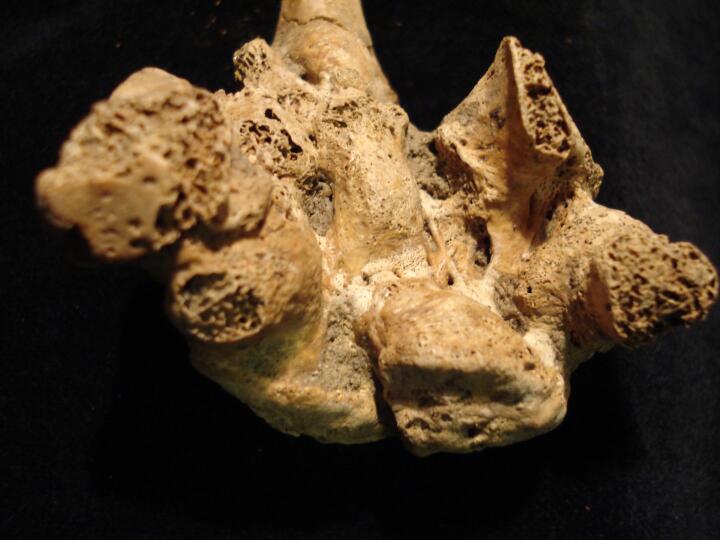
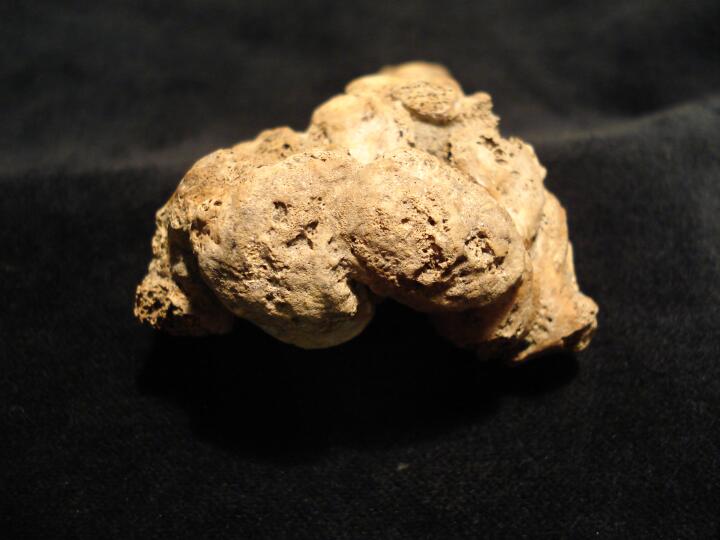
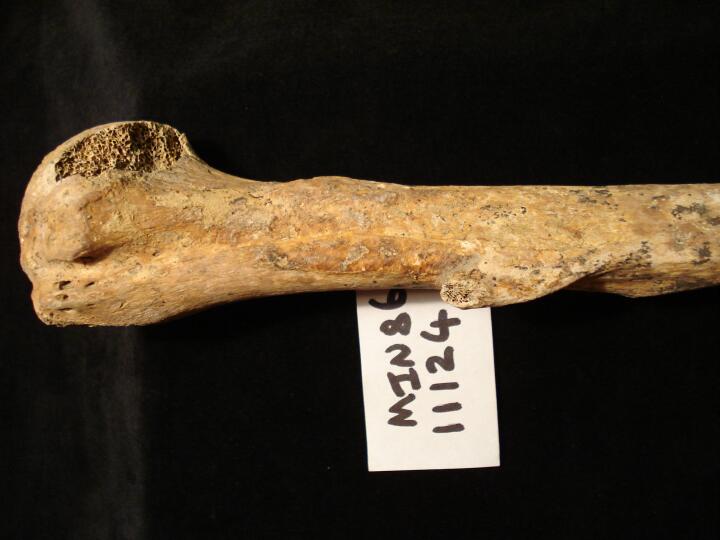
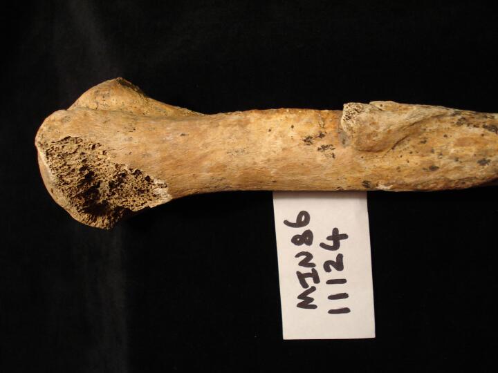
Right humerus (anterior view) osteochondroma
|
| MIN86
|
11124
|
2
|
MIN86_11124_2.jpg
|
Right humerus (lateral aspect) osteochondroma
|
| MIN86
|
11232
|
1
|
MIN86_11232_1.jpg
|
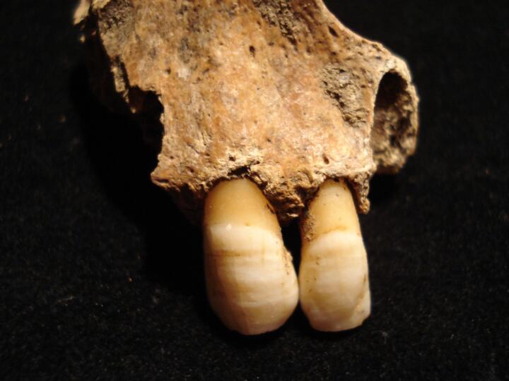
Left maxillary canine & lateral incisor linear hypoplastic defects
|
| MIN86
|
11232
|
2
|
MIN86_11232_2.jpg
|
Left maxillary canine & lateral incisor linear hypoplastic defects
|
| MIN86
|
11249
|
1
|
MIN86_11249_1.jpg
|
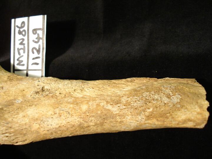
Right tibia possible ulcer, demarcated area of bone surface reaction (medial view)
|
| MIN86
|
11249
|
2
|
MIN86_11249_2.jpg
|
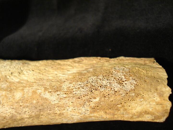
Right tibia possible ulcer, demarcated area of bone surface reaction (medial view/close up)
|
| MIN86
|
11249
|
3
|
MIN86_11249_3.jpg
|
Right tibia possible ulcer, demarcated area of bone surface reaction (anterior view)
|
| MIN86
|
11249
|
4
|
MIN86_11249_4.jpg
|
Right tibia possible ulcer, demarcated area of bone surface reaction (anterior view/close up)
|
| MIN86
|
11252
|
1
|
MIN86_11252_1.jpg
|
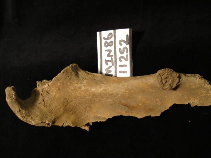
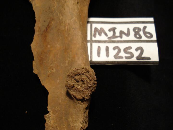
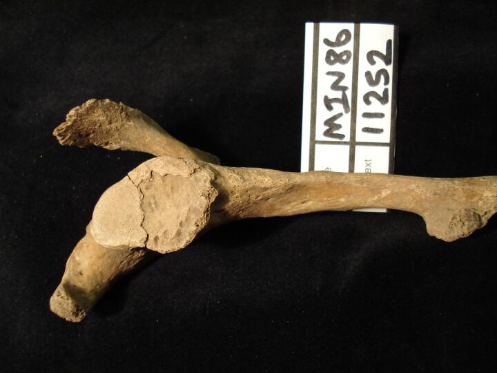
Left scapula (anterior view) osteochondroma
|
| MIN86
|
11252
|
2
|
MIN86_11252_2.jpg
|
Left scapula (anterior view) close up of osteochondroma
|
| MIN86
|
11252
|
3
|
MIN86_11252_3.jpg
|
Left scapula (medial view) of blade with osteochondroma
|
| MIN86
|
11415
|
1
|
MIN86_11415_1.jpg
|
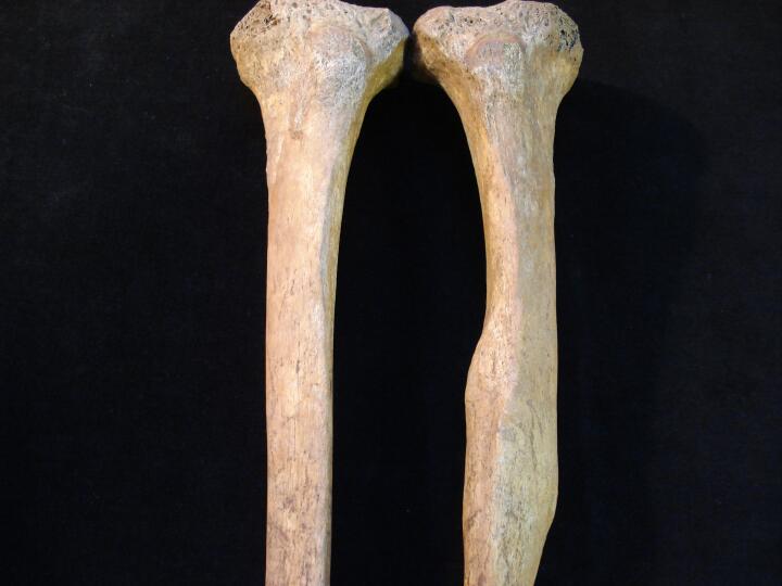
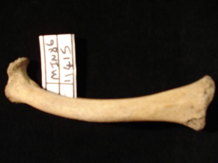
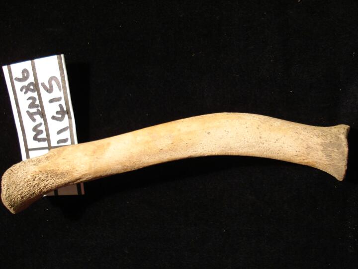
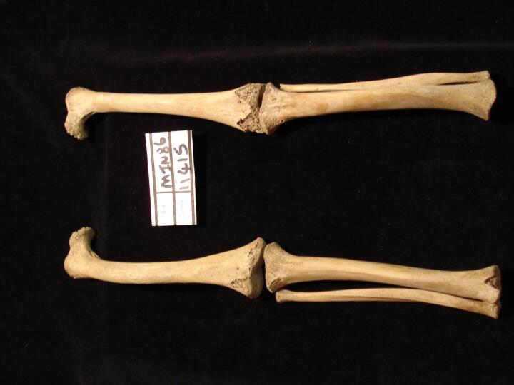
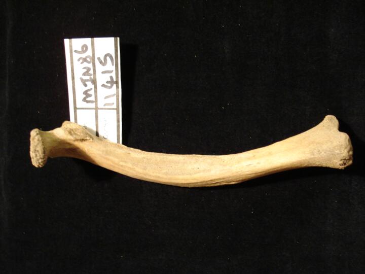
Right femur (anterior view) bowing deformity from rickets
|
| MIN86
|
11415
|
2
|
MIN86_11415_2.jpg
|
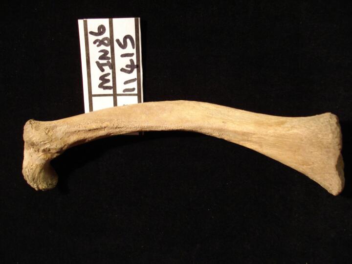
Right femur (posterior view) bowing deformity from rickets
|
| MIN86
|
11415
|
3
|
MIN86_11415_3.jpg
|
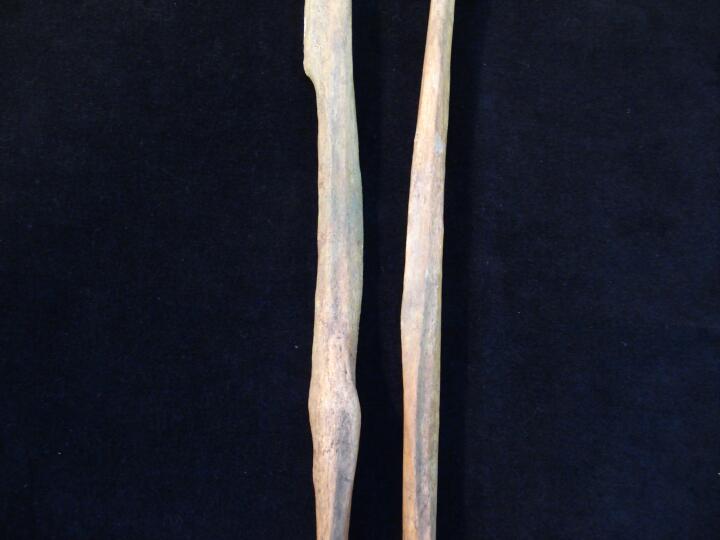
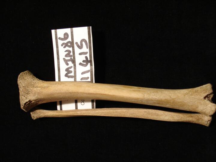
Right tibia & fibula (anterior view) bowing deformity from rickets
|
| MIN86
|
11415
|
4
|
MIN86_11415_4.jpg
|
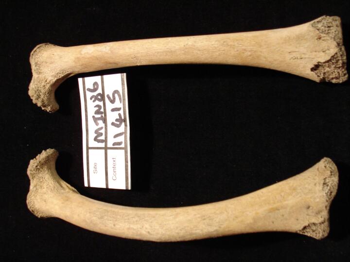
Left & right femur (anterior view) comparing bowing deformity
|
| MIN86
|
11415
|
5
|
MIN86_11415_5.jpg
|
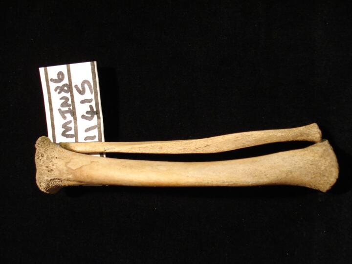
Left tibia & fibula (anterior view) bowing deformity from rickets
|
| MIN86
|
11415
|
6
|
MIN86_11415_6.jpg
|
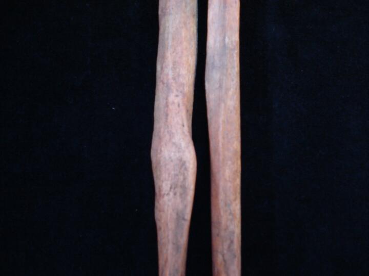
Left tibia (medial view) bowing deformity from rickets
|
| MIN86
|
11415
|
7
|
MIN86_11415_7.jpg
|
Left & right leg (anterior view) comparing racchitic bowing deformity in the long bones of the leg
|
| MIN86
|
11415
|
8
|
MIN86_11415_8.jpg
|
Right femur (medial view) bowing deformity from rickets
|
| MIN86
|
11448
|
1
|
MIN86_11448_1.jpg
|
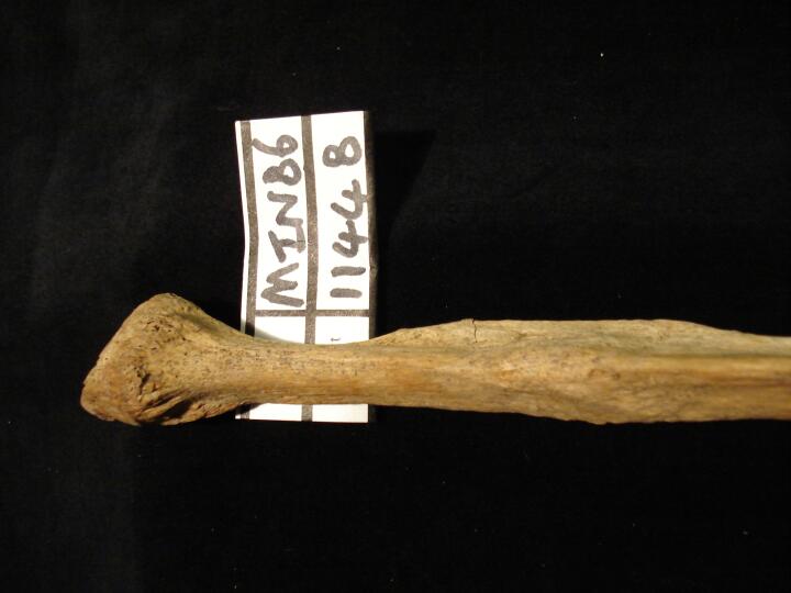
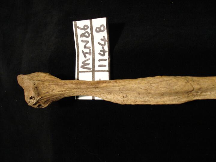
Left fibula, roximal end healed oblique fracture (medial view)
|
| MIN86
|
11448
|
2
|
MIN86_11448_2.jpg
|
Left fibula, roximal end healed oblique fracture (anterior view)
|
| MIN86
|
11448
|
3
|
MIN86_11448_3.jpg
|
Left fibula, roximal end healed oblique fracture (lateral view)
|
| MIN86
|
11480
|
1
|
MIN86_11480_1.jpg
|
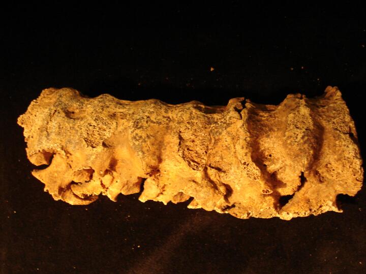
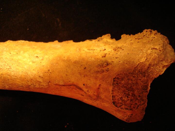
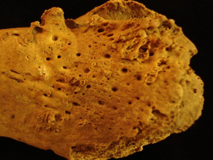
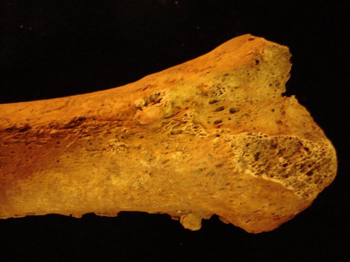
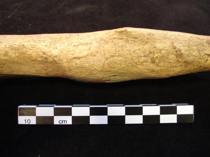
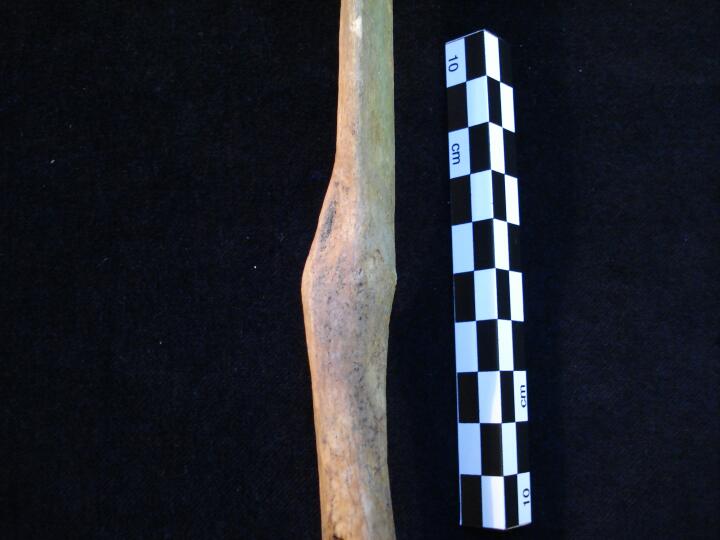
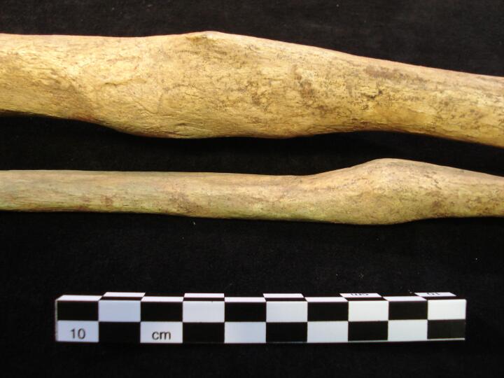
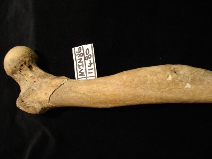
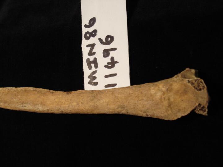
Right femur (anterior view) swelling of the bone possibly attributable to osteomyelitic infection
|
| MIN86
|
11480
|
2
|
MIN86_11480_2.jpg
|
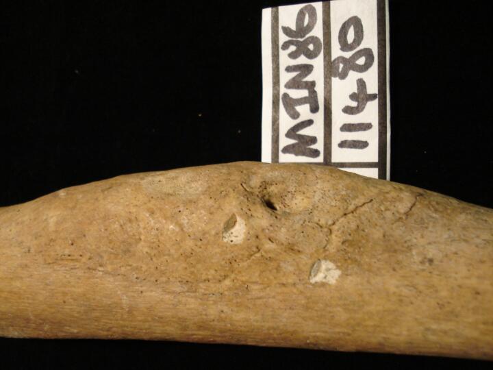
Right femur (anterior view) close up of swelling of the bone possibly attributable to osteomyelitic infection
|
| MIN86
|
11480
|
3
|
MIN86_11480_3.jpg
|
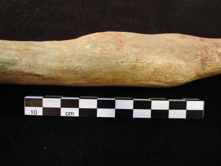
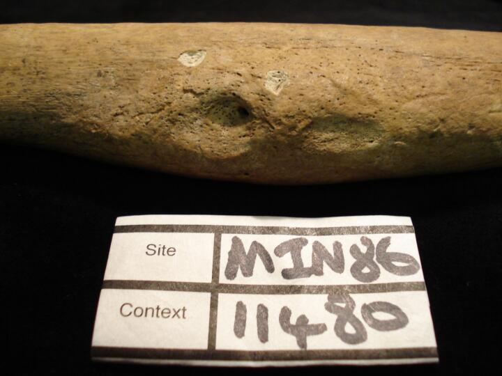
Right femur close of area of swollen bone (medial view)
|
| MIN86
|
11496
|
1
|
MIN86_11496_1.jpg
|
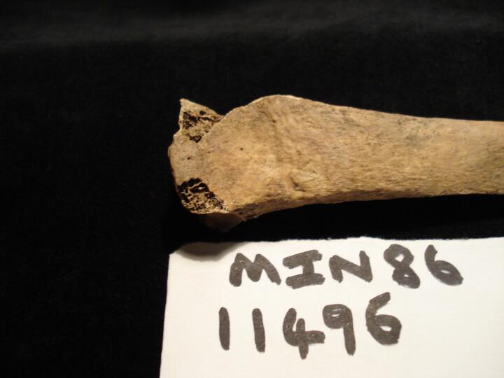
Right radius, very well remodelled & healed 'Colles' fracture (anterior view)
|
| MIN86
|
11496
|
2
|
MIN86_11496_2.jpg
|
Right radius, very well remodelled & healed 'Colles' fracture (medial view)
|
| MIN86
|
11970
|
1
|
MIN86_11970_1.jpg
|
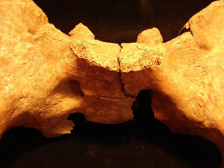
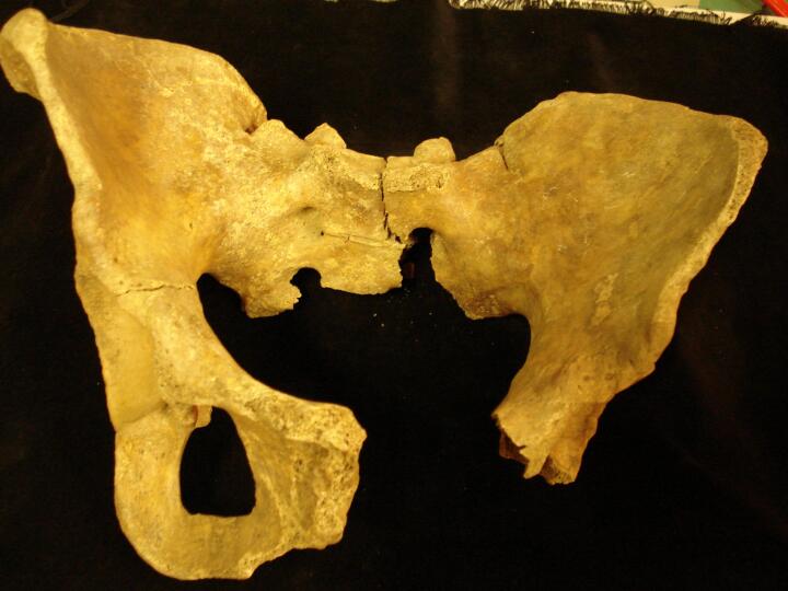
Ankylosing Spondylitis: Complete fusion of the Sacrum to the ossa coxae (Close up)
|
| MIN86
|
11970
|
2
|
MIN86_11970_2.jpg
|
Ankylosing Spondylitis: Complete fusion of the Sacrum to the ossa coxae
|
| MIN86
|
12525
|
1
|
MIN86_12525_1.jpg
|
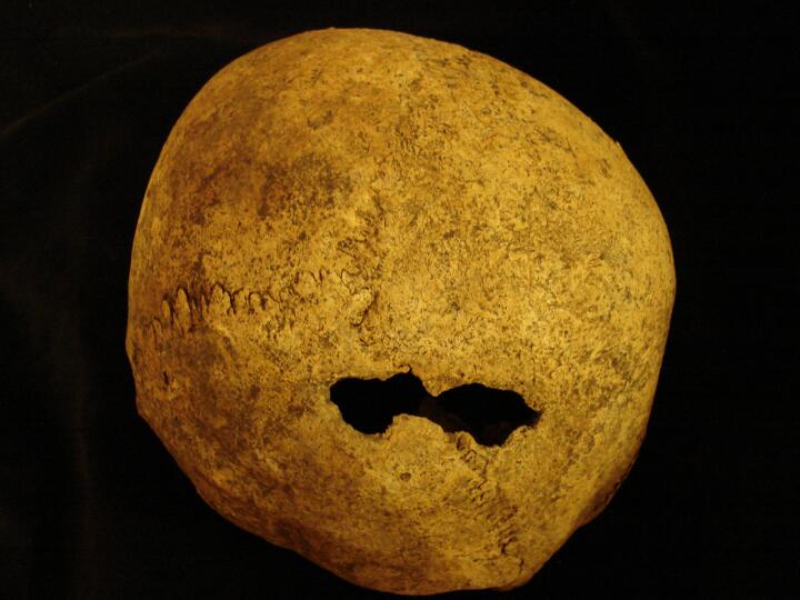
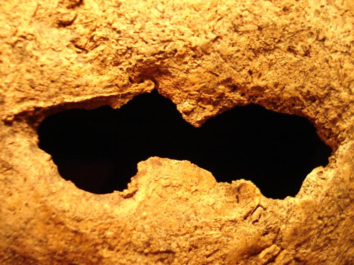
Sharp force trauma wound to the occipital bone. Post mortem damage to the right portion of the wound extending onto the right parietal bone.
|
| MIN86
|
12525
|
2
|
MIN86_12525_2.jpg
|
Close up of a sharp force trauma wound to the occipital bone. Post mortem damage to the right portion of the wound extending onto the right parietal bone.
|
| MIN86
|
12567
|
1
|
MIN86_12567_1.jpg
|
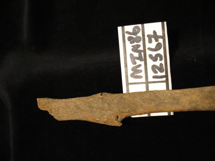
Left fibula proximal end healed fracture (posterior view)
|
| MIN86
|
12567
|
2
|
MIN86_12567_2.jpg
|
Left fibula proximal end healed fracture (lateral view)
|
| MIN86
|
12626
|
1
|
MIN86_12626_1.jpg
|
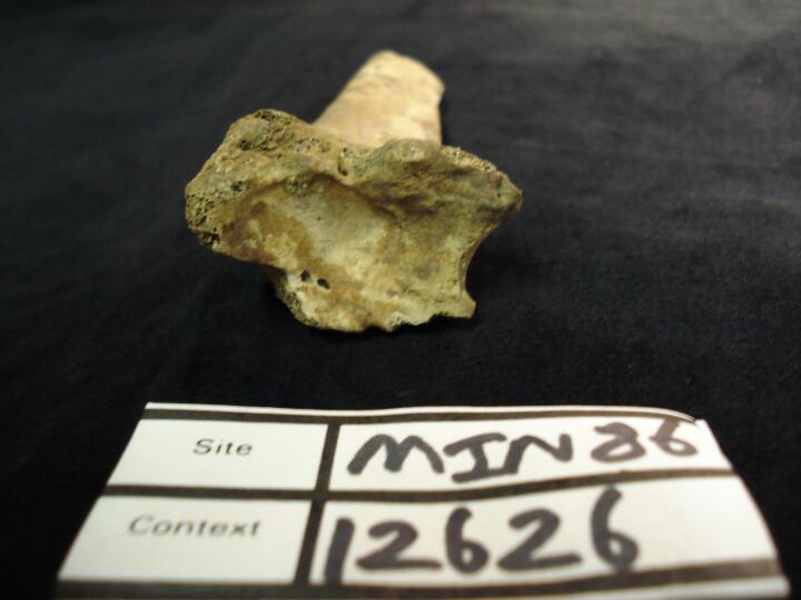
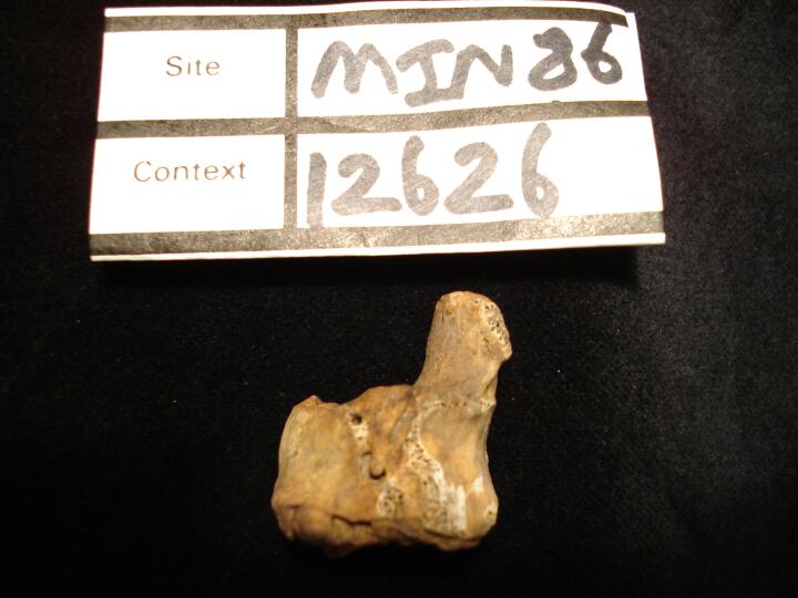
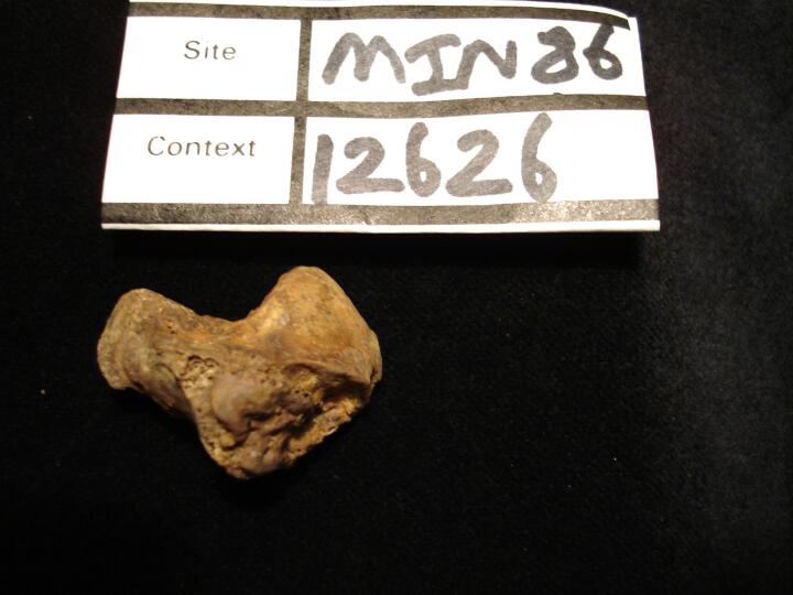
Right radius (distal view) intrarticular fracture line
|
| MIN86
|
12626
|
2
|
MIN86_12626_2.jpg
|
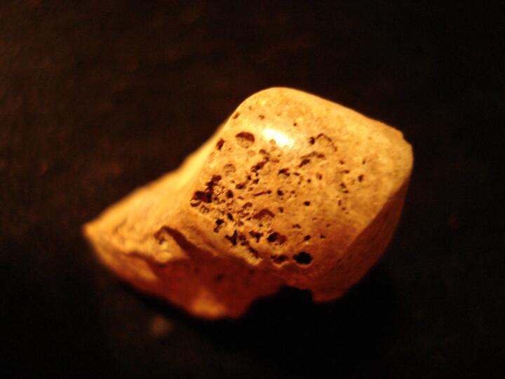
Right hamate (medial view) fracture line
|
| MIN86
|
12626
|
3
|
MIN86_12626_3.jpg
|
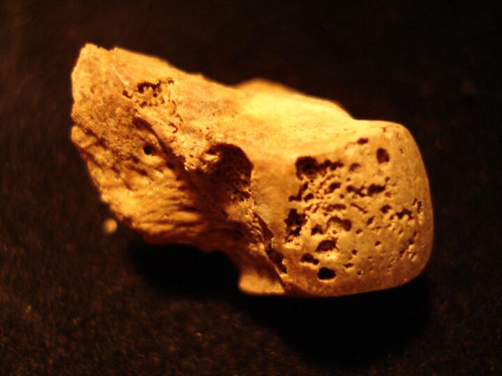
Right hamate (posterior view) fracture line
|
| MIN86
|
12626
|
4
|
MIN86_12626_4.jpg
|
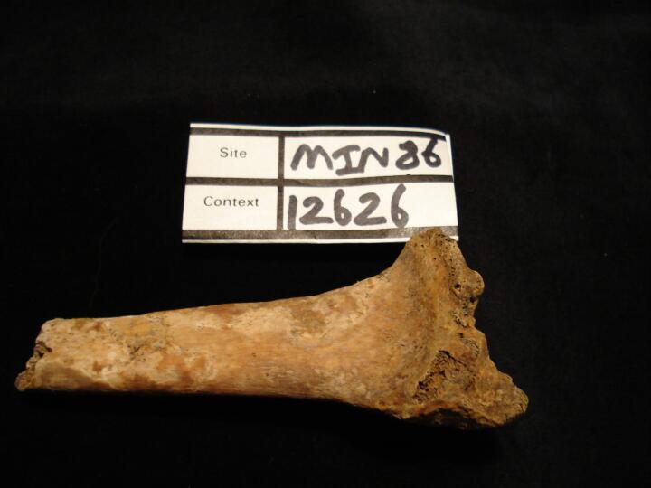
Right radius (anterior view)
|
| MIN86
|
12635
|
1
|
MIN86_12635_1.jpg
|
Subchondral cysts and eburnation of the capitate on the surface of articulation with the scaphoid (view from the scaphoid).
|
| MIN86
|
12635
|
2
|
MIN86_12635_2.jpg
|
Subchondral cysts and eburnation of the capitate on the surface of articulation with the scaphoid (dorsal view).
|
| MIN86
|
12635
|
3
|
MIN86_12635_3.jpg
|
Pronounced and florid build up of calculus on T16. (buccal view)
|
| MIN86
|
12635
|
4
|
MIN86_12635_4.jpg
|
Pronounced and florid build up of calculus on T16. (buccal view)
|
| MIN86
|
12635
|
5
|
MIN86_12635_5.jpg
|
Pronounced and florid build up of calculus on T16. (occlusal view)
|
| MIN86
|
12700
|
1
|
MIN86_12700_1.jpg
|
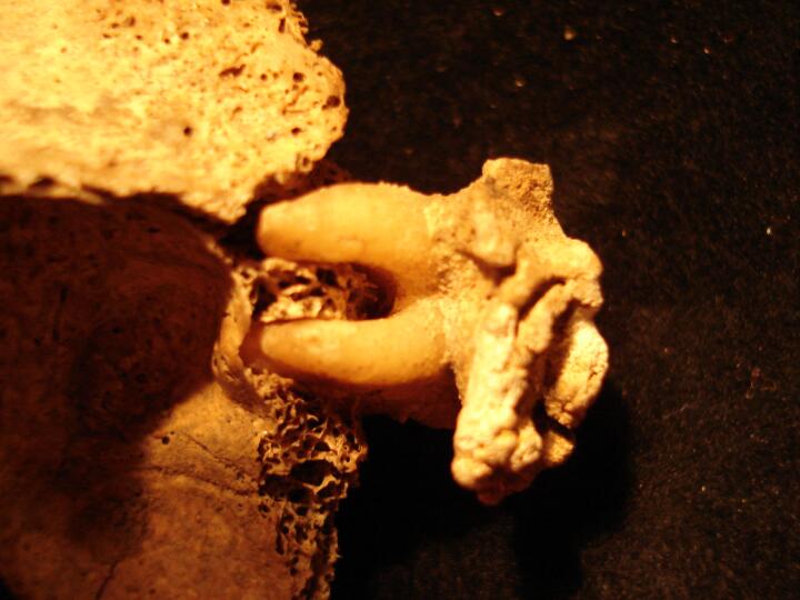
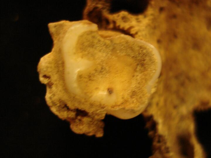
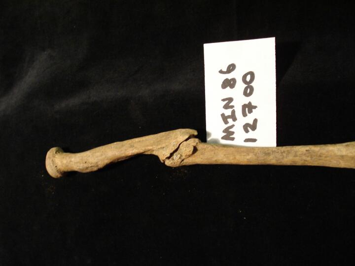
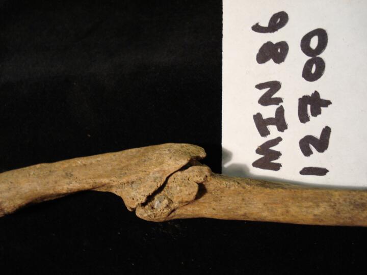
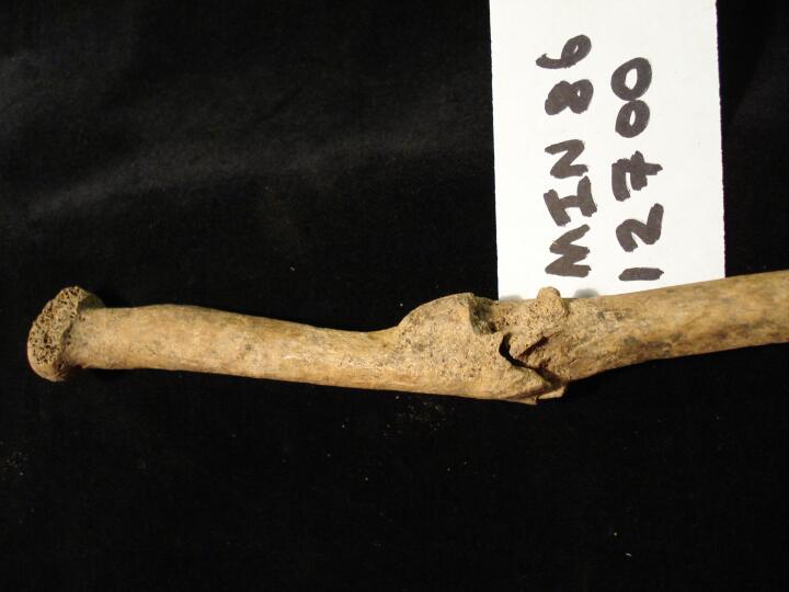
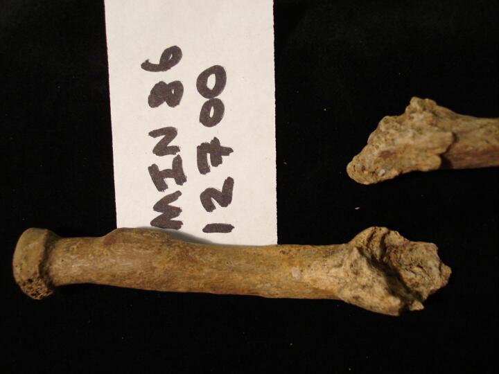
Right radius non-united fracture mid shaft (anterior view)
|
| MIN86
|
12700
|
2
|
MIN86_12700_2.jpg
|
Right radius non-united fracture mid shaft (anterior view) close up of fracture site
|
| MIN86
|
12700
|
3
|
MIN86_12700_3.jpg
|
Right radius non-united fracture mid shaft (posterior view)
|
| MIN86
|
12700
|
4
|
MIN86_12700_4.jpg
|
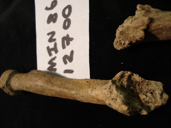
Right radius non-united fracture mid shaft showing the two parts of the fractured radius
|
| MIN86
|
12700
|
5
|
MIN86_12700_5.jpg
|
Right radius close up of the bone surface of the non-united fractured bone ends
|
| MIN86
|
12721
|
1
|
MIN86_12721_1.jpg
|
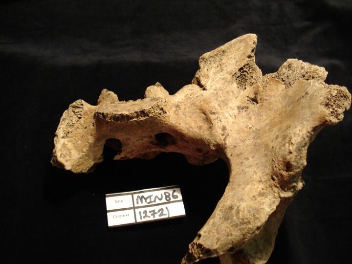
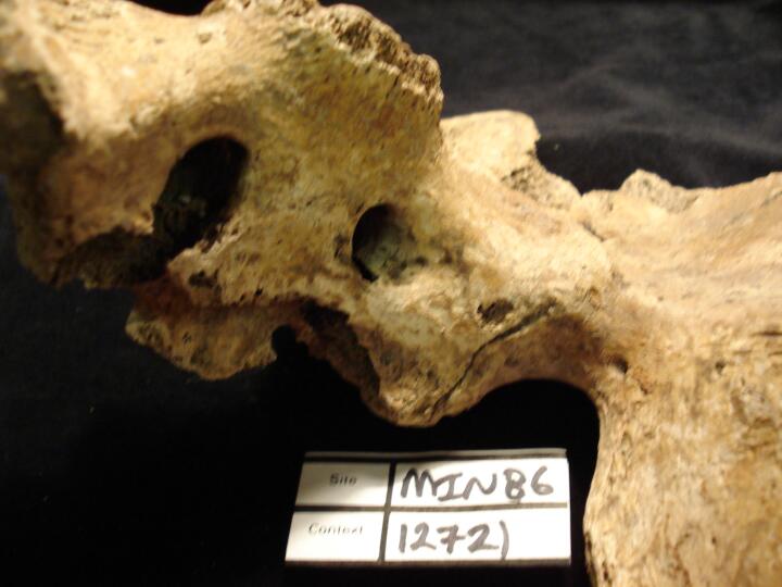
Sacroiliac fusion, ankylosis of left side (anterior view)
|
| MIN86
|
12721
|
2
|
MIN86_12721_2.jpg
|
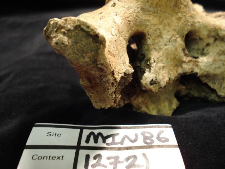
Sacroiliac fusion, ankylosis of left side (anterior view/close up)
|
| MIN86
|
12721
|
3
|
MIN86_12721_3.jpg
|
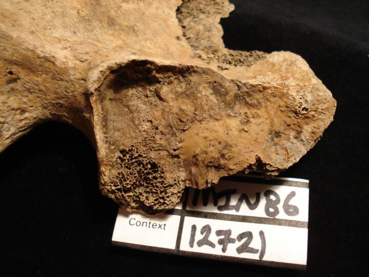
Right sacroiliac joint surface (pelvis) deep depression
|
| MIN86
|
12721
|
4
|
MIN86_12721_4.jpg
|
Right sacroiliac joint surface (sacral ala) bony projection matching deep depression on sacroiliac joint surface of pelvis
|
| MIN86
|
12725
|
1
|
MIN86_12725_1.jpg
|
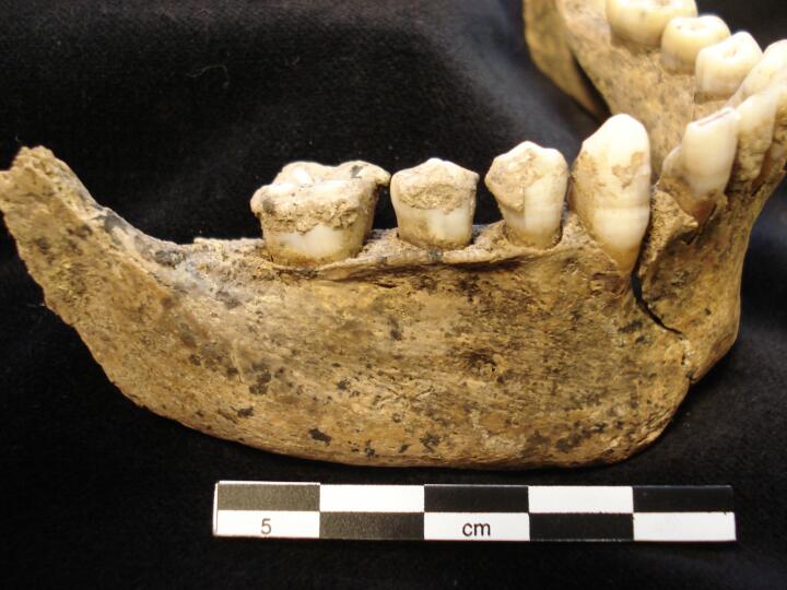
Mandible (buccal view) calculus
|
| MIN86
|
12725
|
2
|
MIN86_12725_2.jpg
|
Mandible lingual view) calculus
|
| MIN86
|
12773
|
1
|
MIN86_12773_1.jpg
|
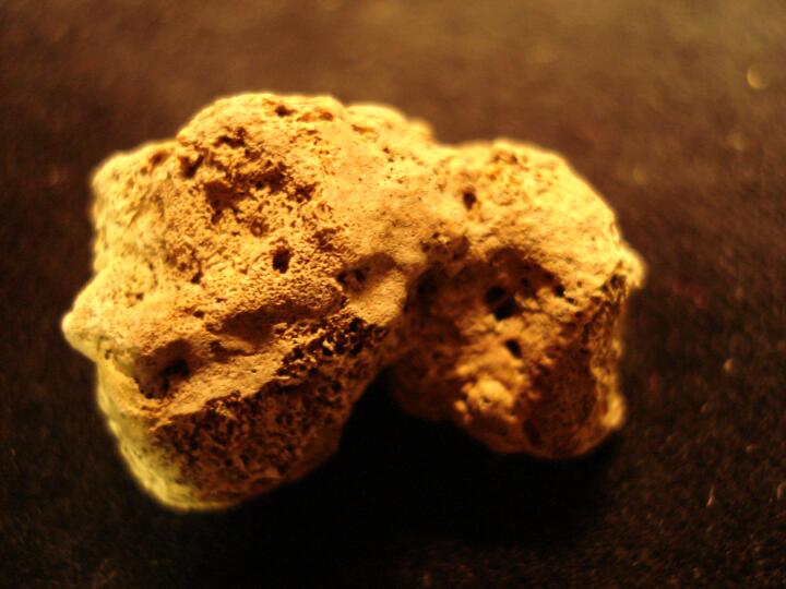
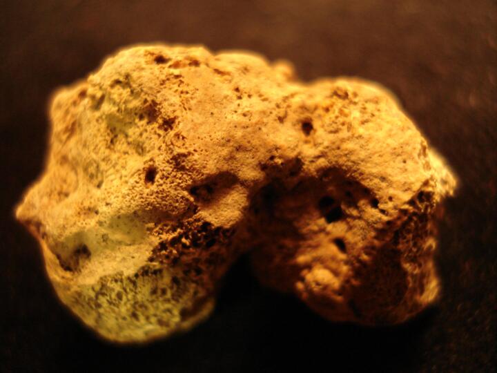
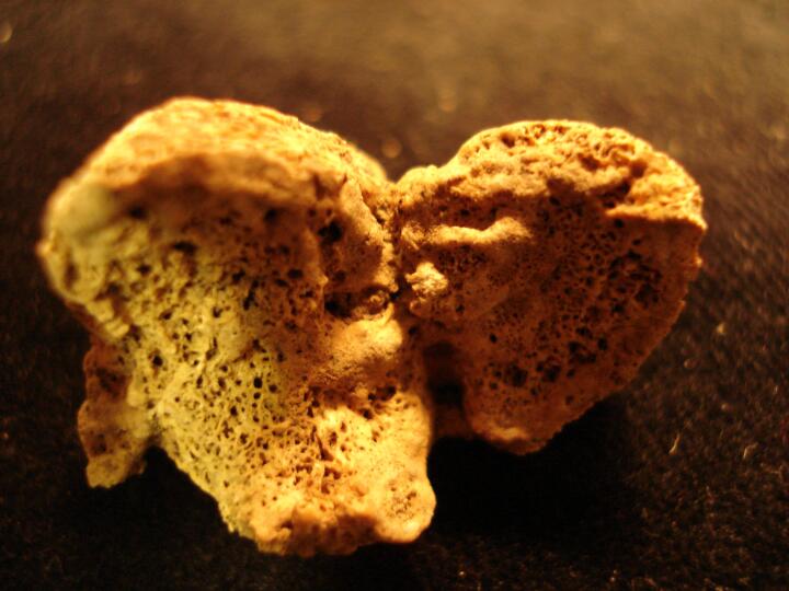
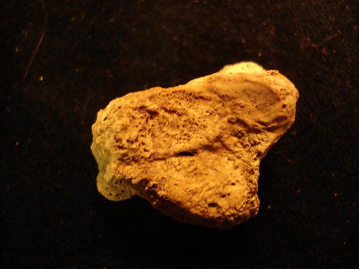
Large button osteoma to the left frontal bone.
|
| MIN86
|
12774
|
1
|
MIN86_12774_1.jpg
|
Osteoarthritis on the left patella. Subchondral cysts and eburnation are both visible.
|
| MIN86
|
12796
|
1
|
MIN86_12796_1.jpg
|
Demarcated lesions to the dorsal surface of a left rib. Characteristic of TB.
|
| MIN86
|
20004
|
1
|
MIN86_20004_1.jpg
|
Vertebrae (C7 & Th1) 'Clay Shoveller's' fracture (posterior view)
|
| MIN86
|
20004
|
2
|
MIN86_20004_2.jpg
|
Vertebrae (C7 & Th1) 'Clay Shoveller's' fracture (posterior view)
|
| MIN86
|
20004
|
3
|
MIN86_20004_3.jpg
|
Vertebrae (C7 & Th1) 'Clay Shoveller's' fracture (posterior view)
|


