| Site code
|
Context
|
Frame number
|
Photo
|
Description
|
| ONE94
|
377
|
1
|
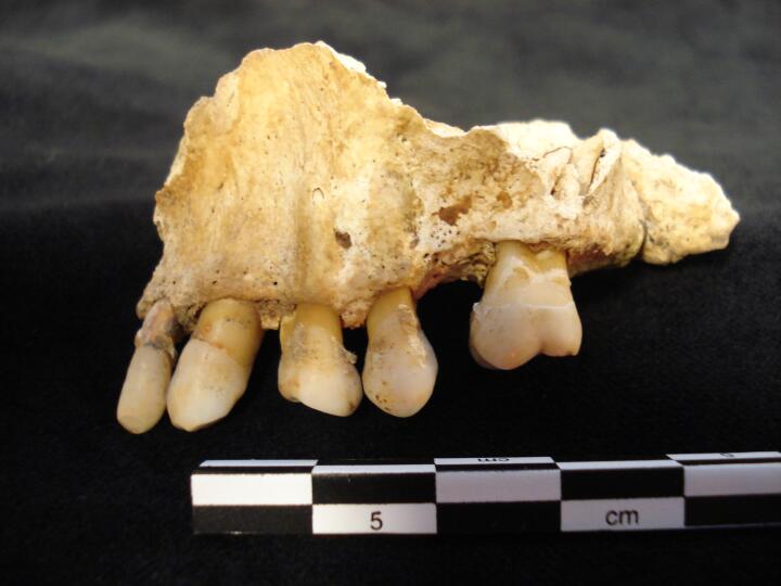
ONE94_377_1.jpg
|
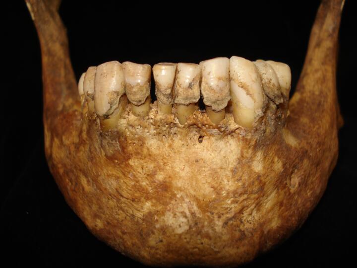
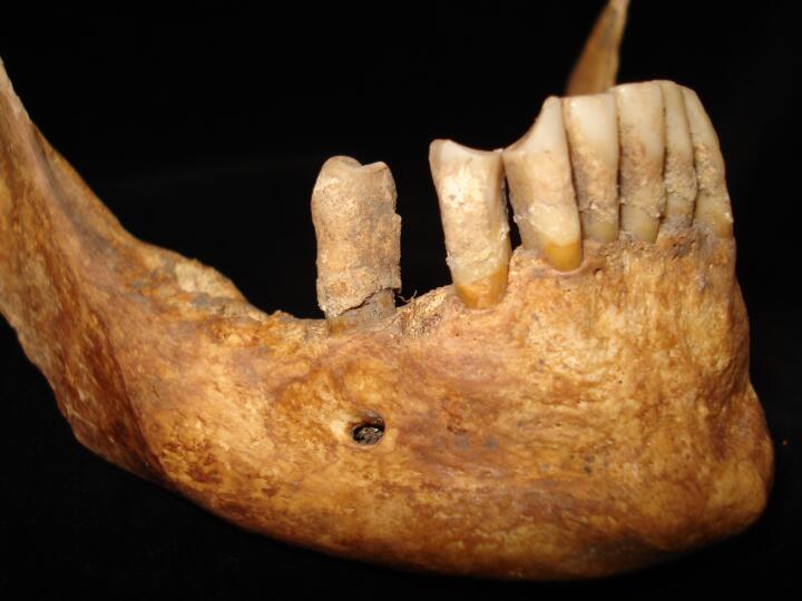
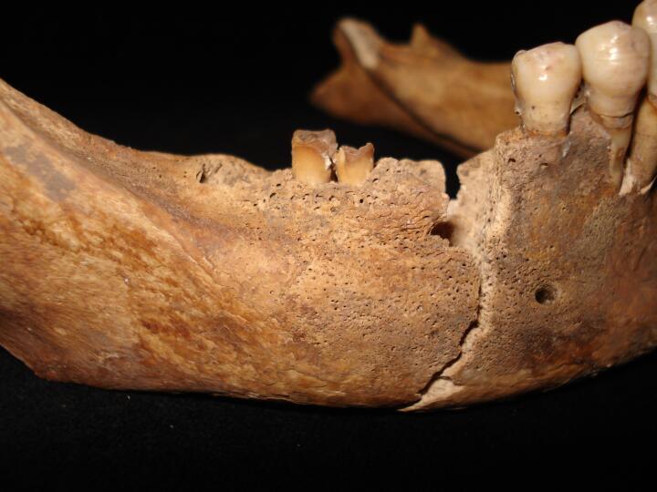
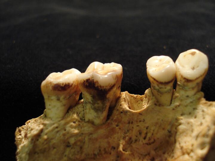
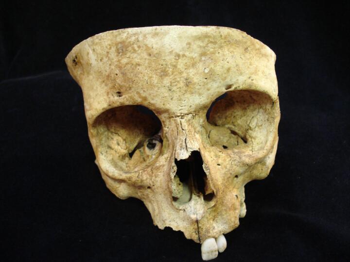
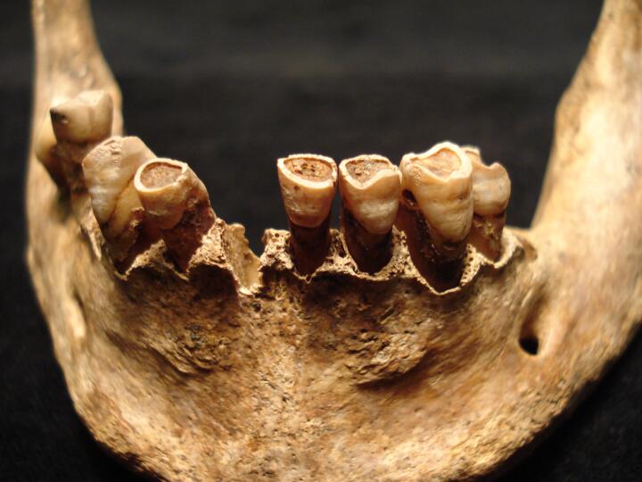
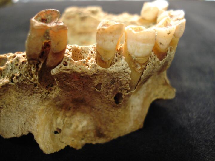
Mandible (buccal view) calculus, hypoplasia & AM tooth loss
|
| ONE94
|
377
|
2
|
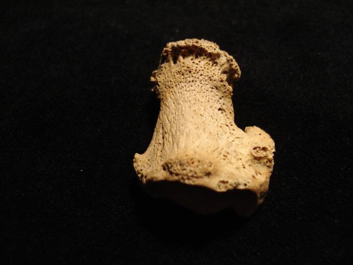
ONE94_377_2.jpg
|
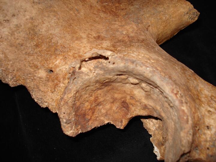
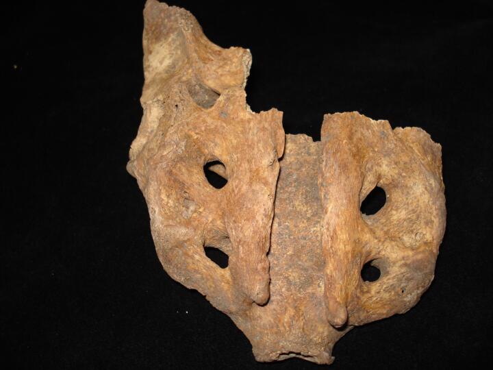
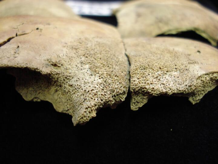
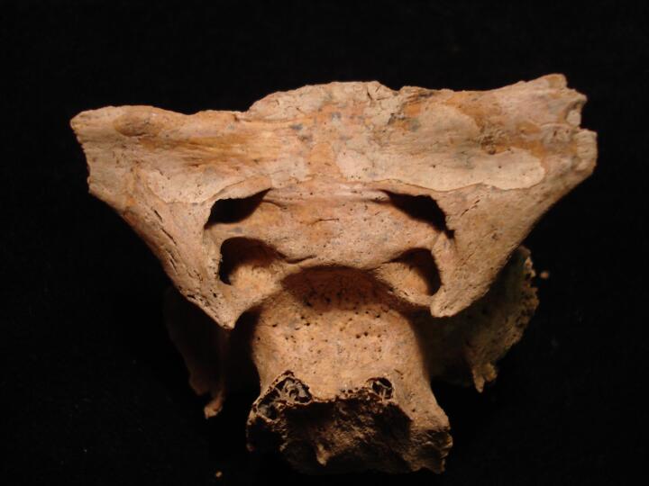
Left pelvis, superior aspect of acetabulum, possible cyst (posterior view)
|
| ONE94
|
377
|
3
|
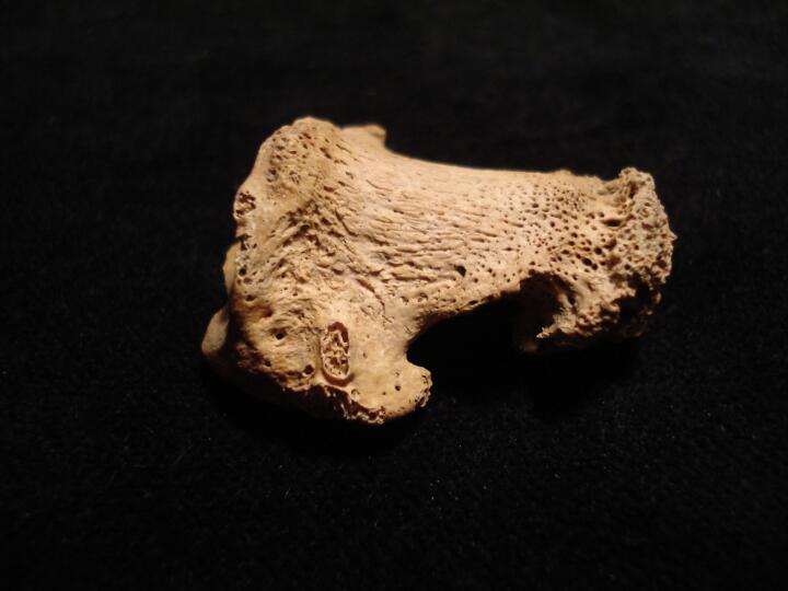
ONE94_377_3.jpg
|
Left pelvis, superior aspect of acetabulum, possible cyst (superior view)
|
| ONE94
|
377
|
4
|
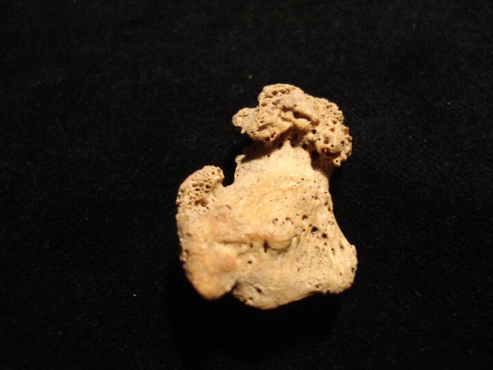
ONE94_377_4.jpg
|
Left pelvis, margin of acetabulum, possible cyst
|
| ONE94
|
20
|
1
|
ONE94_20_1.jpg
|
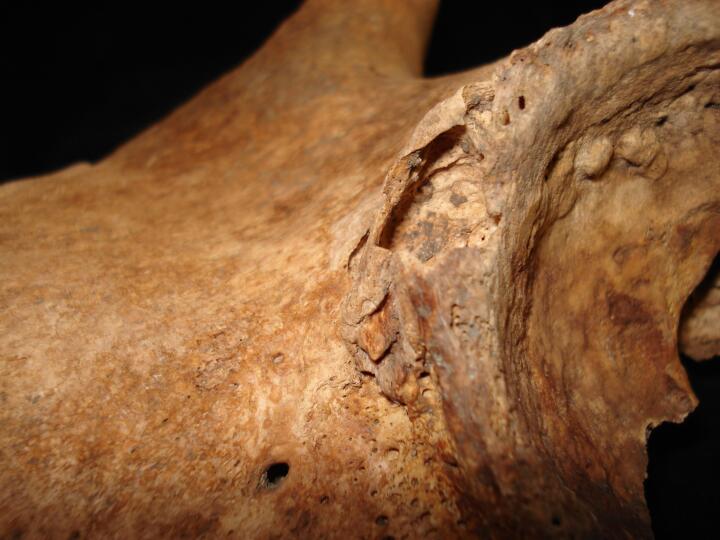
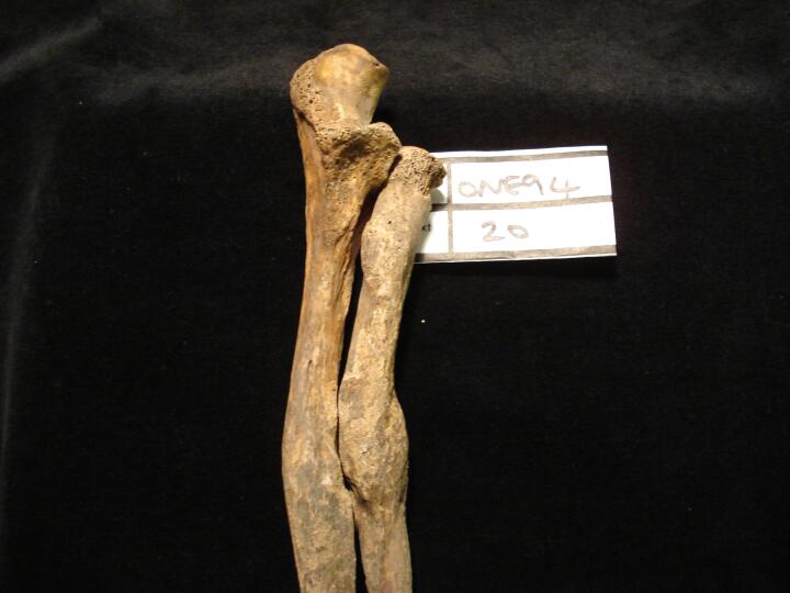
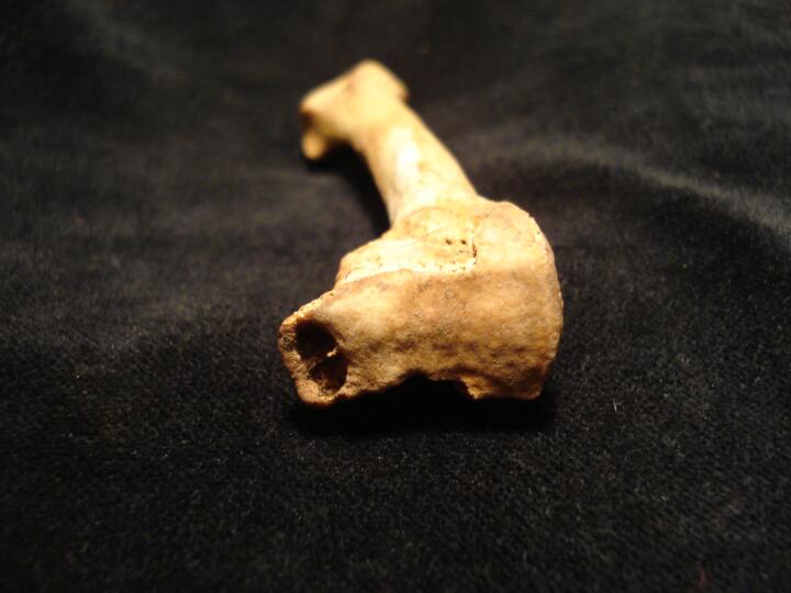
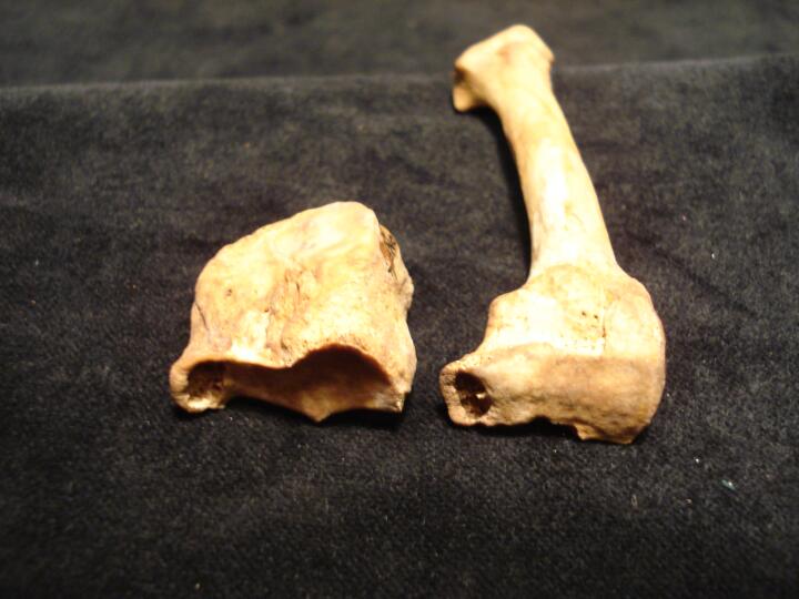
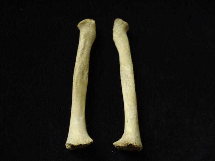
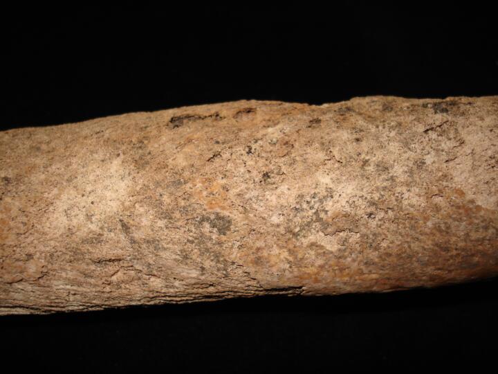
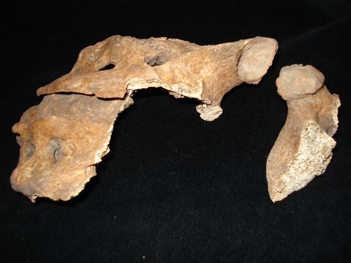
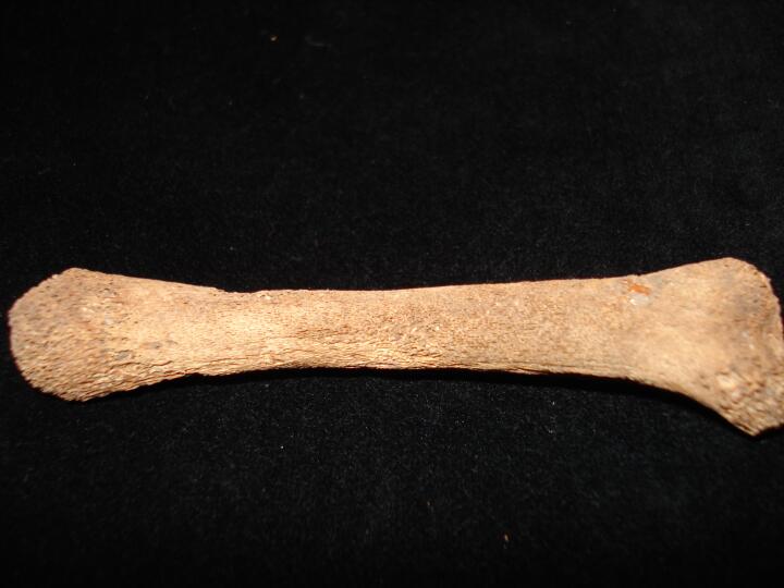
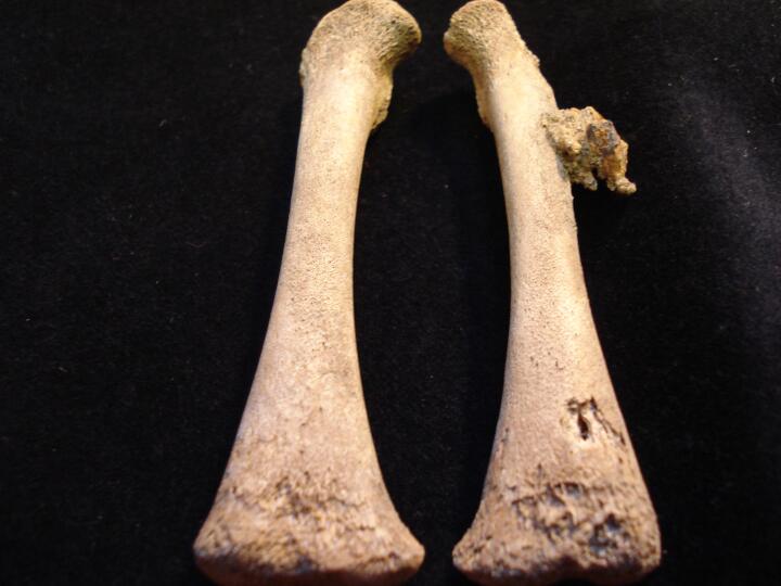
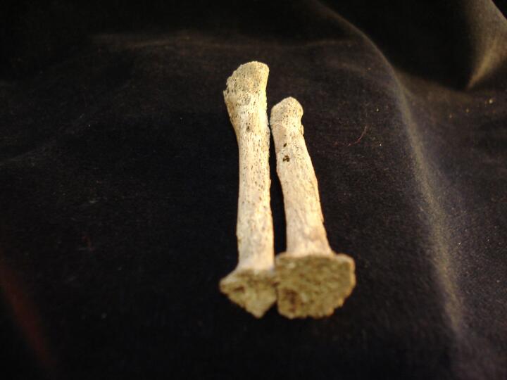
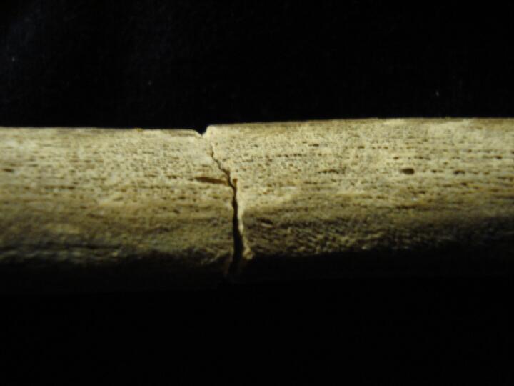
Left ulna, mid shaft, healed fracture (anterior view)
|
| ONE94
|
20
|
2
|
ONE94_20_2.jpg
|
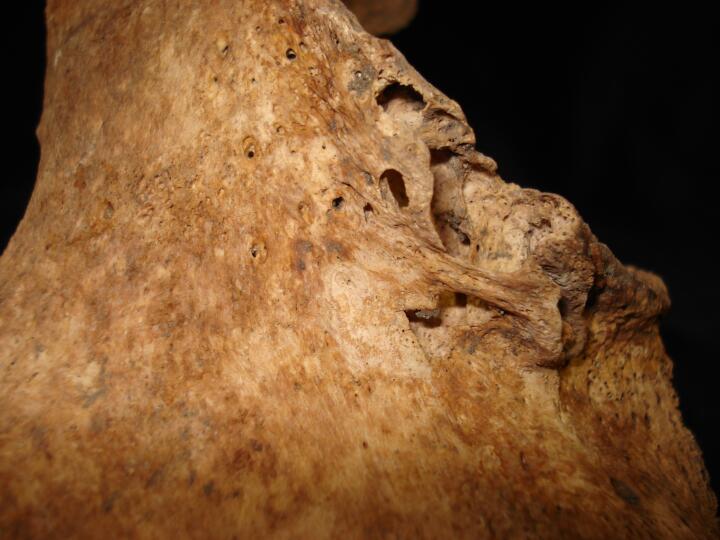
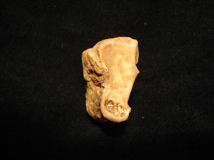
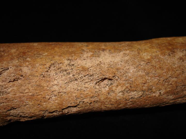
Left radius, mid shaft, healed fracture (anterior view)
|
| ONE94
|
20
|
3
|
ONE94_20_3.jpg
|
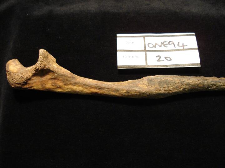
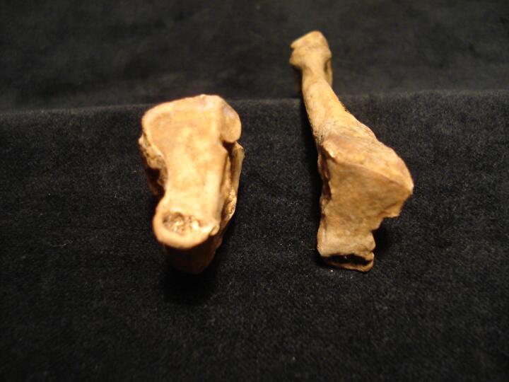
Left ulna & radius articulated, healed fractures mid shaft (anterior view)
|
| ONE94
|
20
|
4
|
ONE94_20_4.jpg
|
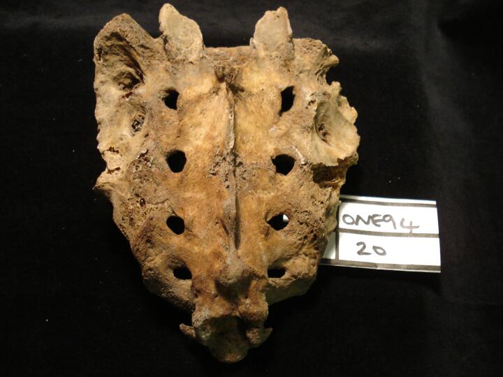
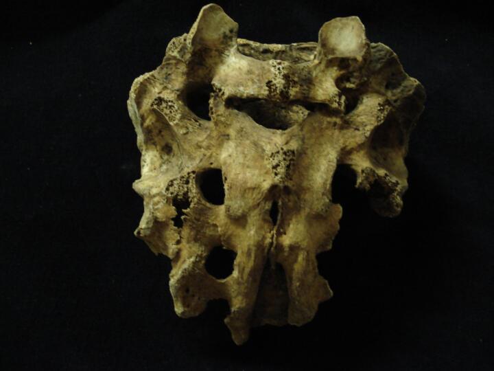
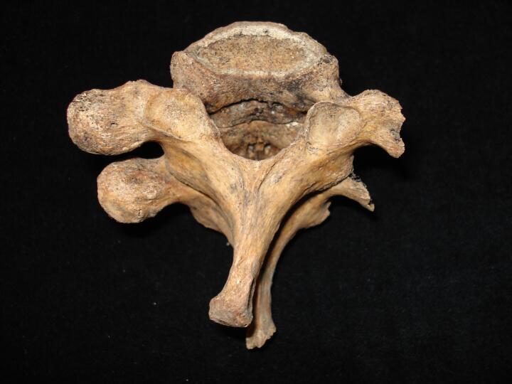
Sacrum, sacralisation of 1st coccygeal vertebra-caudal border shift (anterior view)
|
| ONE94
|
20
|
5
|
ONE94_20_5.jpg
|
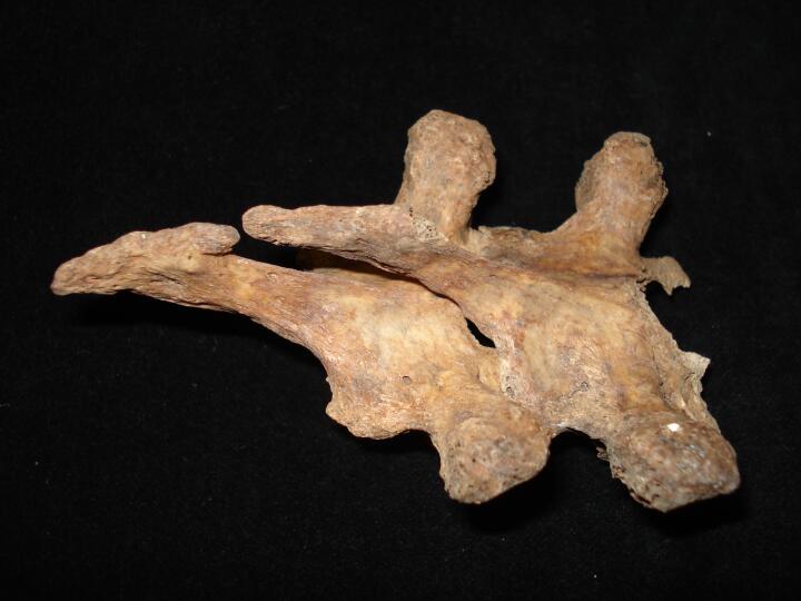
Sacrum, sacralisation of 1st coccygeal vertebra-caudal border shift (posterior view)
|
| ONE94
|
284
|
1
|
ONE94_284_1.jpg
|
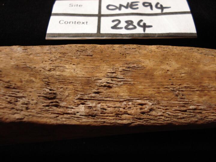
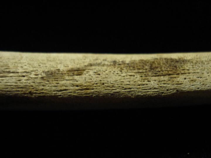
Right tibia, mid shaft healed non-specifi periosteal reaction (lateral surface)
|
| ONE94
|
594
|
1
|
ONE94_594_1.jpg
|
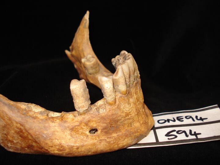
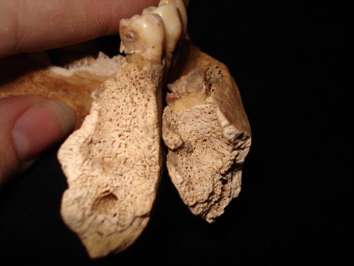
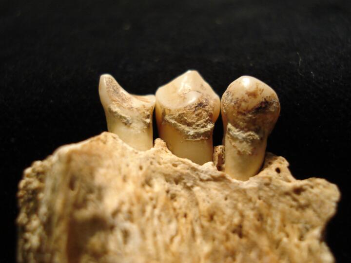
Mandible (buccal view) pipe facets, right lateral incisor & canine
|
| ONE94
|
594
|
2
|
ONE94_594_2.jpg
|
Mandible (buccal view/close up) pipe facets, right lateral incisor & canine
|
| ONE94
|
594
|
3
|
ONE94_594_3.jpg
|
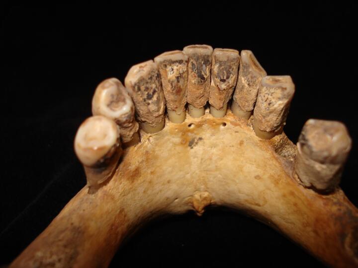
Mandible, anterior teeth (lingual view) ?tar deposits & calculus
|
| ONE94
|
18
|
1
|
ONE94_18_1.jpg
|
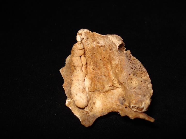
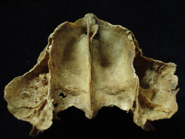
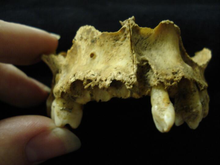
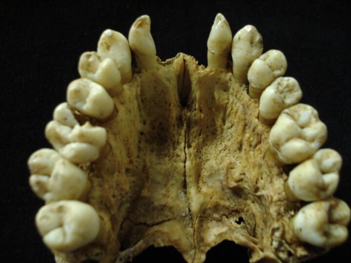
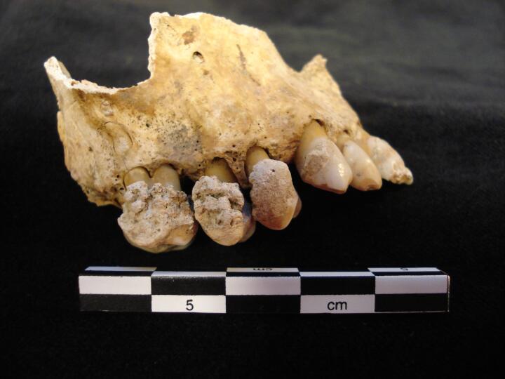
Maxillary process (palatal surface) non metric trait, Torus palatinus
|
| ONE94
|
18
|
2
|
ONE94_18_2.jpg
|
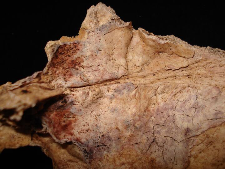
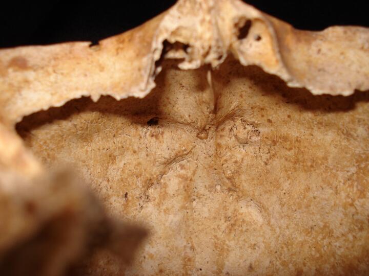
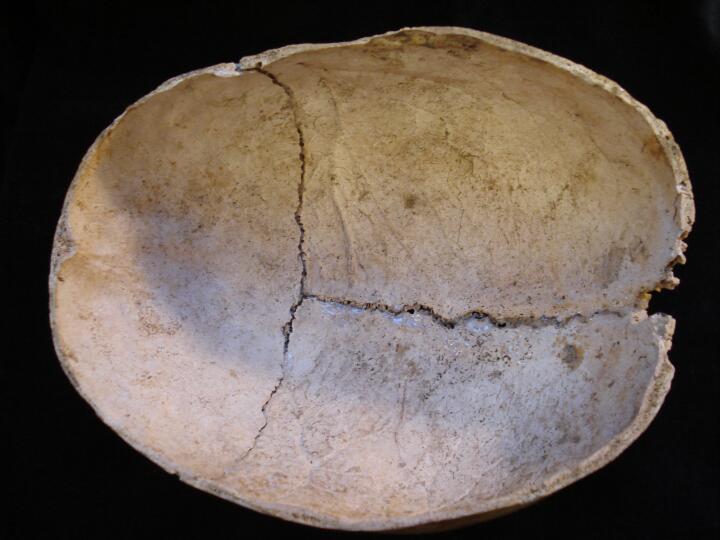
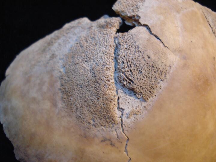
Skull, frontal bone (endocranial surface) HFI type 3-some PM damage
|
| ONE94
|
18
|
3
|
ONE94_18_3.jpg
|
Skull, frontal bone (endocranial surface/close up) HFI stage 3-some PM damage
|
| ONE94
|
18
|
4
|
ONE94_18_4.jpg
|
Skull,frontal bone (cross section) thickening possibly associated with Paget's disease
|
| ONE94
|
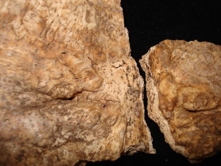
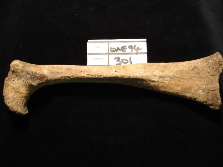
301
|
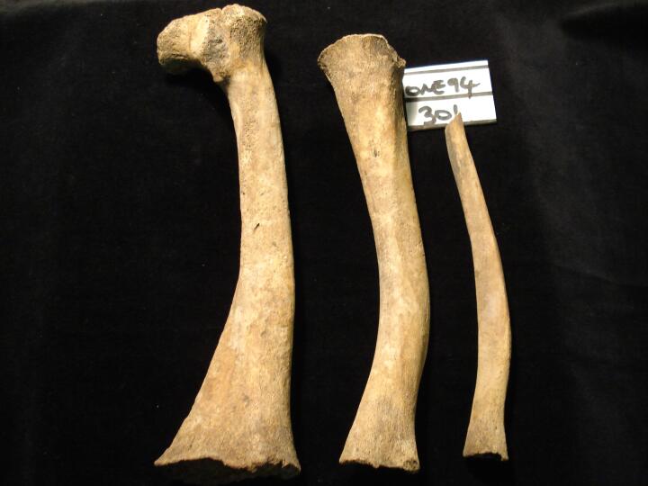
1
|
ONE94_301_1.jpg
|
Right femur (posterior view) healed rickets, bowing of bone & build up of bone on posterior surface
|
| ONE94
|
301
|
2
|
ONE94_301_2.jpg
|
Right tibia &fibula bowing of bone, healed rickets (lateral view)
|
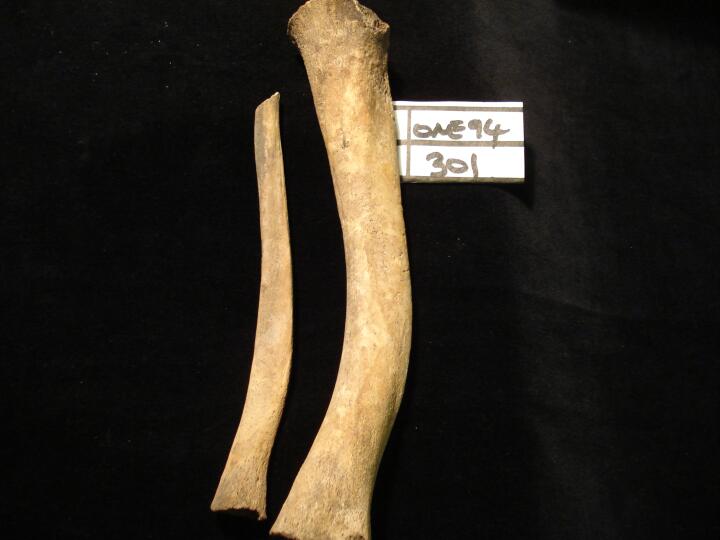
| ONE94
|
301
|
3
|
ONE94_301_3.jpg
|
Left & right tibia bowing from healed rickets (posterior view)
|
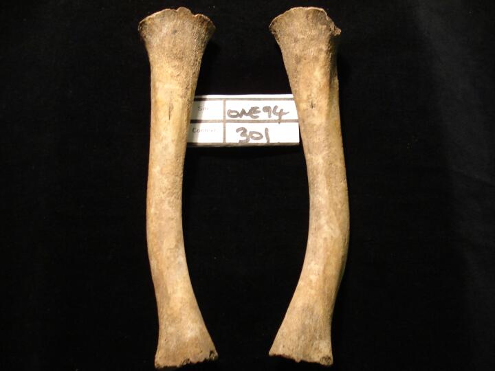
| ONE94
|
301
|
4
|
ONE94_301_4.jpg
|
Right femur, tibia & fibula bowing of bones from healed rickets (posterior view)
|
| ONE94
|
754
|
1
|
ONE94_754_1.jpg
|
Left 2nd metacarpal, smooth raised area of bone on the proximal head, ? Healed trauma
|
| ONE94
|
88
|
1
|
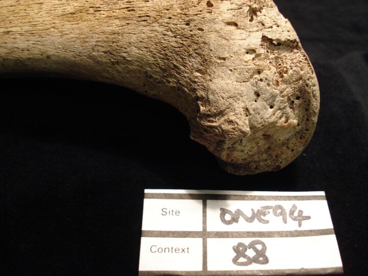
ONE94_88_1.jpg
|
Mandible, left side, heavy wear of dentition, particularly medial aspect of M1
|
| ONE94
|
88
|
2
|
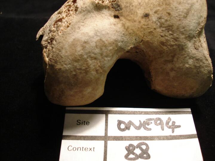
ONE94_88_2.jpg
|
Left femur medial epicondyle, soft tissue trauma associated with muscle (medial view)
|
| ONE94
|
88
|
3
|
ONE94_88_3.jpg
|
Left femur medial epicondyle, soft tissue trauma associated with muscle (inferior view)
|
| ONE94
|
114
|
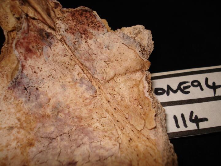
1
|
ONE94_114_1.jpg
|
Skull, frontal bone (endocranial surface) HFI type 2
|
| ONE94
|
114
|
2
|
ONE94_114_2.jpg
|
Skull, frontal bone (endocranial surface/close up) HFI type 2
|
| ONE94
|
114
|
3
|
ONE94_114_3.jpg
|
Ankylosis of right sacroiliac joint
|
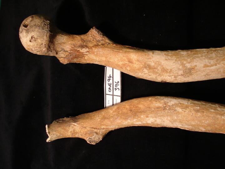
| ONE94
|
396
|
1
|
ONE94_396_1.jpg
|
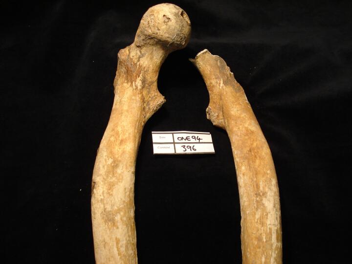
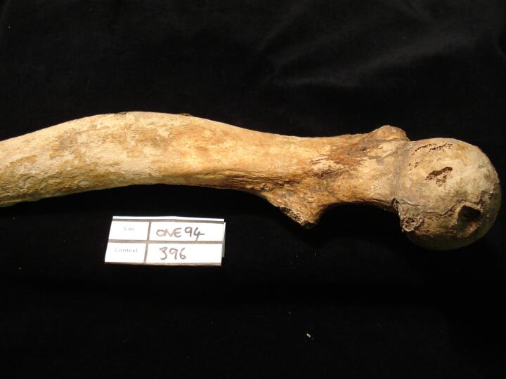
Femora, bowing from healed rickets (anterior view)
|
| ONE94
|
396
|
2
|
ONE94_396_2.jpg
|
Right femur, bowing from healed rickets (medial view)
|
| ONE94
|
396
|
3
|
ONE94_396_3.jpg
|
Femora, bowing from healed rickets (anterior/medial view)
|
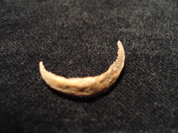
| ONE94
|
396
|
4
|
ONE94_396_4.jpg
|
Tracheal ring (anterior view)
|
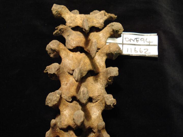
| ONE94
|
11662
|
1
|
ONE94_11662_1.jpg
|
Thoracic vertebrae (posterior view) malaligned spinous processes
|
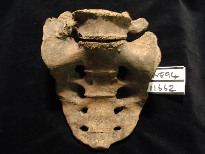
| ONE94
|
11662
|
2
|
ONE94_11662_2.jpg
|
Sacrum & lumbar vertebra L6, partial sacralisation right side (anterior view)
|
| ONE94
|
11662
|
3
|
ONE94_11662_3.jpg
|
Sacrum & lumbar vertebra L6, partial sacralisation right side (posterior view)
|
| ONE94
|
11662
|
4
|
ONE94_11662_4.jpg
|
Sacrum & lumbar vertebra L6, partial sacralisation right side (psuperior view)
|
| ONE94
|
11662
|
5
|
ONE94_11662_5.jpg
|
Sacrum (anterior view) extension of right ala associated with partial sacralisation of L6
|
| ONE94
|
11662
|
6
|
ONE94_11662_6.jpg
|
Sacrum (posterior view) extension of right ala associated with partial sacralisation of L6
|
| ONE94
|
963
|
1
|
ONE94_963_1.jpg
|
Left distal hand phalange (dorsal view) flared & frilly head, ?psoaritic arthropathy
|
| ONE94
|
963
|
2
|
ONE94_963_2.jpg
|
Left distal hand phalange (palmar view) flared & frilly head, ?psoaritic arthropathy
|
| ONE94
|
963
|
3
|
ONE94_963_3.jpg
|
Left distal hand phalange-head (palmar view) flared & frilly head, ?psoaritic arthropathy
|
| ONE94
|
356
|
1
|
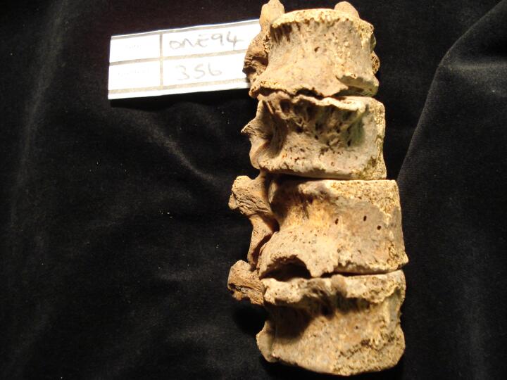
ONE94_356_1.jpg
|
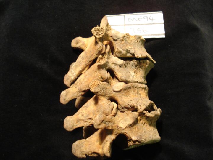
Thoracic vertebrae (anterior view) Th8 to Th11 osteophytic lipping possibly early stage DISH
|
| ONE94
|
356
|
2
|
ONE94_356_2.jpg
|
Thoracic vertebrae (right side view) Th8 to Th11 osteophytic lipping possibly early stage DISH
|
| ONE94
|
156
|
1
|
ONE94_156_1.jpg
|
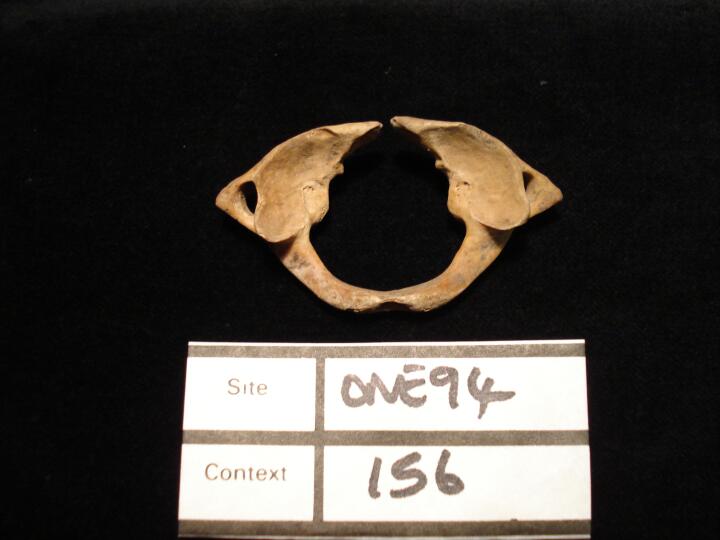
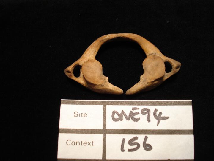
1st cervical vertebra, Atlas,non union between the posterior tubercle (superior view)
|
| ONE94
|
156
|
2
|
ONE94_156_2.jpg
|
1st cervical vertebra, Atlas,non union between the posterior tubercle (inferior view)
|
| ONE94
|
611
|
1
|
ONE94_611_1.jpg
|
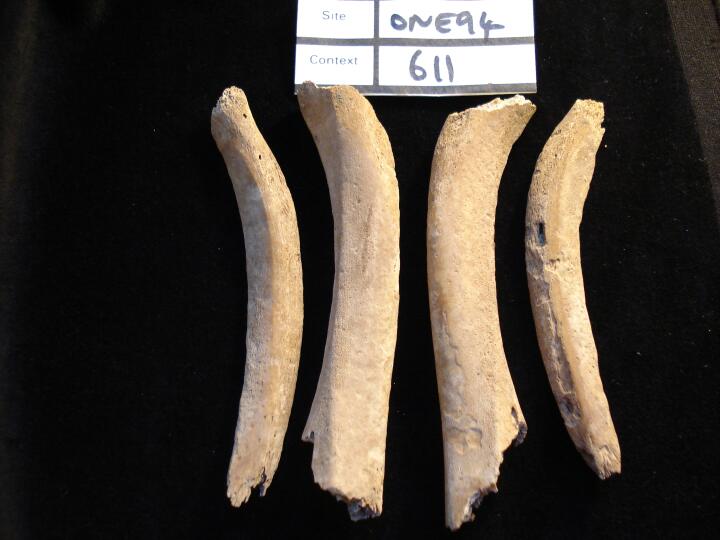
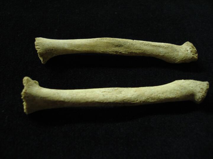
Left & right tibia and fibula bowing from healed rickets (medial view)
|
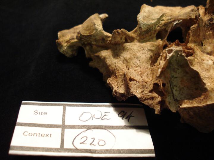
| ONE94
|
220
|
1
|
ONE94_220_1.jpg
|
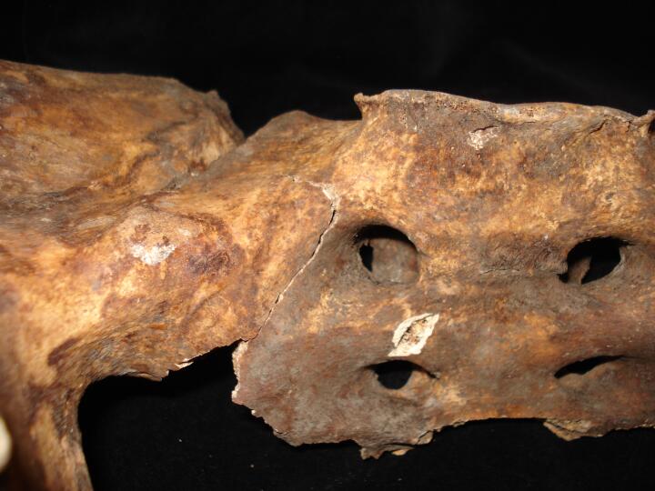
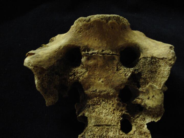
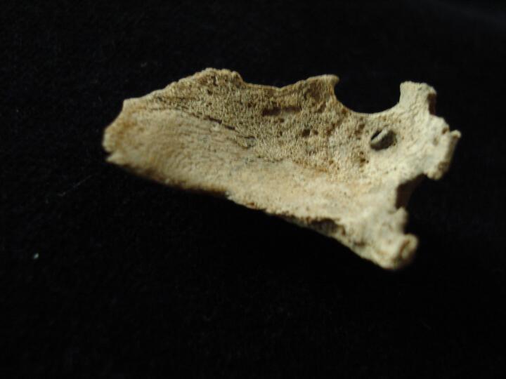
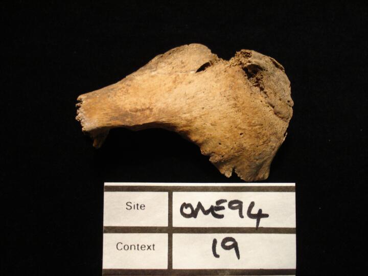
Bony exostosis eminating from the margin of the basioccipital (posterior view)
|
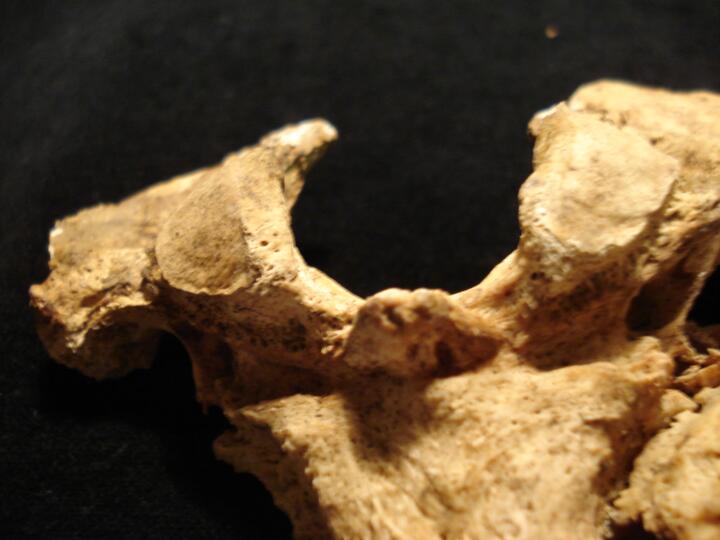
| ONE94
|
220
|
2
|
ONE94_220_2.jpg
|
Bony exostosis eminating from the margin of the basioccipital (posterior view/close up)
|
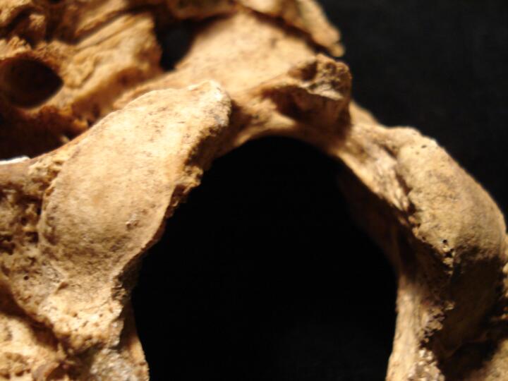
| ONE94
|
220
|
3
|
ONE94_220_3.jpg
|
Bony exostosis eminating from the margin of the basioccipital (inferior view)
|
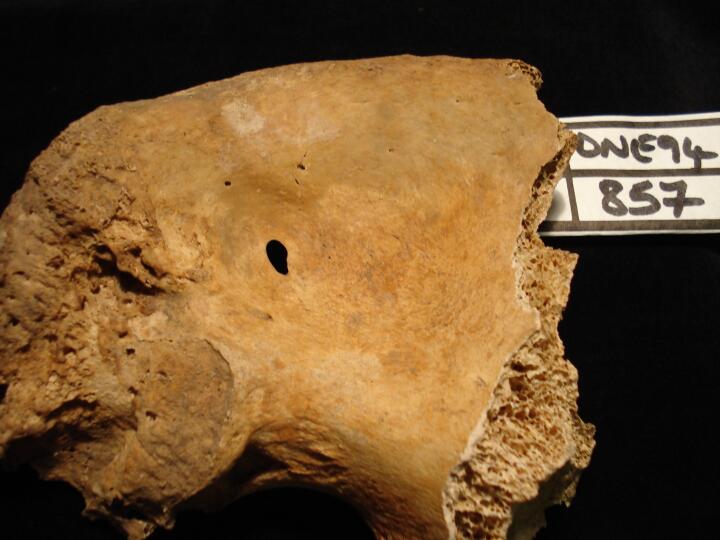
| ONE94
|
857
|
1
|
ONE94_857_1.jpg
|
Left pelvis,iliac fossa thinning producing a smooth edged aperture (medial view)
|
| ONE94
|
857
|
2
|
ONE94_857_2.jpg
|
Left pelvis,iliac fossa thinning producing a smooth edged aperture (medial view/close up)
|
| ONE94
|
857
|
3
|
ONE94_857_3.jpg
|
Left pelvis,iliac fossa thinning producing a smooth edged aperture (lateral view/close up)
|
| ONE94
|
726
|
1
|
ONE94_726_1.jpg
|
Mandibular canines (buccal view) diamond shaped enamel wear, from overbite
|
| ONE94
|
726
|
2
|
ONE94_726_2.jpg
|
Mandibular canines (medial view) calculus & doulbe roots
|
| ONE94
|
726
|
3
|
ONE94_726_3.jpg
|
Mandibular 2nd premolars (buccal/medial view) diamond shaped enamel wear, from overbite
|
| ONE94
|
726
|
4
|
ONE94_726_4.jpg
|
Mandibular 1st premolars (buccal view) diamond shaped enamel wear, from overbite
|
| ONE94
|
782
|
1
|
ONE94_782_1.jpg
|
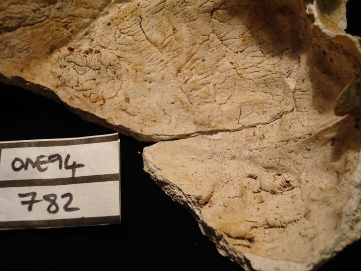
Skull, frontal bone (endocranial surface) left side HFI type 3
|
| ONE94
|
782
|
2
|
ONE94_782_2.jpg
|
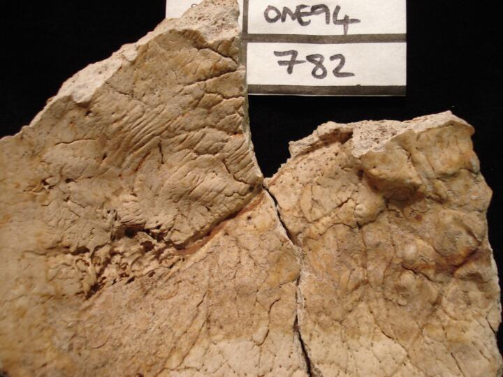
Skull, frontal bone (endocranial surface/close up) left side HFI type 3
|
| ONE94
|
782
|
3
|
ONE94_782_3.jpg
|
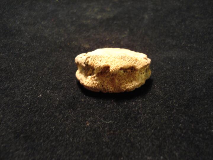
Skull, frontal bone (endocranial surface) left side HFI type 3
|
| ONE94
|
609
|
1
|
ONE94_609_1.jpg
|
Skull, occipital bone (endocranial surface) small oval lesion, ?histiocytosis-X
|
| ONE94
|
609
|
2
|
ONE94_609_2.jpg
|
Skull, occipital bone (endocranial surface/close up) small oval lesion, ?histiocytosis-X
|
| ONE94
|
645
|
1
|
ONE94_645_1.jpg
|
Lumbar L5 centrum (anterior view) scalloped lytic lesion, possibly Tuberculosis
|
| ONE94
|
645
|
2
|
ONE94_645_2.jpg
|
Lumbar L5 centrum (anterior view) scalloped lytic lesion, possibly Tuberculosis
|
| ONE94
|
645
|
3
|
ONE94_645_3.jpg
|
Lumbar L5 centrum (right side view) scalloped lytic lesion, possibly Tuberculosis
|
| ONE94
|
317
|
1
|
ONE94_317_1.jpg
|
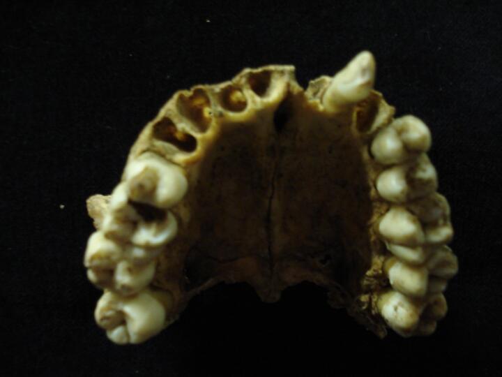
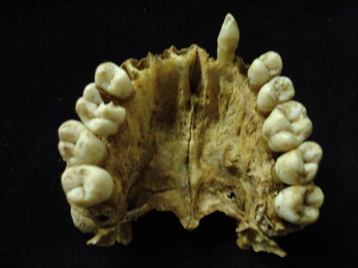
Mandible (occlusal view) carious lesions
|
| ONE94
|
317
|
2
|
ONE94_317_2.jpg
|
Mandible, right side carious lesion 3rd molar (buccal view/close up)
|
| ONE94
|
317
|
3
|
ONE94_317_3.jpg
|
Mandible, left side, carious lesion 1st permanent molar (buccal view/close up)
|
| ONE94
|
118
|
1
|
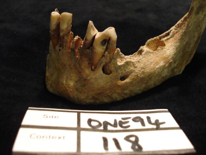
ONE94_118_1.jpg
|
Mandible, left side, pipe facets, lateral incisor & canine (buccal view)
|
| ONE94
|
118
|
2
|
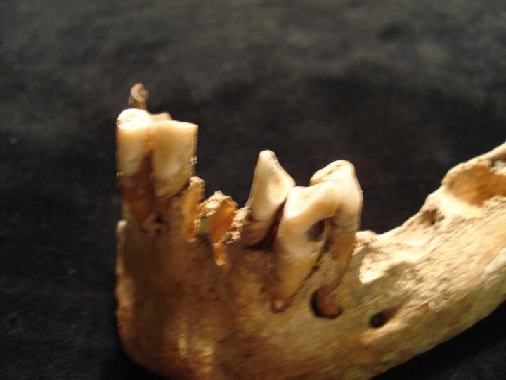
ONE94_118_2.jpg
|
Mandible, left side, pipe facets, lateral incisor & canine (buccal view/close up)
|
| ONE94
|
118
|
3
|
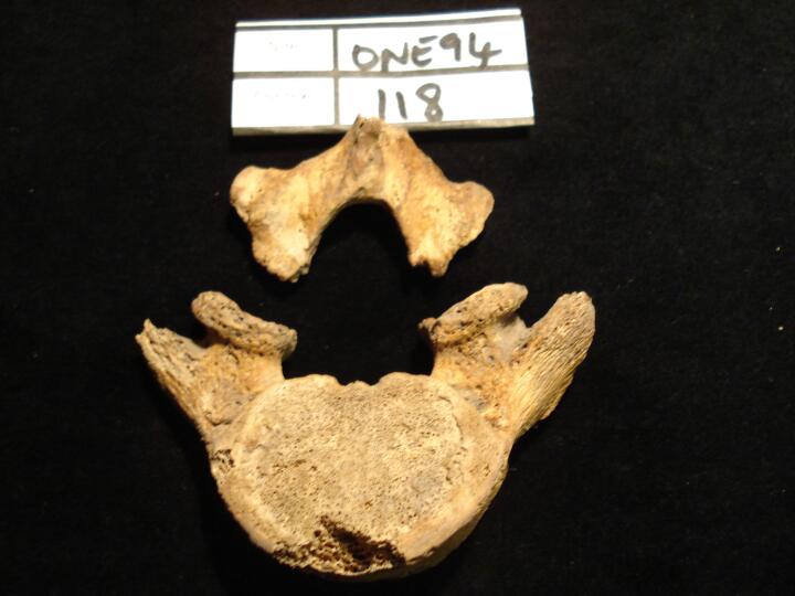
ONE94_118_3.jpg
|
Lumbar vertebra L5, bilateral spondylolisis, body & pars interarticularis (superior view)
|
| ONE94
|
118
|
4
|
ONE94_118_4.jpg
|
Lumbar vertebra L5, bilateral spondylolisis, body & pars interarticularis (inferior view)
|
| ONE94
|
761
|
1
|
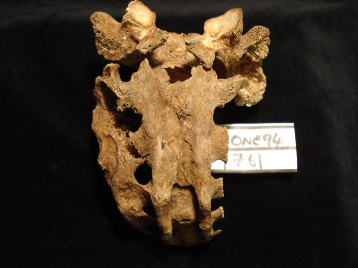
ONE94_761_1.jpg
|
Sacrum,sacralisation & cleft between S3 to S5 (posterior view)
|
| ONE94
|
761
|
2
|
ONE94_761_2.jpg
|
Right 3rd metatarsal, joint lesion, ?circulatory or trauma
|
| ONE94
|
761
|
3
|
ONE94_761_3.jpg
|
Right medial cuneiform, joint lesion associated with 3rd metatarsal, ?circulatory or trauma
|
| ONE94
|
761
|
4
|
ONE94_761_4.jpg
|
Right 3rd metatarsal & medial cuneiform, joint lesions, ?circulatory or trauma (inferior view)
|
| ONE94
|
761
|
5
|
ONE94_761_5.jpg
|
Right 3rd metatarsal & medial cuneiform, joint lesions, ?circulatory or trauma (medial view)
|
| ONE94
|
991
|
1
|
ONE94_991_1.jpg
|
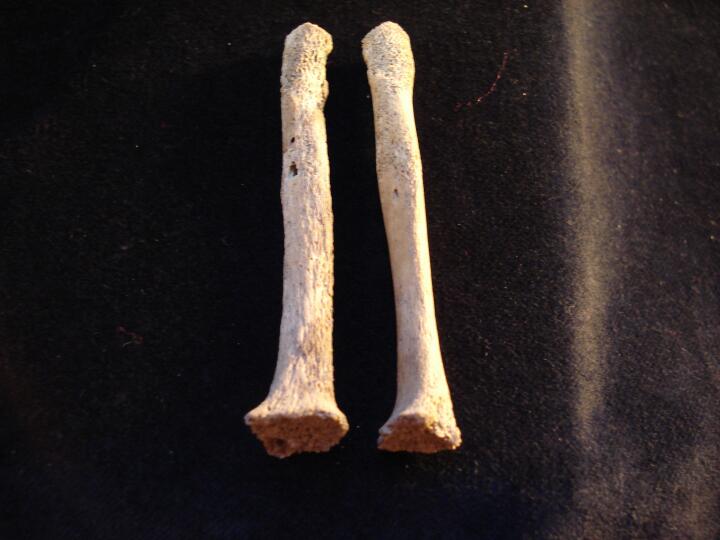
Left & right radius, flared metaphyses, active rickets (anterior view)
|
| ONE94
|
991
|
2
|
ONE94_991_2.jpg
|
Left & right radius, flared metaphyses, active rickets (anterior view/close up)
|
| ONE94
|
991
|
3
|
ONE94_991_3.jpg
|
Left & right tibia, flared metaphyses, active rickets (anterior view)
|
| ONE94
|
991
|
4
|
ONE94_991_4.jpg
|
Left & right tibia, flared metaphyses, active rickets (inferior view/close up)
|
| ONE94
|
991
|
5
|
ONE94_991_5.jpg
|
Left fibula, slight bowing & flared metaphyseal end, active rickets (medial view)
|
| ONE94
|
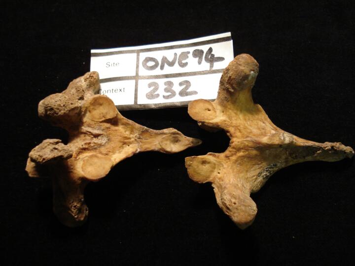
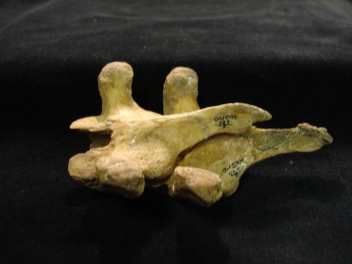
232
|
1
|
ONE94_232_1.jpg
|
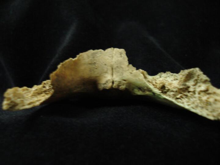
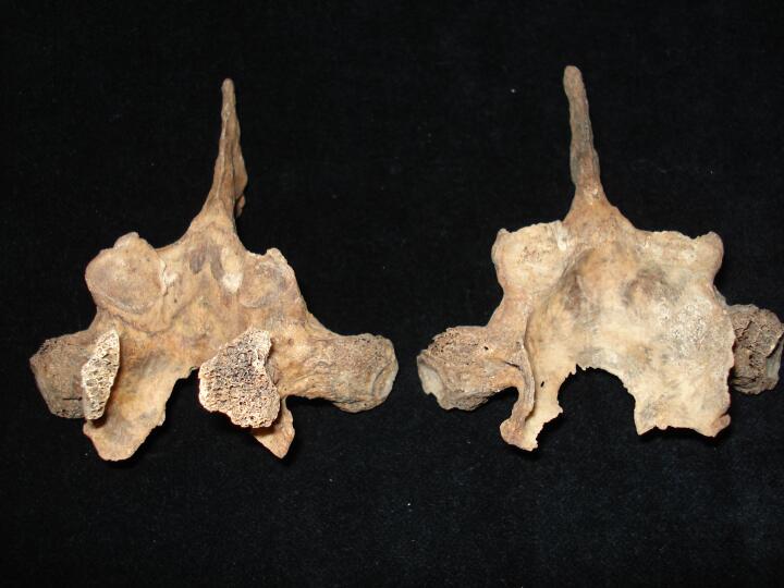
Thoracic vertebrae Th7 (inferior view) & Th8 (superior view) accessory facet between spinous processes
|
| ONE94
|
232
|
2
|
ONE94_232_2.jpg
|
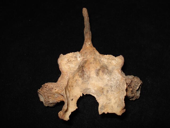
Thoracic vertebrae (articulated) Th7 (inferior view) & Th8 (superior view) accessory facet between spinous processes
|
| ONE94
|
276
|
1
|
ONE94_276_1.jpg
|
Skull occipital bone, increased porostiy & possible transverse cut marks (posterior view)
|
| ONE94
|
276
|
2
|
ONE94_276_2.jpg
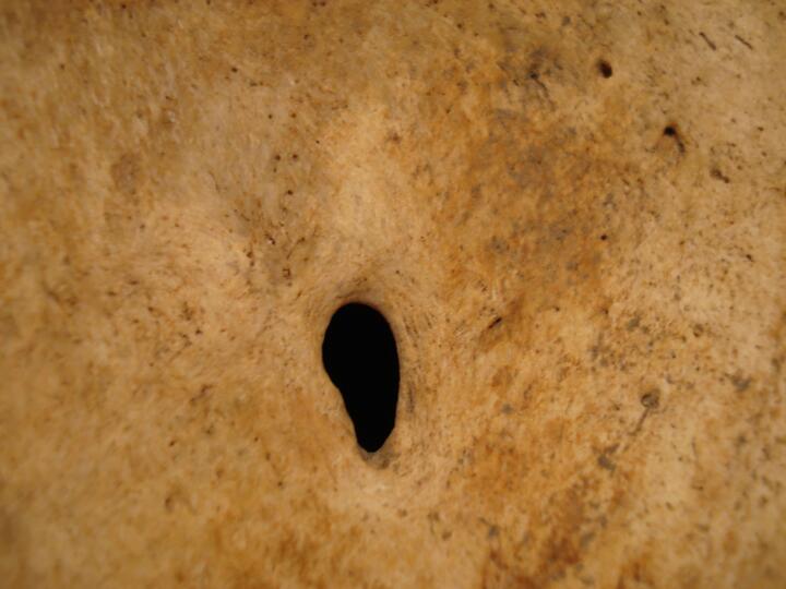
|
Skull occipital bone, increased porostiy & possible transverse cut marks (posterior view/close up)
|
| ONE94
|
276
|
3
|
ONE94_276_3.jpg
|
Maxilla, left sinus, smooth nodule of bone, indication of healed sinusitis (superior view)
|
| ONE94
|
276
|
4
|
ONE94_276_4.jpg
|
Maxillary process (palatal view) left lateral incisor rotated 90 degrees
|
| ONE94
|
276
|
5
|
ONE94_276_5.jpg
|
Maxilla (buccal view) left lateral incisor rotated 90 degrees
|
| ONE94
|
276
|
6
|
ONE94_276_6.jpg
|
Maxilla (palatal view) left lateral incisor rotated 90 degrees
|
| ONE94
|
276
|
7
|
ONE94_276_7.jpg
|
Maxilla (palatal view) left lateral incisor rotated 90 degrees
|
| ONE94
|
276
|
8
|
ONE94_276_8.jpg
|
Sacrum (anterior view) cleft between S1 to S2
|
| ONE94
|
276
|
9
|
ONE94_276_9.jpg
|
Sacrum (posterior view) cleft between S1 to S2
|
| ONE94
|
306
|
1
|
ONE94_306_1.jpg
|
Skull frontal bone (endocranial surface) Possibly early stage of HFI & deep arachnoid granulations
|
| ONE94
|
223
|
1
|
ONE94_223_1.jpg
|
Mandible right side active non-specific infection associated with caries related periapical lesion (buccal view)
|
| ONE94
|
223
|
2
|
ONE94_223_2.jpg
|
Mandible, cross section, active non-specific infection associated with caries related periapical lesion (buccal view) some PM damage
|
| ONE94
|
526
|
1
|
ONE94_526_1.jpg
|
Right zygomatic process (anterior view) smooth circular depression, healed trauma ?interpersonal violence
|
| ONE94
|
526
|
2
|
ONE94_526_2.jpg
|
Right zygomatic process (anterior view/close up) smooth circular depression, healed trauma ?interpersonal violence
|
| ONE94
|
526
|
3
|
ONE94_526_3.jpg
|
Left & right zygomatic process for comparison of change to right process (anterior view)
|
| ONE94
|
557
|
1
|
ONE94_557_1.jpg
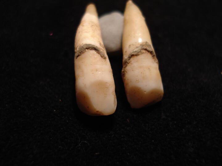
|
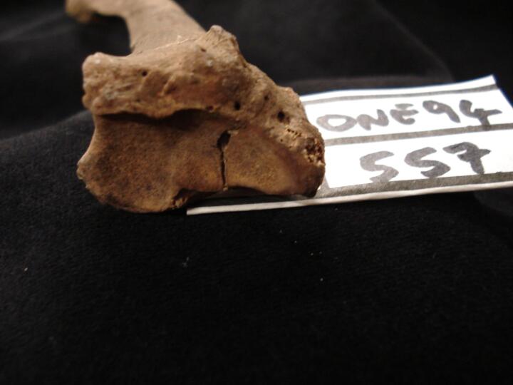
Mandible & maxilla (buccal view/close up) pipe facets in right lateral incisors & canines
|
| ONE94
|
557
|
2
|
ONE94_557_2.jpg
|
Right radius, healed hair line intrarticular fracture across distal articular surface (inferior view)
|
| ONE94
|
557
|
3
|
ONE94_557_3.jpg
|
Left & right 1st metatarsals (medial view) destructive lesion, ?gout
|
| ONE94
|
557
|
4
|
ONE94_557_4.jpg
|
Left & right 1st metatarsals (plantar view) osteoarthritis, eburnation of joint surface
|
| ONE94
|
508
|
1
|
ONE94_508_1.jpg
|
Sacrum (posterior view) sacralisation
|
| ONE94
|
508
|
2
|
ONE94_508_2.jpg
|
Mandible left lateral incisor & canine, pipe facets (buccal view)
|
| ONE94
|
508
|
3
|
ONE94_508_3.jpg
|
Maxilla left lateral incisor & canine, pipe facets (buccal view)
|
| ONE94
|
508
|
4
|
ONE94_508_4.jpg
|
Mandible & maxilla (buccal view/close up) pipe facets in left lateral incisors & canines
|
| ONE94
|
508
|
5
|
ONE94_508_5.jpg
|
Mandible left lateral incisor & canine, pipe facets (lingual view)
|
| ONE94
|
508
|
6
|
ONE94_508_6.jpg
|
Maxilla left lateral incisor & canine, pipe facets (lingual view)
|
| ONE94
|
508
|
7
|
ONE94_508_7.jpg
|
Maxilla (right side/lingual view) black substance adgering to dentition, ?tar depoit from pipe smoking
|
| ONE94
|
508
|
8
|
ONE94_508_8.jpg
|
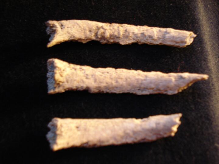
Ribs, shaft fragments with healed & healing fractures (visceral surface)
|
| ONE94
|
508
|
9
|
ONE94_508_9.jpg
|
Two rib shaft fragments (visceral surface/close up) healed & healing fractures
|
| ONE94
|
508
|
10
|
ONE94_508_10.jpg
|
Two rib shaft fragments (visceral surface/close up) healed & healing fractures
|
| ONE94
|
508
|
11
|
ONE94_508_11.jpg
|
Two rib shaft fragments (pleuralsurface/close up) healed & healing fractures
|
| ONE94
|
508
|
12
|
ONE94_508_12.jpg
|
Two rib shaft fragments (pleuralsurface/close up) healed & healing fractures
|
| ONE94
|
508
|
13
|
ONE94_508_13.jpg
|
One rib shaft fragment (visceral surface) line of fracture vsible, healing in progress
|
| ONE94
|
769
|
1
|
ONE94_769_1.jpg
|
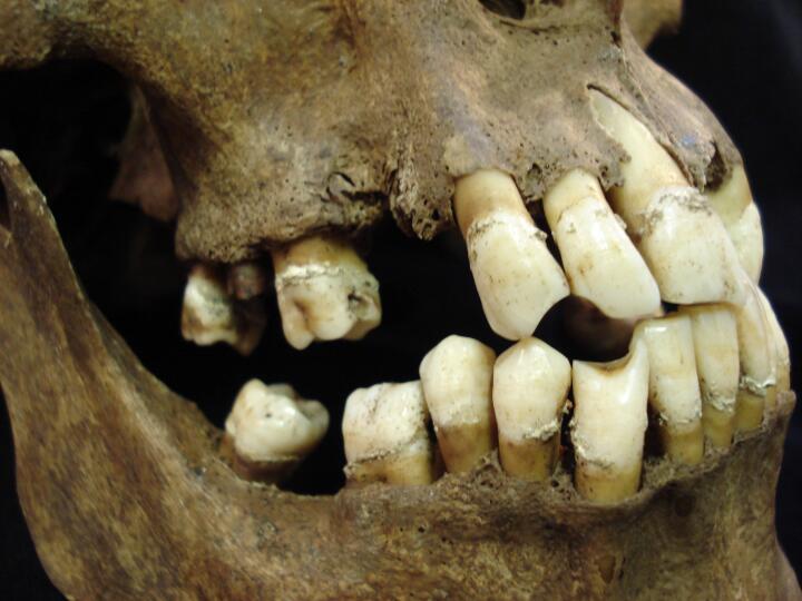
Mandible (occlusal view/some PM damage) ante mortem tooth loss
|
| ONE94
|
769
|
2
|
ONE94_769_2.jpg
|
Skull, parietal fragments (ectocranial surface) pathological bone surface change, ?neoplastic blood disorder
|
| ONE94
|
769
|
3
|
ONE94_769_3.jpg
|
Skull, parietal fragments (ectocranial surface/close up) pathological bone surface change, ?neoplastic blood disorder
|
| ONE94
|
769
|
4
|
ONE94_769_4.jpg
|
Skull, parietal fragments (endocranial surface/close up) pathological bone surface change, ?neoplastic blood disorder
|
| ONE94
|
769
|
5
|
ONE94_769_5.jpg
|
Lumbar vertebra L1 (inferior surface) central focus of destructive lesion & bone surface change, ?neoplastic blood disorder
|
| ONE94
|
769
|
6
|
ONE94_769_6.jpg
|
1st sacral vertebra (superior view) bone surface change & areas of destruction. ?neoplastic blood disorder
|
| ONE94
|
769
|
7
|
ONE94_769_7.jpg
|
Left femur (anterior view) femoral neck, pathological bone surface changes, ?neoplastic blood disorder
|
| ONE94
|
769
|
8
|
ONE94_769_8.jpg
|
Left femur (anterior view) mid shaft, pathological bone surface changes, ?neoplastic blood disorder
|
| ONE94
|
769
|
9
|
ONE94_769_9.jpg
|
Left femur (anterior view/close up) mid shaft, pathological bone surface changes, ?neoplastic blood disorder
|
| ONE94
|
769
|
10
|
ONE94_769_10.jpg
|
Left femur (posterior view/close up) distal 1/3 shaft, pathological bone surface changes, ?neoplastic blood disorder
|
| ONE94
|
128
|
1
|
ONE94_128_1.jpg
|
Skull, frontal bone (anterior view) showing calvarium post mortem cut
|
| ONE94
|
128
|
2
|
ONE94_128_2.jpg
|
Skull, (anterior view) calvarium post mortem cut with two parts of skull & indication of other cut marks
|
| ONE94
|
128
|
3
|
ONE94_128_3.jpg
|
Skull, occipital (posterior view) showing cut marks
|
| ONE94
|
128
|
4
|
ONE94_128_4.jpg
|
Skull, parietal & occipital (endocranial view) showing smooth cut edge from post mortem
|
| ONE94
|
128
|
5
|
ONE94_128_5.jpg
|
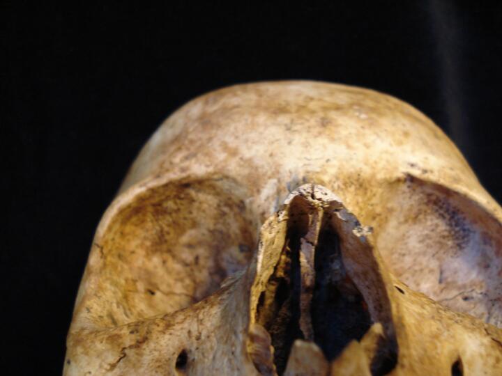
Skull, orbits (anterior view) cribra orbitalia
|
| ONE94
|
325
|
1
|
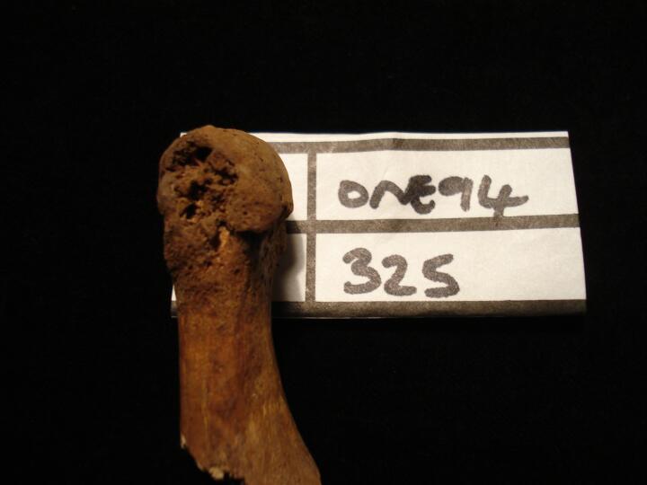
ONE94_325_1.jpg
|
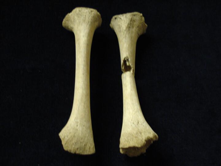
Left 1st metatarsal (medial view) destructive change, ?gout
|
| ONE94
|
257
|
1
|
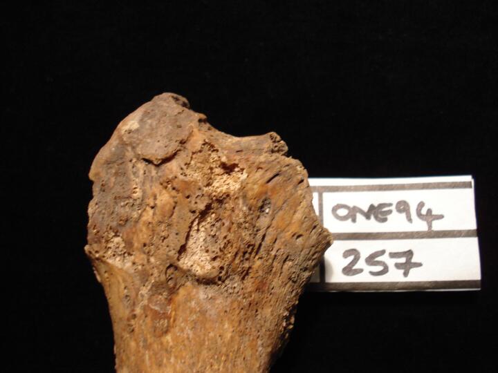
ONE94_257_1.jpg
|
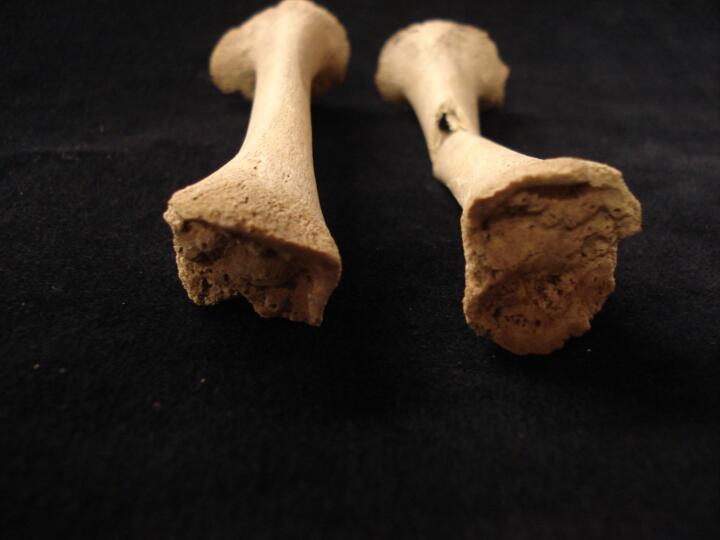
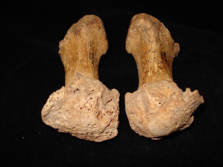
Right femur (anterior view) non- united fracture of the femoral neck (femoral head missing PM)smoothed line of fracture
|
| ONE94
|
257
|
2
|
ONE94_257_2.jpg
|
Right femur (medial view) non- united fracture of the femoral neck (femoral head missing PM) aspect of line of fracture smooth eburnated bone, secondary osteoarthritis
|
| ONE94
|
257
|
3
|
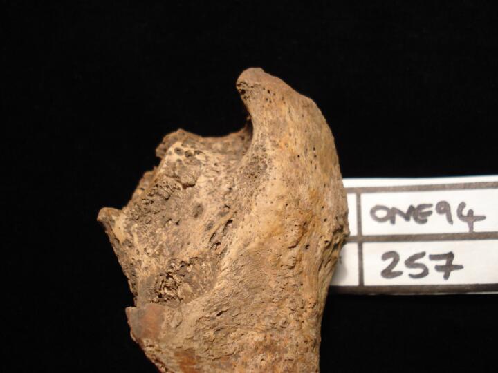
ONE94_257_3.jpg
|
Right femur (posterior view) non- united fracture of the femoral neck (femoral head missing PM) aspect of line of fracture smooth eburnated bone, secondary osteoarthritis
|
| ONE94
|
257
|
4
|
ONE94_257_4.jpg
|
Right tibia proximal end (posterior view), ?osteomyeltic infection
|
| ONE94
|
257
|
5
|
ONE94_257_5.jpg
|
Right tibia mid shaft (posterior view), ?osteomyeltic infection
|
| ONE94
|
257
|
6
|
ONE94_257_6.jpg
|
Right tibia mid shaft (posterior view/close up), ?osteomyeltic infection
|
| ONE94
|
146
|
1
|
ONE94_146_1.jpg
|
Sacrum, post mortem damage (anterior view) 1st sacral vertebra with left side showing partial lumbarisation
|
| ONE94
|
146
|
2
|
ONE94_146_2.jpg
|
Sacrum, post mortem damage (posterior view) cleft opening from S5 to S5
|
| ONE94
|
146
|
3
|
ONE94_146_3.jpg
|
Sacrum (posterior view) post mortem damage, two parts showing partial lumbarisation
|
| ONE94
|
138
|
1
|
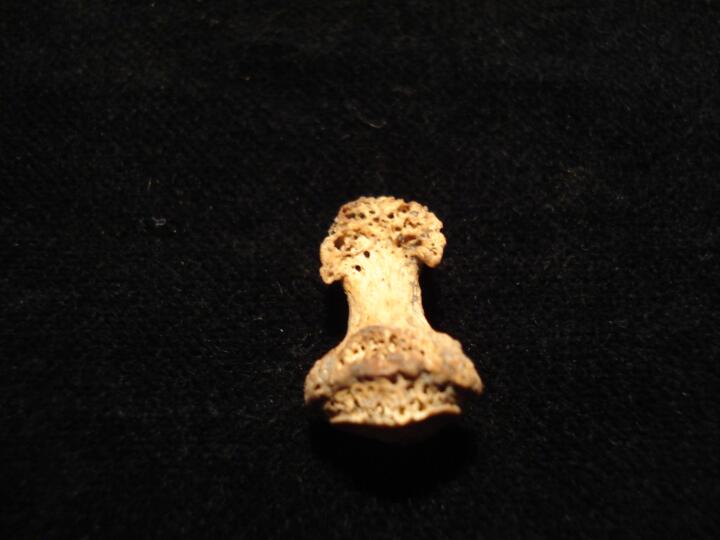
ONE94_138_1.jpg
|
Developing maxillary & mandibular permanent molars (buccal view) with hypoplastic defects, ?indication of congenital syphilis
|
| ONE94
|
138
|
2
|
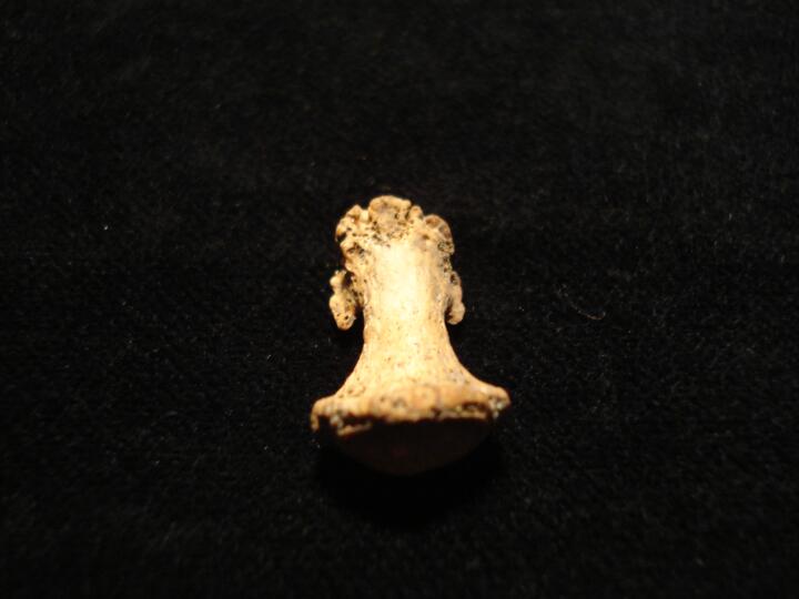
ONE94_138_2.jpg
|
Developing maxillary permanent molar (lingual view) with hypoplastic defects, ?indication of congenital syphilis
|
| ONE94
|
138
|
3
|
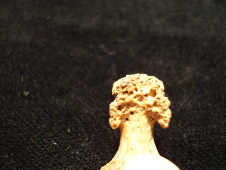
ONE94_138_3.jpg
|
Developing maxillary permanent molar (medial view) with hypoplastic defects, ?indication of congenital syphilis
|
| ONE94
|
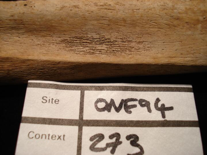
273
|
1
|
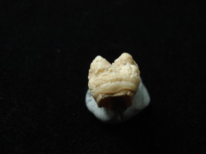
ONE94_273_1.jpg
|
Right tibia (medial view) shaft surface active non-specific perioateal reaction
|
| ONE94
|
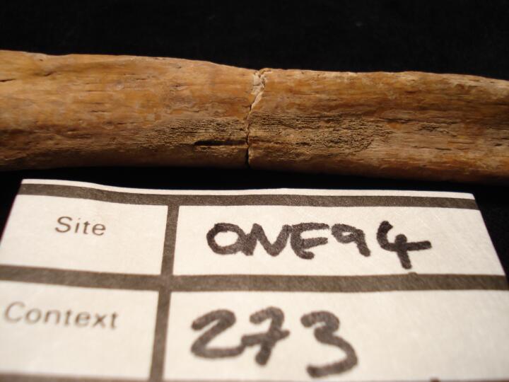
273
|
2
|
ONE94_273_2.jpg
|
Left ulna, mid shaft (medial view) active non-specific periosteal reaction
|
| ONE94
|
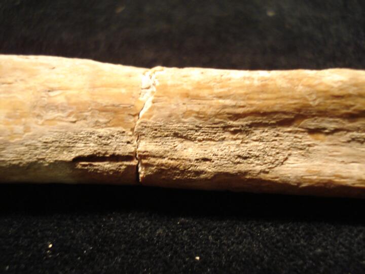
273
|
3
|
ONE94_273_3.jpg
|
Left ulna, mid shaft (medial view/close up) active non-specific periosteal reaction
|
| ONE94
|
846
|
1
|
ONE94_846_1.jpg
|
Skull, parietal fragments (ectocranial surface) increased porosity, ?scurvy
|
| ONE94
|
846
|
2
|
ONE94_846_2.jpg
|
Skull, parietal fragment (ectocranial surface) lesion possibly associated with histiocytosis-X
|
| ONE94
|
846
|
3
|
ONE94_846_3.jpg
|
Skull, parietal fragment (endocranial surface) lesion possibly associated with histiocytosis-X
|
| ONE94
|
846
|
4
|
ONE94_846_4.jpg
|
Body of basioccipital increased porosity, ?scurvy
|
| ONE94
|
846
|
5
|
ONE94_846_5.jpg
|
Right humerus (posterior view) slightly swollen appearance to distal 1/3 of shaft,?scurvy
|
| ONE94
|
846
|
6
|
ONE94_846_6.jpg
|
Right humerus (anterior view) slightly swollen appearance to distal 1/3 of shaft, ?scurvy
|
| ONE94
|
846
|
7
|
ONE94_846_7.jpg
|
Femora (anterior view) increased porosity, ?scurvy & coffin nail adhering to left femur
|
| ONE94
|
846
|
8
|
ONE94_846_8.jpg
|
Right tibia (medial view) mid shaft surface increased porosity, ?scurvy
|
| ONE94
|
902
|
1
|
ONE94_902_1.jpg
|
Skull, parietal fragments (ectocranial surface) reactive surface change associated with ?porotic hyperostosis or scurvy
|
| ONE94
|
164
|
1
|
ONE94_164_1.jpg
|
Skull,left parietal (endocranial surface) deep meningeal vessels
|
| ONE94
|
164
|
2
|
ONE94_164_2.jpg
|
Skull,left parietal (endocranial surface/close up) deep meningeal vessels
|
| ONE94
|
164
|
3
|
ONE94_164_3.jpg
|
Skull,left parietal (endocranial surface) deep meningeal vessels
|
| ONE94
|
164
|
4
|
ONE94_164_4.jpg
|
Mandible edentulous
|
| ONE94
|
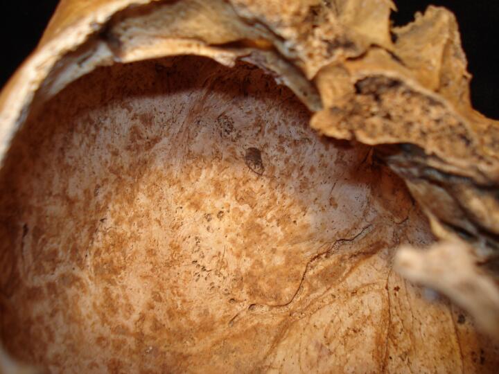
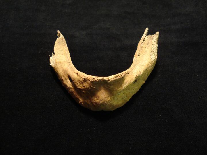
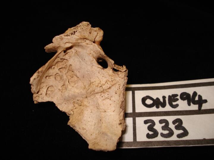
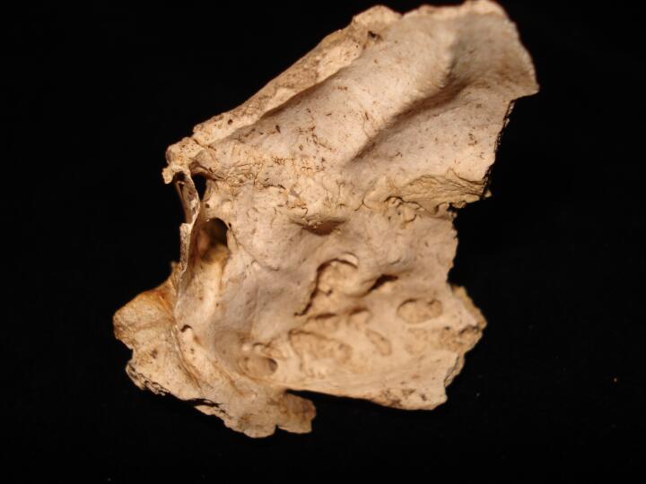
333
|
1
|
ONE94_333_1.jpg
|
Right sphenoid cerebral surface of greater wing smooth edged circular lesions
|
| ONE94
|
333
|
2
|
ONE94_333_2.jpg
|
Right sphenoid cerebral surface of greater wing smooth edged circular & oval lesions
|
| ONE94
|
333
|
3
|
ONE94_333_3.jpg
|
Right & left shenoid cerebral surface of greater wings smooth edged circular/oval lesions
|
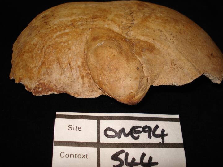
| ONE94
|
544
|
1
|
ONE94_544_1.jpg
|
Skull, right side frontal & parietal healed blunt force trauma across coronal suture large smooth lobule of bone (right side view)
|
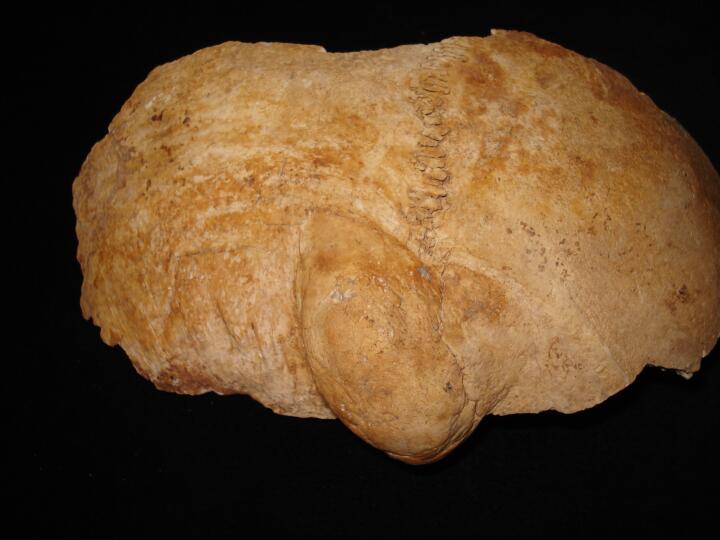
| ONE94
|
544
|
2
|
ONE94_544_2.jpg
|
Skull, right side frontal & parietal healed blunt force trauma across coronal suture large smooth lobule of bone (posterior view)
|
| ONE94
|
544
|
3
|
ONE94_544_3.jpg
|
Skull, right side frontal & parietal healed blunt force trauma across coronal suture large smooth lobule of bone (frontal view)
|
| ONE94
|
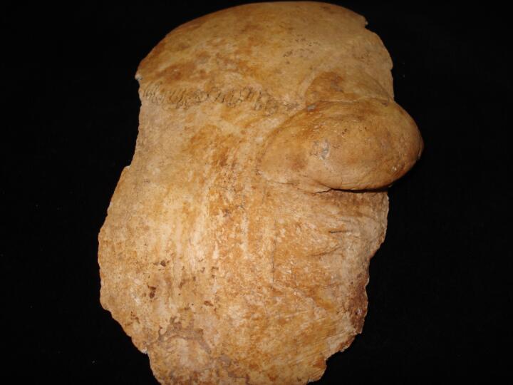
544
|
4
|
ONE94_544_4.jpg
|
Skull, right side frontal & parietal healed blunt force trauma across coronal suture large smooth lobule of bone (superior view/ectocranial surface)
|
| ONE94
|
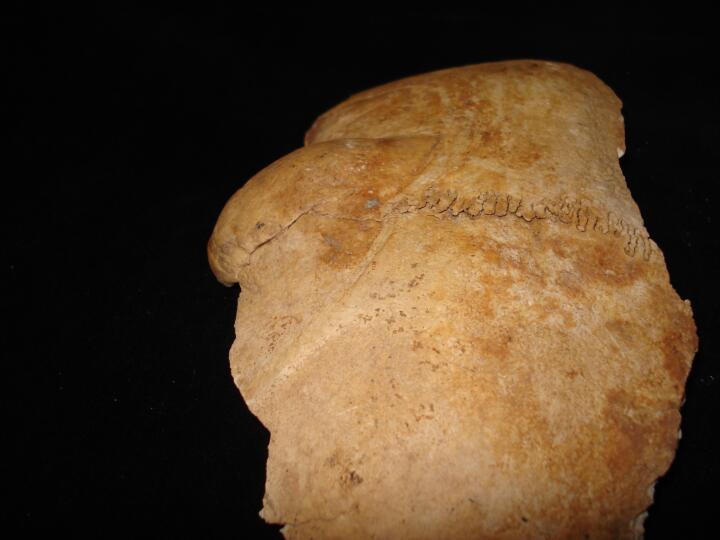
544
|
5
|
ONE94_544_5.jpg
|
Skull, right side frontal & parietal healed blunt force trauma across coronal suture large smooth lobule of bone (superior view/endocranial surface)
|
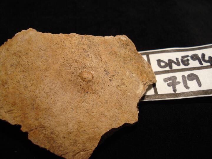
| ONE94
|
719
|
1
|
ONE94_719_1.jpg
|
Skull, right parietal (ectocranial surface) button osteoma
|
| ONE94
|
672
|
1
|
ONE94_672_1.jpg
|
Skull, right parietal (ectocranial surface) porotic hyperostosis
|
| ONE94
|
672
|
2
|
ONE94_672_2.jpg
|
Skull, right parietal (ectocranial surface/medial view) porotic hyperostosis
|
| ONE94
|
672
|
3
|
ONE94_672_3.jpg
|
Skull, left parietal fragment (ectocranial surface) porotic hyperostosis
|
| ONE94
|
672
|
4
|
ONE94_672_4.jpg
|
Skull, right parietal (ectocranial surface/close up) porotic hyperostosis
|
| ONE94
|
712
|
1
|
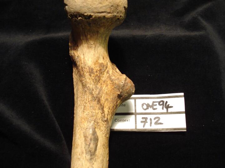
ONE94_712_1.jpg
|
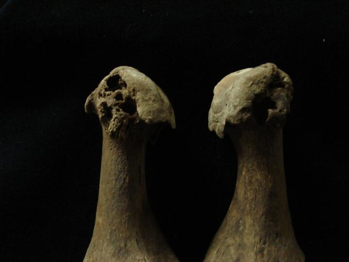
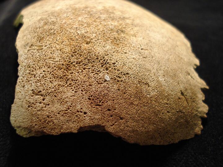
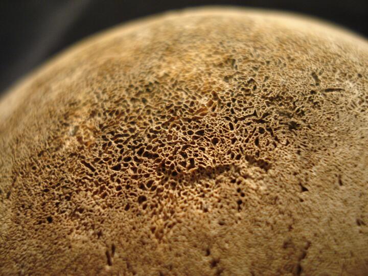
Right femur (anterior view) ?ossified haematoma
|
| ONE94
|
712
|
2
|
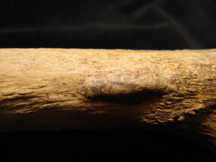
ONE94_712_2.jpg
|
Right femur (medial view) ?ossified haematoma
|
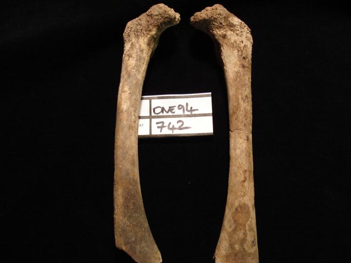
| ONE94
|
742
|
1
|
ONE94_742_1.jpg
|
Femora (anterior view) slight bowing of right femur, ?indication of healed rickets
|
| ONE94
|
96
|
1
|
ONE94_96_1.jpg
|
Right tibia (posterior view) pronounced enthesopathy fro Soleus
|
| ONE94
|
96
|
2
|
ONE94_96_2.jpg
|
Right tibia (posterior view) pronounced enthesopathy fro Soleus
|
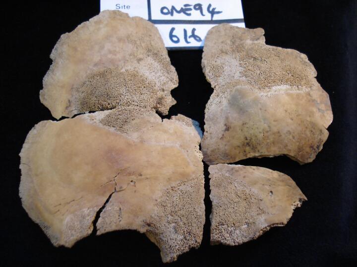
| ONE94
|
616
|
1
|
ONE94_616_1.jpg
|
Skull, frontal bone porotic hyperostosis, indication of active rickets (superior view)
|
| ONE94
|
616
|
2
|
ONE94_616_2.jpg
|
Skull, frontal bone - glabella & orbits, porotic hyperostosis, indication of active rickets (anterior view/close up)
|
| ONE94
|
616
|
3
|
ONE94_616_3.jpg
|
Skull, right side of frontal & parietal (superior view) porotic hyperostosis, indication of active rickets
|
| ONE94
|
616
|
4
|
ONE94_616_4.jpg
|
Left ulna & radius flaring of metaphyseal ends (anterior view) indication of active rickets
|
| ONE94
|
616
|
5
|
ONE94_616_5.jpg
|
Left & right radius (anterior view) flaring of meatphyseal ends, indication of active rickets
|
| ONE94
|
616
|
6
|
ONE94_616_6.jpg
|
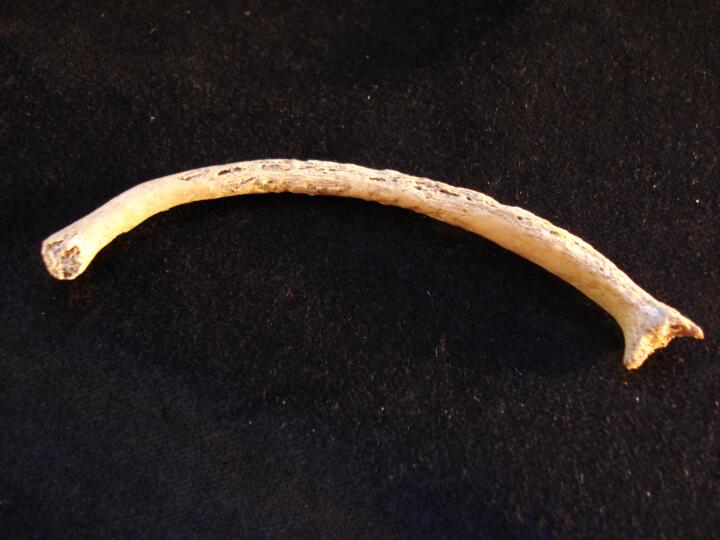
Ribs, shaft fragments with flaring of the sternal ends (visceral surface) indication of active rickets
|
| ONE94
|
616
|
7
|
ONE94_616_7.jpg
|
One left rib (superior view) flaring of sternal end, indication of active rickets
|
| ONE94
|
562
|
1
|
ONE94_562_1.jpg
|
Mandible (lingual view) fusion line of two halve of mandible still visible
|
| ONE94
|
553
|
1
|
ONE94_553_1.jpg
|
Right sphenoid cerebral surface of greater wing new fine bone
|
| ONE94
|
553
|
2
|
ONE94_553_2.jpg
|
Left & right sphenoid cerebral surface of greater wings, comparison of surfaces
|
| ONE94
|
492
|
1
|
ONE94_492_1.jpg
|
Right femur (medial view) mid shaft, active non-specific periosteal infection
|
| ONE94
|
492
|
2
|
ONE94_492_2.jpg
|
Right tibia (lateral view) proximal 1/3, active non-specific infection
|
| ONE94
|
492
|
3
|
ONE94_492_3.jpg
|
Left tibia (lateral view) proximal to mid shaft, active non-specific periosateal infection
|
| ONE94
|
13
|
1
|
ONE94_13_1.jpg
|
Skull, frontal bone (ectocranial surface) two butoon osteomas
|
| ONE94
|
19
|
1
|
ONE94_19_1.jpg
|
Left pubic bone (dorsal view) healed fracture
|
| ONE94
|
19
|
2
|
ONE94_19_2.jpg
|
Left pubic bone (dorsal view/close up) healed fracture
|
| ONE94
|
19
|
3
|
ONE94_19_3.jpg
|
Left pubic bone (ventral view) healed fracture
|
| ONE94
|
54
|
1
|
ONE94_54_1.jpg
|
Six lumbar vertebrae (right side view)
|
| ONE94
|
54
|
2
|
ONE94_54_2.jpg
|
Lumbar vertebra L6 (inferior surface) facet on right side for partial sacralisation
|
| ONE94
|
90
|
1
|
ONE94_90_1.jpg
|
Skull, parietal fragments (endocranial surface/close up) deep meningeal vessels with smooth undulated surface
|
| ONE94
|
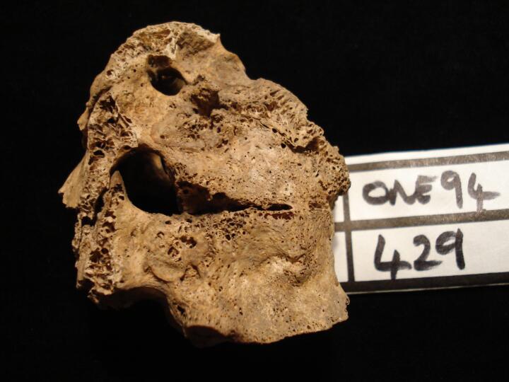
429
|
1
|
ONE94_429_1.jpg
|
Thoracic vertebrae (Th5 -Th6) collapse from probable tuberculosis (right side view)
|
| ONE94
|
429
|
2
|
ONE94_429_2.jpg
|
Thoracic vertebrae (Th5 -Th6) collapse from probable tuberculosis (left side view)
|
| ONE94
|
429
|
3
|
ONE94_429_3.jpg
|
Thoracic vertebrae (Th5 -Th6) collapse from probable tuberculosis (right side view)
|
| ONE94
|
429
|
4
|
ONE94_429_4.jpg
|
Thoracic vertebrae (Th5 -Th6) collapse from probable tuberculosis (left side view)
|
| ONE94
|
429
|
5
|
ONE94_429_5.jpg
|
Thoracic vertebrae (Th5 -Th6) collapse from probable tuberculosis (anterior view)
|
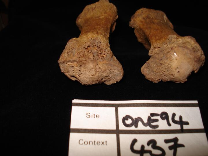
| ONE94
|
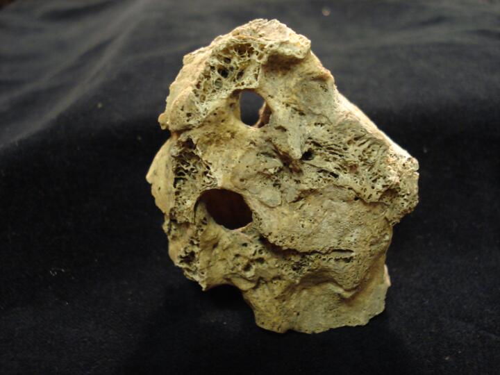
437
|
1
|
ONE94_437_1.jpg
|
Left & right 1st metatarsals heads (dorsal view) osteoarthritis
|
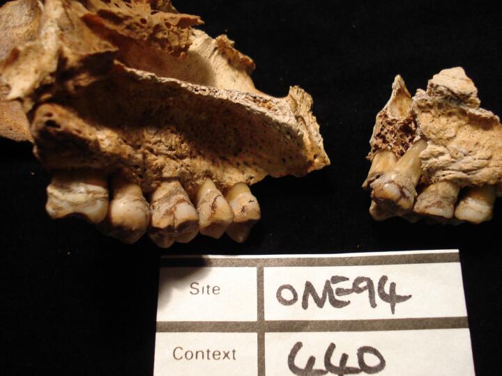
| ONE94
|
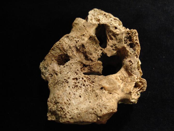
437
|
2
|
ONE94_437_2.jpg
|
Left & right 1st metatarsals heads (plantar view) osteoarthritis
|
| ONE94
|
440
|
1
|
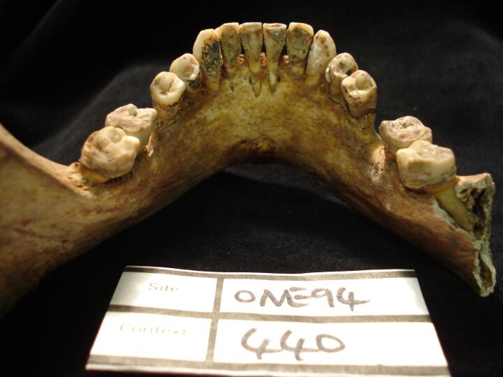
ONE94_440_1.jpg
|
Mandible (lingual view) black substance adhering to dentition, ? Tar deposit from smoking
|
| ONE94
|
440
|
2
|
ONE94_440_2.jpg
|
Mandible (lingual view/close up) black substance adhering to dentition, ? Tar deposit from smoking
|
| ONE94
|
440
|
3
|
ONE94_440_3.jpg
|
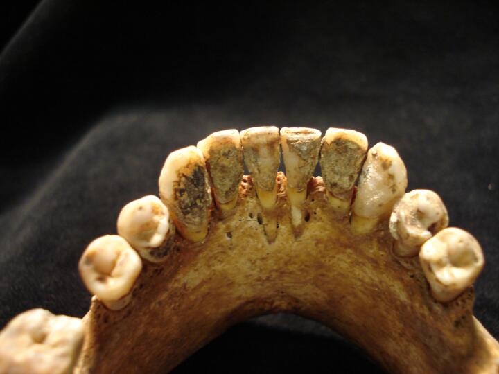
Mandible anterior teeth (buccal view surface) calculus deposits
|
| ONE94
|
440
|
4
|
ONE94_440_4.jpg
|
Maxilla (lingual view) black substance adhering to dentition, ? Tar deposit from smoking
|
| ONE94
|
601
|
1
|
ONE94_601_1.jpg
|
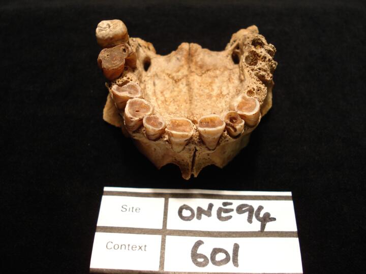
Mandible (occlusal view) heavy wear of dentition & ante mortem tooth loss
|
| ONE94
|
601
|
2
|
ONE94_601_2.jpg
|
Mandible (buccal view/close up) heavy wear of dentition
|
| ONE94
|
601
|
3
|
ONE94_601_3.jpg
|
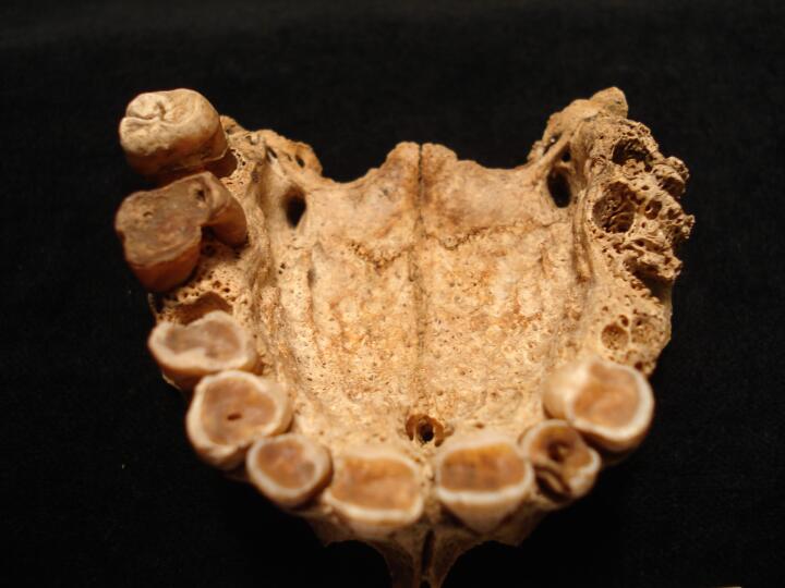
Maxilla (palatal view) heavy wear of dentition
|
| ONE94
|
601
|
4
|
ONE94_601_4.jpg
|
Maxilla (palatal view/close up) heavy wear of dentition
|
| ONE94
|
601
|
5
|
ONE94_601_5.jpg
|
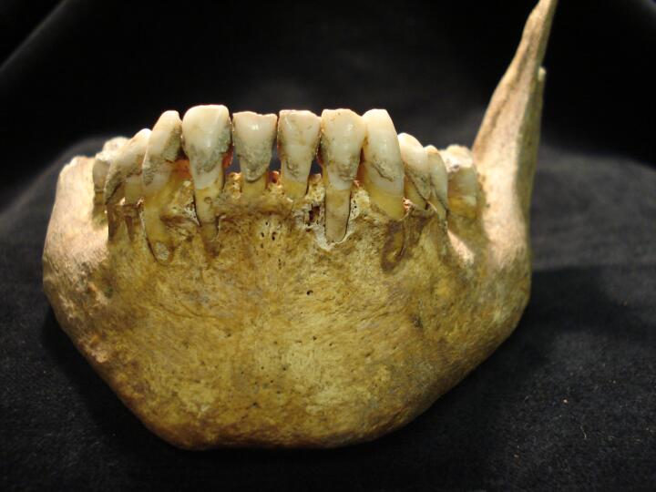
Mandible (lingual view) heavy wear of dentition & ante mortem tooth loss
|
| ONE94
|
601
|
6
|
ONE94_601_6.jpg
|
Maxilla (left side) external draining peripaical lesion
|
| ONE94
|
691
|
1
|
ONE94_691_1.jpg
|
Skull, frontal bone (endocranial surface) fine plaque like layer of bone
|
| ONE94
|
691
|
2
|
ONE94_691_2.jpg
|
Skull, occipital bone (endocranial surface) fine plaque like layer of bone
|
| ONE94
|
691
|
3
|
ONE94_691_3.jpg
|
Skull, parietal fragments (endocranial surface) fine plaque like layer of bone
|
| ONE94
|
691
|
4
|
ONE94_691_4.jpg
|
Sphenoid lesser wings plaque like layer of bone
|
| ONE94
|
894
|
1
|
ONE94_894_1.jpg
|
Thoracic vertebrae Th1 & Th2, deflection of spinous processes, ?congenital
|
| ONE94
|
894
|
2
|
ONE94_894_2.jpg
|
Mid thoracic vertebrae, bony 'bridging' extensions of spinus processes
|
| ONE94
|
894
|
3
|
ONE94_894_3.jpg
|
Mid thoracic vertebrae, vertebral arches only (inferior view) smooth scooped appearance of arch
|
| ONE94
|
894
|
4
|
ONE94_894_4.jpg
|
Mid thoracic vertebra, vertebral arch only (inferior view) smooth scooped appearance of arch
|
| ONE94
|
1693
|
1
|
ONE94_1693_1.jpg
|
Left foot proximal phalange of 1st metatarsal (dorsal view) bony exostoses, porosity, ?inflammation
|
| ONE94
|
1693
|
2
|
ONE94_1693_2.jpg
|
Left foot proximal phalange of 1st metatarsal (dorsal view) bony exostoses, porosity, ?inflammation
|
| ONE94
|
1693
|
3
|
ONE94_1693_3.jpg
|
Left foot proximal phalange of 1st metatarsal (plantar view)smooth nodular bony exostoses
|
| ONE94
|
96
|
1
|
ONE94_96_1.jpg
|
Left tibia (anterior view) mid shaft, healed fracture
|
| ONE94
|
96
|
2
|
ONE94_96_2.jpg
|
Left tibia (posterior view) mid shaft, healed fracture
|
| ONE94
|
96
|
3
|
ONE94_96_3.jpg
|
Left tibia (anterior view/close up) mid shaft, healed fracture
|
| ONE94
|
96
|
4
|
ONE94_96_4.jpg
|
Left tibia (posterior view/close up) mid shaft, healed fracture
|
| ONE94
|
526
|
1
|
ONE94_526_1.jpg
|
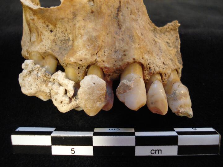
Maxilla (right side/buccal view) heavy nondular calculus
|
| ONE94
|
526
|
2
|
ONE94_526_2.jpg
|
Maxilla (right side/buccal view) close up of heavy nondular calculus & hypolplastic defects of anterior teeth
|
| ONE94
|
526
|
3
|
ONE94_526_3.jpg
|
Maxilla left side (buccal view) 'peg' like lateral incisor
|
| ONE94
|
526
|
4
|
ONE94_526_4.jpg
|
Maxilla left side (buccal view/close up) 'peg' like lateral incisor
|
| ONE94
|
526
|
5
|
ONE94_526_5.jpg
|
Maxilla (anterior/buccal view) comparison of the lateral incisors
|
| ONE94
|
526
|
6
|
ONE94_526_6.jpg
|
External surface of possible Gallstone
|
| ONE94
|
526
|
7
|
ONE94_526_7.jpg
|
Internal surface of possible Gallstone
|
| ONE94
|
526
|
8
|
ONE94_526_8.jpg
|
Internal (close up) surface of possible Gallstone
|
| ONE94
|
792
|
1
|
ONE94_792_1.jpg
|
Maxilla (buccal view) conjoined central & lateral incisors
|
| ONE94
|
792
|
2
|
ONE94_792_2.jpg
|
Maxilla (buccal view/close up) conjoined central & lateral incisors
|
| ONE94
|
792
|
3
|
ONE94_792_3.jpg
|
Maxilla (lingual view) conjoined central & lateral incisors
|
| ONE94
|
792
|
4
|
ONE94_792_4.jpg
|
Maxilla (buccal view) close up of conjoined anterior teeth
|


